

Synapse stability in the precuneus early in the progression of Alzheimer's disease
- PMID: 23478309
- PMCID: PMC4000262
- DOI: 10.3233/JAD-122353
Synapse stability in the precuneus early in the progression of Alzheimer's disease
Abstract
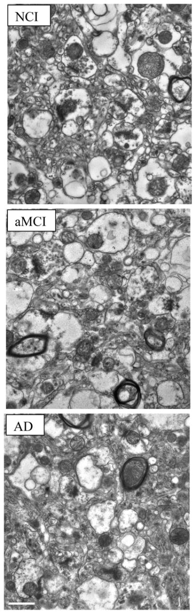
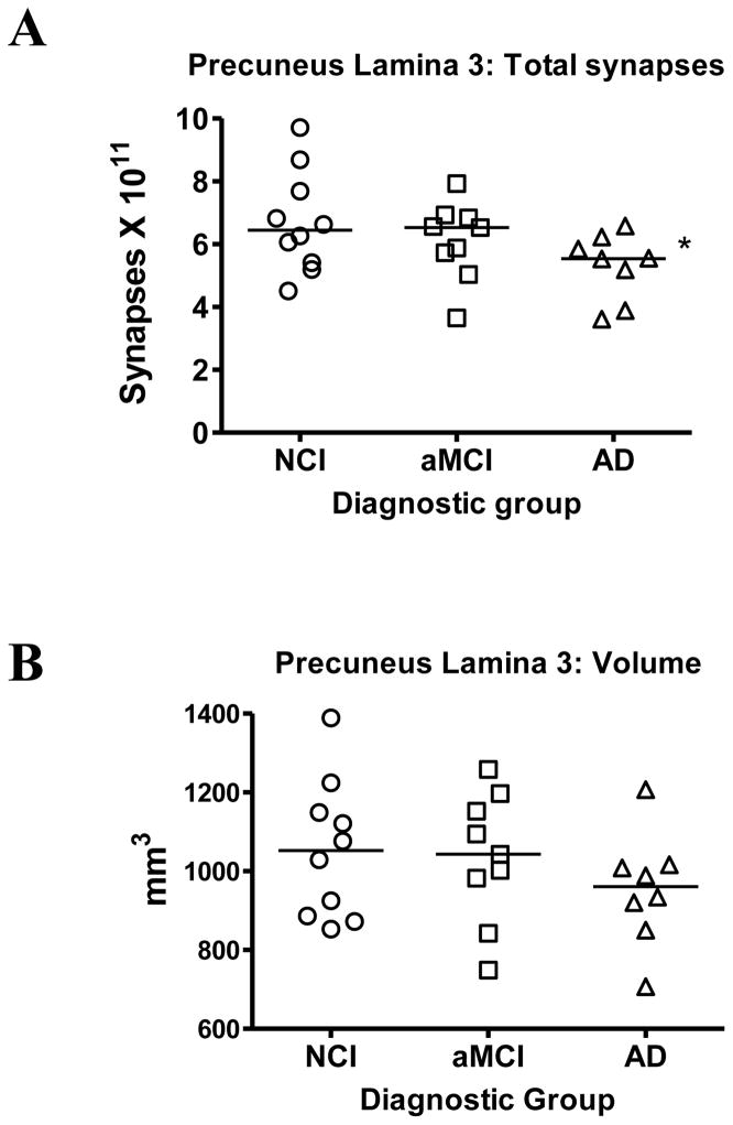
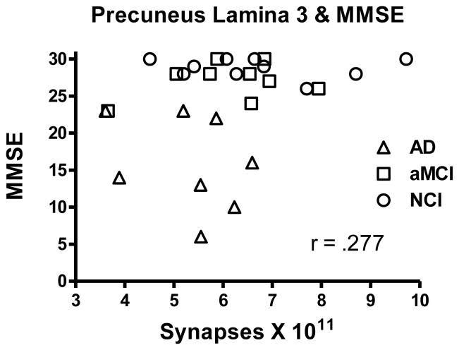
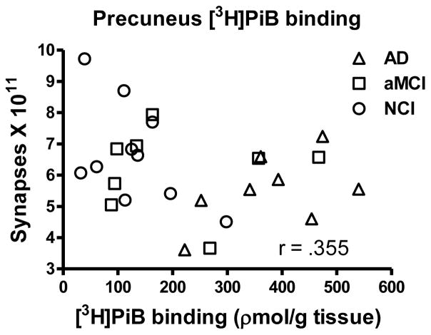
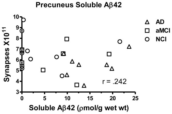
Amnestic mild cognitive impairment (aMCI) is considered to be one of the early stages in the progression from no cognitive impairment (NCI) to Alzheimer's disease (AD). Individuals with aMCI have increased levels of AD-type neuropathology in multiple regions of the neocortex and hippocampus and demonstrate a loss of synaptic connectivity. Recent neuroimaging studies have reported increased levels of 11C-PiB (Pittsburgh, compound B) in regions of the neocortex including the precuneus region of the medial parietal lobe. This cortical region has been implicated in episodic memory, which is disrupted early in the progression of AD. In this study, unbiased stereology coupled with electron microscopy was used to quantify total synaptic numbers in lamina 3 of the precuneus from short postmortem autopsy tissue harvested from subjects who died at different cognitive stages during the progression of AD. Individuals with aMCI did not reveal a statistically significant decline in total synapses compared to the NCI cohort while the AD group did show a modest but significant decline. Synaptic numbers failed to correlate with several different cognitive tasks including the Mini-Mental State Examination scores and episodic memory scores. Although levels of [3H]PiB binding were elevated in both the aMCI and AD groups, it did not strongly correlate with synaptic counts. These results support the idea that despite increased amyloid load, the precuneus region does not show early changes in synaptic decline during the progression of AD.
Figures
References
-
- Consensus recommendations for the postmortem diagnosis of Alzheimer’s disease. The National Institute on Aging, and reagan Institute Working Group on Diagnostic Criteria for the Neuropathological Assessment of Alzheimer’s Disease. Neurobiol Aging. 1997;18:S1–S2. - PubMed
-
- Montine TJ, Phelps CH, Beach TG, Bigio EH, Cairns NJ, Dickson DW, Duyckaerts C, Frosch MP, Masliah E, Mirra SS, Nelson PT, Schneider JA, Thal DR, Trojanowski JQ, Vinters HV, Hyman BT. National Institute on Aging-Alzheimer’s Association guidelines for the neuropathologic assessment of Alzheimer’s disease: a practical approach. Acta Neuropathol. 2012;123:1–11. - PMC - PubMed
-
- Scheff SW, Price DA. Alzheimer’s disease-related alterations in synaptic density: neocortex and hippocampus. J Alzheimers Dis. 2006;9:101–115. - PubMed
-
- Terry RD, Masliah E, Salmon DP, Butters N, DeTeresa R, Hill R, Hansen LA, Katzman R. Physical basis of cognitive alterations in Alzheimer’s disease: synapse loss is the major correlate of cognitive impairment. Ann Neurol. 1991;30:572–580. - PubMed
-
- Scheff SW, Price DA, Schmitt FA, DeKosky ST, Mufson EJ. Synaptic alterations in CA1 in mild Alzheimer disease and mild cognitive impairment. Neurology. 2007;68:1501–1508. - PubMed
Publication types
MeSH terms
Grants and funding
LinkOut - more resources
Full Text Sources
Other Literature Sources
Medical
