

The gastrointestinal mucus system in health and disease
- PMID: 23478383
- PMCID: PMC3758667
- DOI: 10.1038/nrgastro.2013.35
The gastrointestinal mucus system in health and disease
Abstract
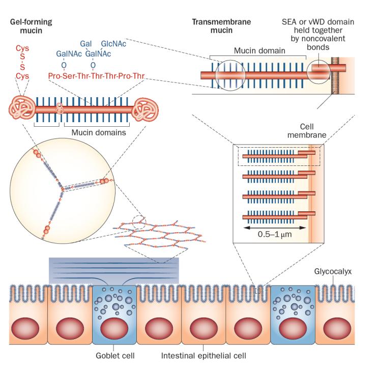
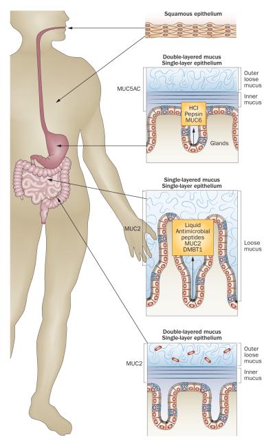
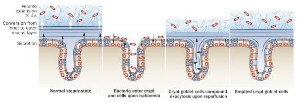
Mucins--large, highly glycosylated proteins--are important for the luminal protection of the gastrointestinal tract. Enterocytes have their apical surface covered by transmembrane mucins and goblet cells produce the secreted gel-forming mucins that form mucus. The small intestine has a single unattached mucus layer, which in cystic fibrosis becomes attached, accounting for the intestinal manifestations of this disease. The stomach and colon have two layers of mucus; the inner layer is attached and the outer layer is less dense and unattached. In the colon, the outer mucus layer is the habitat for commensal bacteria. The inner mucus layer is impervious to bacteria and is renewed every hour by surface goblet cells. The crypt goblet cells have the ability to restitute the mucus layer by secretion, for example after an ischaemic challenge. Proteases of certain parasites and some bacteria can cleave mucins and dissolve the mucus as part of their pathogenicity. The inner mucus layer can, however, also become penetrable to bacteria by several other mechanisms, including aberrations in the immune system. When bacteria reach the epithelial surface, the immune system is activated and inflammation is triggered. This mechanism might occur in some types of ulcerative colitis.
Figures

Similar articles
-
The mucus and mucins of the goblet cells and enterocytes provide the first defense line of the gastrointestinal tract and interact with the immune system.Immunol Rev. 2014 Jul;260(1):8-20. doi: 10.1111/imr.12182. Immunol Rev. 2014. PMID: 24942678 Free PMC article. Review.
-
Mucus and the goblet cell.Dig Dis. 2013;31(3-4):305-9. doi: 10.1159/000354683. Epub 2013 Nov 14. Dig Dis. 2013. PMID: 24246979 Free PMC article. Review.
-
Studies of mucus in mouse stomach, small intestine, and colon. I. Gastrointestinal mucus layers have different properties depending on location as well as over the Peyer's patches.Am J Physiol Gastrointest Liver Physiol. 2013 Sep 1;305(5):G341-7. doi: 10.1152/ajpgi.00046.2013. Epub 2013 Jul 5. Am J Physiol Gastrointest Liver Physiol. 2013. PMID: 23832518 Free PMC article.
-
Mucus and mucins in diseases of the intestinal and respiratory tracts.J Intern Med. 2019 May;285(5):479-490. doi: 10.1111/joim.12910. Epub 2019 Apr 22. J Intern Med. 2019. PMID: 30963635 Free PMC article. Review.
-
An intercrypt subpopulation of goblet cells is essential for colonic mucus barrier function.Science. 2021 Apr 16;372(6539):eabb1590. doi: 10.1126/science.abb1590. Science. 2021. PMID: 33859001 Free PMC article.
Cited by
-
Adaptive response activated by dietary cis9, trans11 conjugated linoleic acid prevents distinct signs of gliadin-induced enteropathy in mice.Eur J Nutr. 2016 Mar;55(2):729-740. doi: 10.1007/s00394-015-0893-2. Epub 2015 Apr 4. Eur J Nutr. 2016. PMID: 25840667
-
MUC4 is negatively regulated through the Wnt/β-catenin pathway via the Notch effector Hath1 in colorectal cancer.Genes Cancer. 2016 May;7(5-6):154-168. doi: 10.18632/genesandcancer.108. Genes Cancer. 2016. PMID: 27551331 Free PMC article.
-
IL-22 Restrains Tapeworm-Mediated Protection against Experimental Colitis via Regulation of IL-25 Expression.PLoS Pathog. 2016 Apr 7;12(4):e1005481. doi: 10.1371/journal.ppat.1005481. eCollection 2016 Apr. PLoS Pathog. 2016. PMID: 27055194 Free PMC article.
-
Activated sympathetic nerve post stroke downregulates Toll-like receptor 5 and disrupts the gut mucosal barrier.Cell Rep Med. 2024 Oct 15;5(10):101754. doi: 10.1016/j.xcrm.2024.101754. Epub 2024 Oct 8. Cell Rep Med. 2024. PMID: 39383869 Free PMC article.
-
The immunology of the vermiform appendix: a review of the literature.Clin Exp Immunol. 2016 Oct;186(1):1-9. doi: 10.1111/cei.12821. Epub 2016 Jul 19. Clin Exp Immunol. 2016. PMID: 27271818 Free PMC article. Review.
References
-
- Neutra MR, O’Malley LJ, Specian RD. Regulation of intestinal goblet cell secretion. II. A survey of potentid secretugogues. Am. J. Physiol. Gastroint. Liver Physiol. 1982;242:G380–G387. - PubMed
-
- Ito S. Structure and function of the glycocalyx. Fed. Proc. 1969;28:12–25. - PubMed
-
- Soergel KH, Ingelfinger FJ. Composition of rectal mucus in normal subjects and patients with ulcerative colitis. Gastroenterology. 1964;47:610–616. - PubMed
Publication types
MeSH terms
Grants and funding
LinkOut - more resources
Full Text Sources
Other Literature Sources
Medical
