. 2013 Apr 11;50(1):56-66.
doi: 10.1016/j.molcel.2013.02.001. Epub 2013 Mar 7.

Splicing-factor oncoprotein SRSF1 stabilizes p53 via RPL5 and induces cellular senescence

Affiliations

Splicing-factor oncoprotein SRSF1 stabilizes p53 via RPL5 and induces cellular senescence

Oliver I Fregoso et al. Mol Cell. .

Abstract

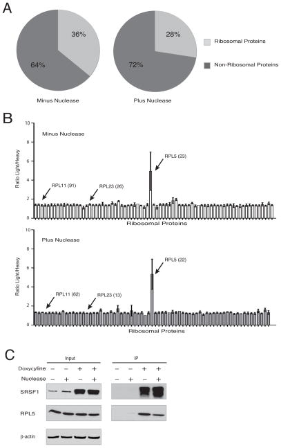
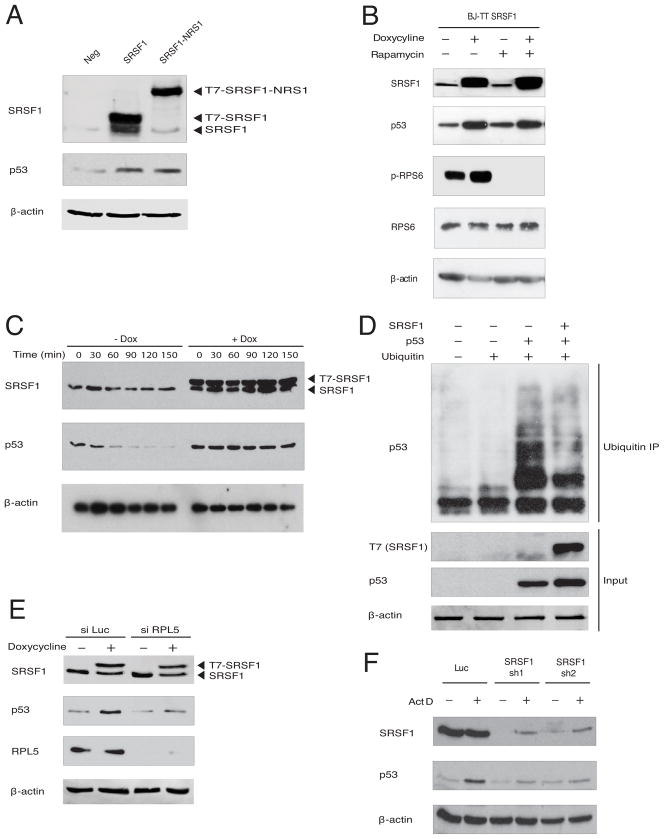
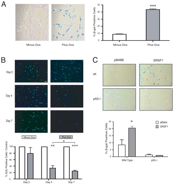
Splicing and translation are highly regulated steps of gene expression. Altered expression of proteins involved in these processes can be deleterious. Therefore, the cell has many safeguards against such misregulation. We report that the oncogenic splicing factor SRSF1, which is overexpressed in many cancers, stabilizes the tumor suppressor protein p53 by abrogating its MDM2-dependent proteasomal degradation. We show that SRSF1 is a necessary component of an MDM2/ribosomal protein complex, separate from the ribosome, that functions in a p53-dependent ribosomal-stress checkpoint pathway. Consistent with the stabilization of p53, increased SRSF1 expression in primary human fibroblasts decreases cellular proliferation and ultimately triggers oncogene-induced senescence (OIS). These findings underscore the deleterious outcome of SRSF1 overexpression and identify a cellular defense mechanism against its aberrant function. Furthermore, they implicate the RPL5-MDM2 complex in OIS and demonstrate a link between spliceosomal and ribosomal components, functioning independently of their canonical roles, to monitor cellular physiology and cell-cycle progression.


Figures

Figure 1
Figure 1. SRSF1 Interacts Specifically with RPL5
(A) Percentage of detected SRSF1 interactions that are ribosomal (light gray). T7-SRSF1 was immunoprecipitated from lysates of doxycycline-induced HeLa cells (HeLa TT7-SRSF1) with or without nuclease treatment, and interacting proteins were identified by MudPIT MS. (B) I-DIRT ratios of light/heavy peptides are shown for all RPs co-immunoprecipitated with T7-SRSF1, with or without nuclease treatment. Bars show average value (+/− s.d.) for each protein, based on individual peptides identified, and total number of peptides for the indicated proteins are shown in parentheses. (C) HeLa TT7-SRSF1 cells were left untreated or induced with doxycycline for 36 h. Lysates were immunoprecipitated with T7 monoclonal antibody, with or without nuclease treatment. Whole-cell lysates (Input) and IPs were analyzed by immunoblotting with the indicated antibodies. See also Figure S1 and Table S1.
Figure 2
Figure 2. SRSF1 is a Component of an RP-MDM2 Complex and Induces p53 Protein Expression
(A) BJ fibroblasts were either left untreated or treated with 5 nM actinomycin D for 8 h, followed by 50 μM of proteasomal inhibitor MG132 for 8 h. Lysates were immunoprecipitated with either control IgG or AK96 monoclonal antibody against SRSF1. Whole-cell lysates (Input) and IPs were analyzed with the indicated antibodies. (B) BJ TT7-SRSF1 fibroblasts or empty-vector control (BJ TT7) were treated with doxycycline for 36 h and 5 nM actinomycin D for 8 h and analyzed by immunoblotting, as indicated. Values represent fold change in protein levels, relative to a loading control. Representative western blots are shown. Data are means +/− s.d. (n=3), *P<0.05, **P<0.01, *** P<0.001. (C) Total RNA from BJ TT7-SRSF1 cells was amplified by radioactive RT-PCR and analyzed by native PAGE. Values represent fold change in mRNA levels relative to β-actin. Data are means +/− s.d. (n=3), *P<0.05, **P<0.01, *** P<0.001. See also Figure S2.
Figure 3
Figure 3. SRSF1 Blocks the Ubiquitylation of p53 and is a Necessary Component of the RPL5-MDM2 Complex
(A) BJ cells were transduced with empty vector, T7-SRSF1, or T7-NRS-SRSF1. Post puromycin selection, whole-cell lysates were analyzed by immunoblotting with the indicated antibodies. (B) BJ TT7-SRSF1 cells were treated with or without doxycycline for 36 h, followed by rapamycin (200 nM) for 8 h. Lysates were analyzed by immunoblotting with the indicated antibodies. (C) BJ TT7-SRSF1 cells were treated with or without doxycycline for 36 h, followed by cycloheximide (10 μg/mL) for the indicated times. Lysates were analyzed by immunoblotting. (D) H1299 cells lacking endogenous p53 were transfected with His-Ub, Flag-p53, and/or T7-SRSF1 plasmids, or the corresponding empty vectors. Cells were lysed under denaturing conditions and incubated with nickel-agarose beads for 3 h. Input and nickel-bound proteins were analyzed by immunoblotting with the indicated antibodies. (E) BJ TT7-SRSF1 cells were transfected with luciferase or a pool of RPL5 siRNA for 36 h, followed by doxycycline induction for 36 h, as indicated, and analyzed by immunoblotting. (F) U2OS cells were transduced with luciferase or SRSF1 shRNA, selected with puromycin, followed by actinomycin D (5 nM) treatment for 8 h, as indicated, and analyzed by immunoblotting. See also Figure S3.
Figure 4
Figure 4. Overexpression of SRSF1 Leads to Senescence of Primary Fibroblasts through p53 Induction
(A) BJ TT7-SRSF1 cells were induced with doxycycline for 7 d, fixed, stained with X-gal, and observed at 20× magnification (left panels). 200 cells were counted for each condition (right panel); n=6. Means +/− s.d. are shown; ***P = 0.0003. (B) BJ TT7-SRSF1 cells were induced with doxycycline for 2, 4, or 7 d, incubated with 10 μM EdU, and observed at 20× magnification (top panels). 100 cells were counted for each condition (lower panel); n=6. Means +/− s.d. are shown; *P = 0.05, **P = 0.005, ***P = 0.0008. (C) Wild-type or p53-null MEFs transduced with control or T7-SRSF1-expressing retroviruses were fixed and stained with X-gal (top panels). 200 cells were counted for each condition (lower panels); n=3. Means +/− s.d. are shown; *P = 0.05. See also Figure S4.
Figure 5
Figure 5. RRM1 of SRSF1 is Required for Interaction with the RPL-MDM2 Complex, p53 Induction, and OIS
(A) HeLa cells were transfected with wild-type SRSF1 and domain-deletion mutants. Lysates were immunoprecipitated with T7 monoclonal antibody, with nuclease treatment. Whole-cell lysates (Input) and IPs were analyzed by immunoblotting with the indicated antibodies. (B) BJ TT7-SRSF1, BJ TT7-SRSF1-ΔRRM1, BJ TT7-SRSF1-ΔRRM2 and BJ TT7-SRSF1-ΔRS cells were treated with doxycycline for 36 h and analyzed by immunoblotting, as indicated. (C) BJ TT7-SRSF1, BJ TT7-SRSF1-ΔRRM1, BJ TT7-SRSF1-ΔRRM2 and BJ TT7-SRSF1-ΔRS cells were induced with doxycycline for 2, 4, or 7 d, and incubated with 10 μM EdU. One hundred cells were counted for each condition; n=3. Means +/− s.d. are shown, *P = 0.05, **P = 0.005. (D) BJ TT7-SRSF1, BJ TT7-SRSF1-ΔRRM1, BJ TT7-SRSF1-ΔRRM2 and BJ TT7-SRSF1-ΔRS cells were treated with doxycycline for 7 d, fixed, and stained with X-gal. 200 cells were counted for each condition; n=2. Ranges are shown. See also Figure S5.
Figure 6
Figure 6. A model for SRSF1’s Role in the Ribosomal-stress Pathway and Oncogene-Induced Senescence
We have identified SRSF1 as a critical component of the RP-MDM2 complex, which is formed in response to induction of ribosomal stress. Sequestration of the E3 ligase MDM2 in this complex results in decreased ubiquitylation and increased stability of the tumor-supressor p53 protein. Moreover, we have identified and characterized an anti-tumorigenic response that primary cells mount in response to overexpression of the SRSF1 oncoprotein, which triggers the formation of a nuclear ternary SRSF1-RPL5-MDM2 complex, leading to activation of the p53-mediated tumor-suppressive pathway and OIS.

Comment in

Similar articles

Cited by

References

    1. Alimonti A, Nardella C, Chen Z, Clohessy JG, Carracedo A, Trotman LC, Cheng K, Varmeh S, Kozma SC, Thomas G, et al. A novel type of cellular senescence that can be enhanced in mouse models and human tumor xenografts to suppress prostate tumorigenesis. J Clin Invest. 2010;120:681–93. - PMC - PubMed
    1. Allemand E, Guil S, Myers M, Moscat J, Cáceres JF, Krainer AR. Regulation of heterogenous nuclear ribonucleoprotein A1 transport by phosphorylation in cells stressed by osmotic shock. Proc Natl Acad Sci USA. 2005;102:3605–3610. - PMC - PubMed
    1. Anczuków O, Rosenberg AZ, Akerman M, Das S, Zhan L, Karni R, Muthuswamy S, Krainer AR. The splicing-factor SRSF1 regulates apoptosis and proliferation to promote mammary epithelial cell transformation. Nat Struct Mol Biol. 2012;19:220–228. - PMC - PubMed
    1. Astle MV, Hannan KM, Ng PY, Lee RS, George AJ, Hsu AK, Haupt Y, Hannan RD, Pearson RB. AKT induces senescence in human cells via mTORC1 and p53 in the absence of DNA damage: implications for targeting mTOR during malignancy. Oncogene. 2012;31:1949–1962. - PMC - PubMed
    1. Bartkova J, Rezaei N, Liontos M, Karakaidos P, Kletsas D, Issaeva N, Vassiliou LV, Kolettas E, Niforou K, Zoumpourlis VC, et al. Oncogene-induced senescence is part of the tumorigenesis barrier imposed by DNA damage checkpoints. Nature. 2006;444:633–637. - PubMed

Publication types

MeSH terms