. 2013 Mar 28;49(6):1147-58.
doi: 10.1016/j.molcel.2013.02.003. Epub 2013 Mar 7.

CRL1-FBXO11 promotes Cdt2 ubiquitylation and degradation and regulates Pr-Set7/Set8-mediated cellular migration

Affiliations

CRL1-FBXO11 promotes Cdt2 ubiquitylation and degradation and regulates Pr-Set7/Set8-mediated cellular migration

Tarek Abbas et al. Mol Cell. .

Abstract

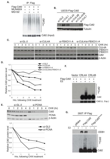
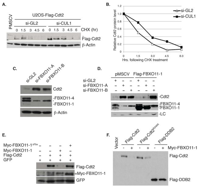
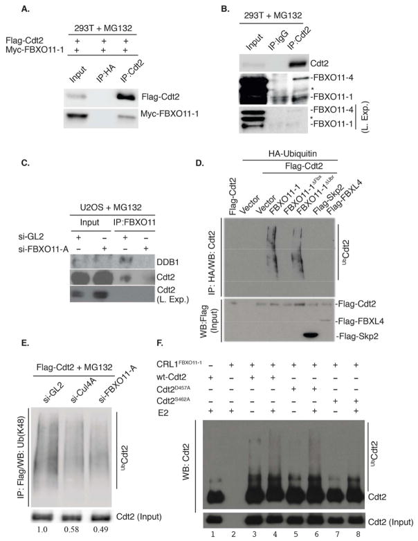
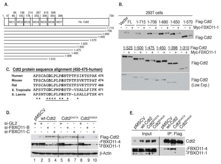
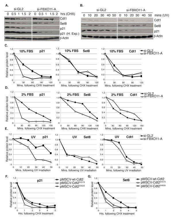
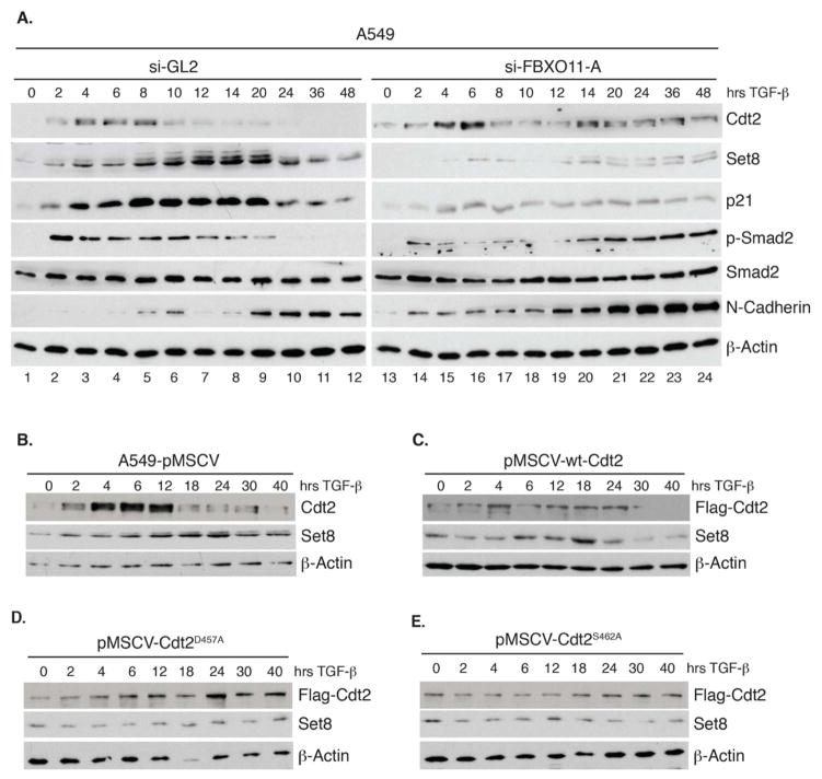
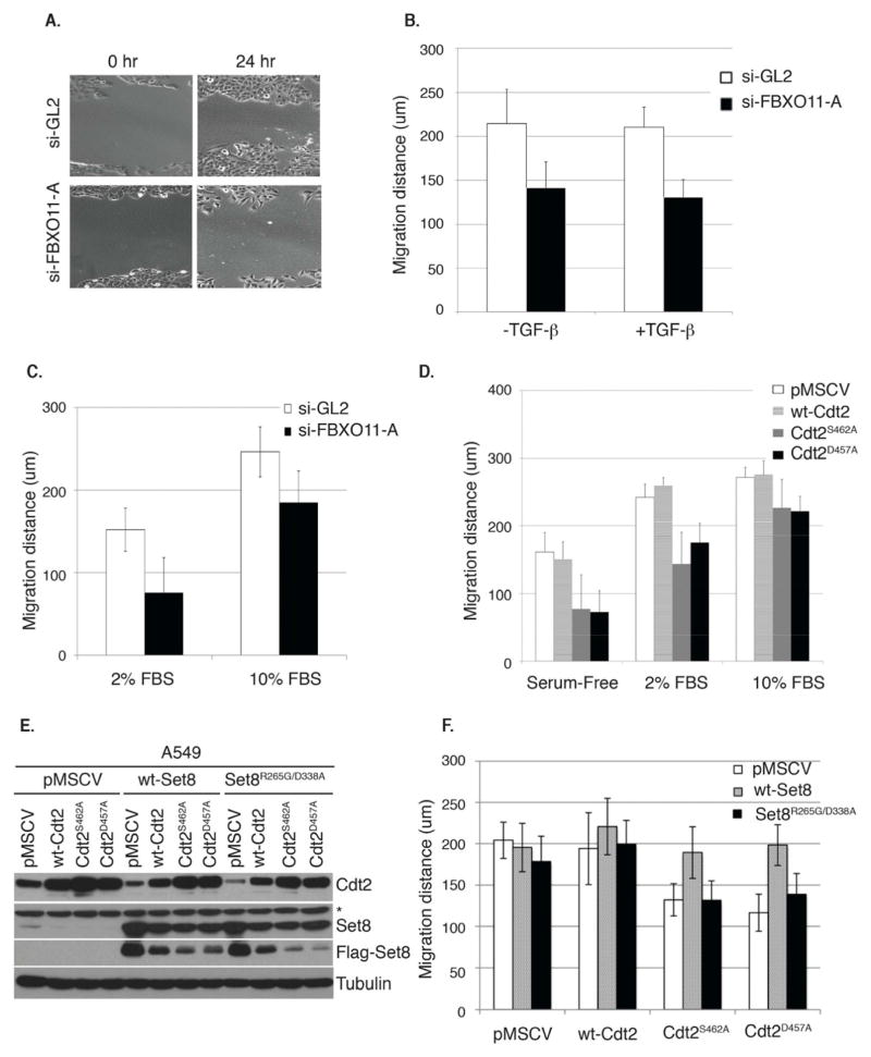
The Cul4-Cdt2 (CRL4(Cdt2)) E3 ubiquitin ligase is a master regulator of cell-cycle progression and genome stability. Despite its central role in the degradation of many cell-cycle regulators, e.g., Cdt1, p21, and Pr-Set7/Set8, little is known about the regulation of its activity. We report that Cdt2 is autoubiquitylated by the CRL4A E3 ubiquitin ligase. Cdt2 is additionally polyubiquitylated and degraded by Cul1-FBXO11 (CRL1(FBXO11)). CRL1(FBXO11)-mediated degradation of Cdt2 stabilizes p21 and Set8, and this is important during the response to TGF-β, with the Set8 induction being important for turning off the activation of Smad2. The migration of epithelial cells is also stimulated by CRL1(FBXO11)-mediated downregulation of Cdt2 and the consequent stabilization of Set8. This is an interesting example of cross-regulation between specific Cullin 4 and Cullin 1 E3 ubiquitin ligases and highlights the role of ubiquitylation in regulating cellular responses to TGF-β and the migration of epithelial cells.


Figures

Figure 1
Figure 1. CRL4A promotes the autoubiquitylation and degradation of Cdt2
(A) Cdt2 is polyubiquitylated in vivo by cullin-dependent mechanism. Western blot of immunoprecipitated Cdt2 (IP: Flag). Both basal and MG132-stimulated polyubiquitylation of Cdt2 are repressed by MLN4924. (B) Cul4A and Cul1 decrease the steady-state levels of Cdt2. Western blot of ectopic Cdt2 in U2OS depleted of the indicated cullins by si-RNA. Loading control: tubulin. (C, D) Cul4A and FBXO11 destabilize Cdt2 protein (Also see Figure 2). (C) Western blot of Cdt2 and FBXO11 in U2OS cells transfected with si-GL2 (luciferase gene, control) si-Cul4A or si-FBXO11-A as indicated. Cells harvested at indicated times following inhibition of new protein synthesis by cyclohexamide (CHX). Loading control: β-actin. (D) Quantitation of Cdt2 from (C) normalized to the loading control and expressed relative to the 0 hr time point. (E) PCNA is not required for the degradation of Cdt2. Top: Immunoblots of U2OS cells transfected with si-GL2 or si-PCNA and harvested at the indicated times following CHX. Bottom: Plot showing the quantitation of Cdt2 protein normalized to β-actin. Rest as in C and D. (F, G) CRL4A, but not CRL4B promotes the autoubiquitylation of Cdt2 in vitro. (F) Immunoblot of Cdt2 following incubation in vitro of immunopurified CRL4ACdt2 or CRL4BCdt2 from 293T cells with ATP and ubiquitin ± E2 ubiquitin-conjugating enzyme. (G) Similar to (F) except Flag-Cdt2 or -Cdt2R246A was immunopurified with anti-Flag from 293T cells before incubation for in vitro ubiquitylation. Immunoblot of Cdt2 (bottom) and co-precipitated endogenous DDB1 (top).
Figure 2
Figure 2. Cul1 and FBXO11 regulate the steady-state level of Cdt2
(A) Immunoblot of ectopic Flag-Cdt2 from U2OS cells transfected with control si-GL2 or si-Cul1. Cells harvested at the indicated times following cycloheximide (CHX). Asterisk represents a cross-reactive band in the anti-Flag immunoblot. β-actin: loading control. (B) Quantitation of the Cdt2 protein from (A) normalized to β-actin and expressed relative to the 0 hr time point. (C) FBXO11 decreases the steady-state levels of Cdt2 (see also Figure 1C, D, and Figure S1). Immunoblot of Cdt2 and FBXO11 following transfection of two si-RNAs targeting distinct regions of the FBXO11 (si-FBXO11-A and si-FBXO11-B). Two isoforms of FBXO11 are detected (FBXO11-4 and FBXO11-1), with a cross-reactive band indicated by an asterisk. β-actin: loading control. (D) Rescue of Cdt2 degradation by si-RNA resistant, ectopic FBXO11. Control U2OS (pMSCV) or U2OS stably expressing Flag-FBXO11-1 mutated at the target site of si-FBXO11-A, but not of si-FBXO11-B. Cells were transfected with indicated si-RNAs. Immunoblot of Flag-FBXO11-1 shows its sensitivity to si-FBXO11-B, but not si-FBXO11-A. The anti-Cdt2 blot shows restoration of Cdt2 degradation by Flag- FBXO11-1 in cells transfected with si-FBXO11-A but not si-FBXO11-B. Cross-reactive bands in the anti-Cdt2 blot (LC; loading control) and in the anti-FBXO11 blot (indicated with an asterisk) serve as loading controls. (E)FBXO11 requires the F-box motif to destabilize Cdt2. Immunoblot of Flag-Cdt2 in 293T cells transiently transfected with the indicated plasmids. Cells were cotransfected with a plasmid expressing GFP to control for transfection efficiency. (F) FBXO11 specifically destabilizes Cdt2 even when it is not associated with the rest of the CRL4 complex. Similar to (E): immunoblot of flag-wt-Cdt2, flag-Cdt2R246A and flag-DDB2, another CRL4 substrate receptor, in the absence or presence of co-transfected myc-FBXO11-1.
Figure 3
Figure 3. Cdt2 is a direct polyubiquitylation substrate of the CRL1FBXO11 E3 ligase
(A) Immunoblot of Flag-FBXO11-1 co-immunoprecipitated with exogenous Cdt2 by anti-Cdt2 antibody from 293T cells transfected with plasmids expressing the indicated proteins. Cells were treated with MG132 for 3 hr prior to lysis. (B) Similar to (A), except the interaction is observed with endogenous proteins. Both isoforms of FBXO11 (FBXO11-4 (upper) and FBXO11-1 (lower)) are detected in the anti-Cdt2 immunoprecipitates. Asterisk indicates a cross-reactive band in the anti-FBXO11 immunoblot. Two exposures are shown of the FBXO11 blot for clarity. (C) Western blot showing that Cdt2 is detected in the anti-FBXO11 immunoprecipitates in control U2OS cells (si-GL2), but not in cells depleted of FBXO11 by si-FBXO11-A. DDB1, another component of the CRL4Cdt2 ubiquitin ligase, also co-immunoprecipitates with anti-FBXO11 in control cells, but not in cells depleted of FBXO11. (D) FBXO11 promotes Cdt2 polyubiquitylation in vivo. Western blot analysis of polyubiquitylated Cdt2 (anti-Cdt2) in immunoprecipitated ubiquitylated proteins (anti-HA) from lysates of 293T cells transfected with the indicated F-box proteins, Flag-Cdt2 and HA-ubiquitin (see experimental procedure for details). Immunoblot of lysates with anti-Flag detects the expression of Flag-Cdt2, Flag-FBXL4 and Flag-Skp2 in the input. (E) Depletion of 293T cells of FBXO11 or Cul4A decreases K-48-linked polyubiquitylation of Cdt2. 293T cells were transfected with flag-Cdt2 plasmid and subsequently transfected with the indicated si-RNA and treated with MG132 for 3 hr before lysis. Western blotting of anti-Flag-Cdt2 immunoprecipitates with anti-K48-linkage-specific-polyubiquitin antibody shows that both Cul4A and FBXO11 are required for maximal Cdt2 polyubiquitylation in vivo. Quantitation of polyubiquitylated Cdt2 relative to nonubiquitylated Cdt2 is shown below each lane. (F) CRL1FBXO11 promotes the polyubiquitylation of Cdt2 in vitro. Immunoblot of Cdt2 immunoprecipitated from 293T cells transiently transfected with wt-Cdt2, Cdt2D457A or Cdt2S462A and incubated in an in vitro ubiquitylation reaction with separately immunopurified CRL1FBXO11-1 E3 ligase complex (prepared as described in Methods). Incubation was in the presence or absence of E2 ubiquitin-conjugating enzyme.
Figure 4
Figure 4. Identification of a 9 amino acid region within Cdt2 conferring sensitivity to FBXO11-mediated polyubiquitylation and degradation
(A) A schematic of the human Cdt2 protein and various Cdt2 C-terminal truncations analyzed. (B) A central fragment (amino acids 450–475) in human Cdt2 is required for FBXO11-mediated degradation. Ectopic expression of Cdt2 (wt and mutants) ± FBXO11-1 in 293T cells. Immunoblot of ectopic Cdt2. Low exposure (L. Exp.) of the lower immunoblot is also shown for clarity. (C) Sequence alignment of the central fragment of human Cdt2 (450–475) with Cdt2 from the indicated species shows conservation of 9 amino acids (underlined). The two residues mutated in the subsequent studies are shown in bold. (D) D457 and S462 of Cdt2 are both required for degradation by FBXO11. U2OS cells stably expressing wild type (wt) or mutated Cdt2 were transfected with indicated si-RNAs. si-FBXO11-B and si-FBXO11-C (targeting different regions of FBXO11) stabilize wt-Cdt2 compared to si-GL2. This stabilization is not seen for Cdt2D457A or Cdt2S462A. Asterisk represents a cross-reactive band in the anti-FBXO11 immunoblot. Loading control: β-Actin. (E) Endogenous FBXO11 protein interacts with wt-Cdt2 or Cdt2D457A, but not with Cdt2S462A. U2OS cells stably expressing indicated ectopic Flag-Cdt2 proteins. Lysates (input) or anti-Flag immunoprecipitates of U2OS cells immunoblotted with anti-Cdt2 or anti-FBXO11. Asterisk represents a cross-reactive band in the anti-FBXO11 immunoblot.
Figure 5
Figure 5. Inactivation of FBXO11-mediated degradation of Cdt2 increases the turnover of the Cdt2 substrates p21 and Set8
(A–E) Measurement of the half-life (t1/2) of various CRL4Cdt2 substrate proteins following the depletion of FBXO11 from asynchronously proliferating U2OS cells cultured in 10% (A, C) or 2% serum (D) or in U2OS cells cultured in 10% serum and irradiated with 30J/m2 UV (B, E). (A, B) Immunoblot of Cdt1, Set8 and p21 in extracts of U2OS cells cultured in 10% serum and transfected with indicated siRNAs following CHX addition for the indicated hours (A), or at the indicated time following UV irradiation (B). β-actin: loading control. (C–E) t1/2 of p21, Set8 or Cdt1 in cells grown in 10% serum (C), 2% serum (D) or following UV irradiation (E). (C and E) Quantitation of indicated proteins shown in (A and B, respectively), normalized to β-actin and expressed relative to the 0 time point in each case. (D) Same as (C), except U2OS cells were cultured in 2% serum for 24 hr prior to the CHX addition. Western blots not shown. See also Figure S3. (F, G) FBXO11-resistant Cdt2 proteins destabilize p21 and Set8. Quantitation of p21 (F) and Set8 (G) proteins in extracts of U2OS cells stably expressing wt-Cdt2, Cdt2D457A or Cdt2S462A from pMSCV-based retrovirus vectors. Values are normalized to those of β-actin and expressed relative to the 0 time point when CHX is added.
Figure 6
Figure 6. FBXO11 promotes the degradation of Cdt2 and the accumulation of p21 and Set8 and downregulates P-Smad2 in TGF-beta treated epithelial cells
(A) Immunoblots of A549 lung epithelial cancer cells growing in 2% FBS and transfected with si-GL2 or si-FBXO11-A. Cells were harvested at indicated times following TGF-beta addition at 5 ng/ml. Lysates were immunoblotted with the indicated antibodies. A lighter exposure of the P-Smad2 is shown for the FBXO11-depleted cells to demonstrate further induction of the signal late in the time course. (B–E) Similar to (A) except control A549 (pMSCV) or A549 cells stably expressing wt-Cdt2 or Cdt2 proteins resistant to FBXO11 (Cdt2D457A and Cdt2S462A) were used. Quantitation of the Set8 protein expression for this experiment is shown in Figure S4E. See also Figure S4.
Figure 7
Figure 7. FBXO11 promotes Set8-dependent migration of epithelial cancer cells through the targeted degradation of Cdt2
(A) Representative images of scratch or wound healing assay showing the migration of A549 cells transfected with si-GL2 or si-FBXO11. Cells migrate more efficiently in the presence of FBXO11 as demonstrated 24 hrs following the scratch. (B) Quantitation of the migration distance traveled (in micrometers) in si-GL2 or si-FBXO11-trasnfected cells without or with TGF-beta for 48 hrs. Error bars represent the average +/− SD from 12 independent scratches (three fields per scratch). (C) Similar to (B) except that the analysis was performed on A549 cells growing in 2% or 10% serum concentrations. Error bars represent the average +/− SD from 12 independent scratches (three fields per scratch). (D) Similar to (B, C), but in control A549 cells (pMSCV) or in cells stably expressing wt-Cdt2 or Cdt2 proteins resistant to degradation by FBXO11 (Cdt2D457A and Cdt2S462A). See also Figure S5. Error bars represent the average +/− SD from 12 independent scratches (three fields per scratch). (E, F) Set8 is required for the efficient migration of epithelial cells. (E) Immunoblot of lysates from A549 cell lines from (D) infected with a second retrovirus expressing either wt-Set8 or catalytically inactive Set8 (Set8R265G/D338A) to show the expression of the indicated Set8 proteins. Anti-flag immunoblot shows the expression of ectopic Set8 proteins. Asterisk represents a cross-reactive band in the anti-Set8 immunoblot. Loading control: tubulin. (F) Similar to (D) except the analysis was performed in the indicated A549 cell lines as described and shown in (E). Migration distance (in micrometers) was determined 48 hrs following the initial scratches. Error bars represent the average +/− SD from 12 independent scratches (three fields per scratch).

References

    1. Abbas T, Dutta A. CRL4Cdt2: master coordinator of cell cycle progression and genome stability. Cell Cycle. 2011;10:241–249. - PMC - PubMed
    1. Abbas T, Shibata E, Park J, Jha S, Karnani N, Dutta A. CRL4(Cdt2) regulates cell proliferation and histone gene expression by targeting PR-Set7/Set8 for degradation. Mol Cell. 2010;40:9–21. - PMC - PubMed
    1. Abbas T, Sivaprasad U, Terai K, Amador V, Pagano M, Dutta A. PCNA-dependent regulation of p21 ubiquitylation and degradation via the CRL4Cdt2 ubiquitin ligase complex. Genes Dev. 2008;22:2496–2506. - PMC - PubMed
    1. Angers S, Thorpe CJ, Biechele TL, Goldenberg SJ, Zheng N, MacCoss MJ, Moon RT. The KLHL12-Cullin-3 ubiquitin ligase negatively regulates the Wnt-beta-catenin pathway by targeting Dishevelled for degradation. Nat Cell Biol. 2006;8:348–357. - PubMed
    1. Baraniskin A, Birkenkamp-Demtroder K, Maghnouj A, Zollner H, Munding J, Klein-Scory S, Reinacher-Schick A, Schwarte-Waldhoff I, Schmiegel W, Hahn SA. MiR-30a-5p suppresses tumor growth in colon carcinoma by targeting DTL. Carcinogenesis. 2012;33:732–739. - PubMed

Publication types

MeSH terms

Substances