. 2013 May;57(5):2352-61.
doi: 10.1128/AAC.00001-13. Epub 2013 Mar 11.

Extracellular DNA shields against aminoglycosides in Pseudomonas aeruginosa biofilms

Affiliations

Extracellular DNA shields against aminoglycosides in Pseudomonas aeruginosa biofilms

Wen-Chi Chiang et al. Antimicrob Agents Chemother. 2013 May.

Abstract

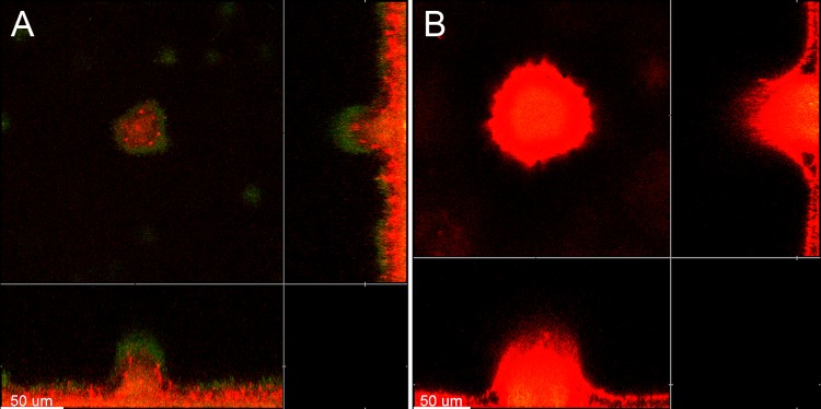
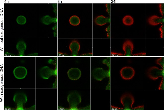
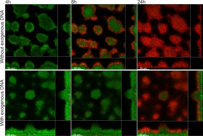
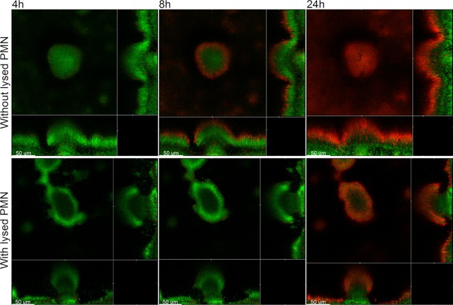
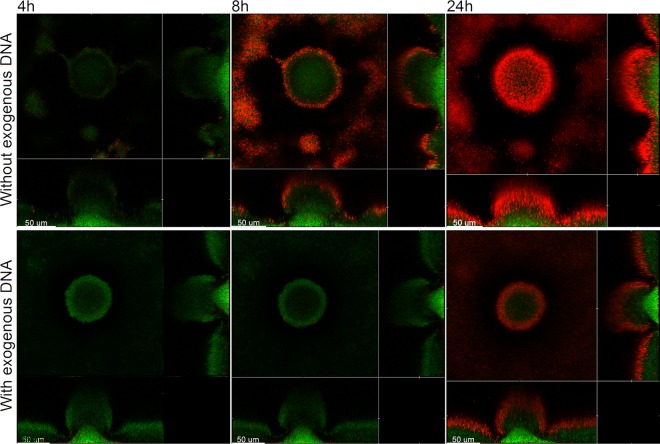
Within recent years, it has been established that extracellular DNA is a key constituent of the matrix of microbial biofilms. In addition, it has recently been demonstrated that DNA binds positively charged antimicrobials such as aminoglycosides and antimicrobial peptides. In the present study, we provide evidence that extracellular DNA shields against aminoglycosides in Pseudomonas aeruginosa biofilms. We show that exogenously supplemented DNA integrates into P. aeruginosa biofilms and increases their tolerance toward aminoglycosides. We provide evidence that biofilms formed by a DNA release-deficient P. aeruginosa quorum-sensing mutant are more susceptible to aminoglycoside treatment than wild-type biofilms but become rescued from the detrimental action of aminoglycosides upon supplementation with exogenous DNA. Furthermore, we demonstrate that exposure to lysed polymorphonuclear leukocytes, which are thought to be a source of extracellular DNA at sites of infections, increases the tolerance of P. aeruginosa biofilms toward aminoglycosides. Although biofilm-associated aminoglycoside tolerance recently has been linked to extracellular DNA-mediated activation of the pmr genes, we demonstrate that the aminoglycoside tolerance mediated by the presence of extracellular DNA is not caused by activation of the pmr genes in our P. aeruginosa biofilms but rather by a protective shield effect of the extracellular DNA.


Figures

Fig 1
Fig 1
Visualization of extracellular DNA in 4-day-old P. aeruginosa PAO1-GFP biofilms grown in flow chambers without addition of exogenous DNA (A) or with addition of salmon sperm DNA (B) to the medium irrigated to the flow chambers after 2 days of cultivation. The bacteria appear green due to expression of GFP, whereas the extracellular DNA surrounding the bacteria appears red due to staining with propidium iodide and visualization with ultrasensitive confocal laser scanning microscopy. Scale bars, 50 μm.
Fig 2
Fig 2
Spatiotemporal distribution of live and dead bacteria in tobramycin-treated P. aeruginosa PAO1-GFP biofilms that were grown with or without exogenous DNA. Biofilms were grown for 4 days and were then continuously exposed to tobramycin (25 μg/ml) and propidium iodide. In the case of the biofilm shown in the lower panel, the medium irrigated to the flow chambers was supplied with salmon sperm DNA after 2 days of cultivation. Confocal laser scanning micrographs were acquired 4, 8, and 24 h after the beginning of tobramycin treatment. The images show a horizontal xy section, with two flanking images representing sections in the xz and yz planes. Live cells appear green due to expression of GFP, and dead cells appear red due to staining with the dead-cell indicator propidium iodide. Scale bars, 50 μm.
Fig 3
Fig 3
Spatiotemporal distribution of live and dead bacteria in tobramycin-treated P. aeruginosa lasR rhlR-GFP biofilms that were grown with or without exogenous DNA. Biofilms were grown for 4 days and were then continuously exposed to tobramycin (25 μg/ml) and propidium iodide. In the case of the biofilm shown in the lower panel, the medium irrigated to the flow chambers was supplied with salmon sperm DNA after 2 days of cultivation. Confocal laser scanning micrographs were acquired 4, 8, and 24 h after the beginning of tobramycin treatment. The images show a horizontal xy section, with two flanking images representing sections in the xz and yz planes. Live cells appear green due to expression of GFP, and dead cells appear red due to staining with the dead-cell indicator propidium iodide. Scale bars, 50 μm.
Fig 4
Fig 4
Spatiotemporal distribution of live and dead bacteria in tobramycin-treated P. aeruginosa PAO1-GFP biofilms that were grown with or without addition of lysed PMNs. Biofilms were grown for 4 days and were then continuously exposed to tobramycin (25 μg/ml) and propidium iodide. In the case of the biofilm shown in the lower panel, the medium irrigated to the flow chambers was supplied with lysed PMNs after 2 days of cultivation. Confocal laser scanning micrographs were acquired 4, 8, and 24 h after the beginning of tobramycin treatment. The images show a horizontal xy section, with two flanking images representing sections in the xz and yz planes. Live cells appear green due to expression of GFP, and dead cells appear red due to staining with the dead-cell indicator propidium iodide. Scale bars, 50 μm.
Fig 5
Fig 5
Spatiotemporal distribution of live and dead bacteria in tobramycin-treated P. aeruginosa pmrF-GFP biofilms that were grown with or without exogenous DNA. Biofilms were grown for 4 days and were then continuously exposed to tobramycin (25 μg/ml) and propidium iodide. In the case of the biofilm shown in the lower panel, the medium irrigated to the flow chambers was supplied with salmon sperm DNA after 2 days of cultivation. Confocal laser scanning micrographs were acquired 4, 8, and 24 h after the beginning of tobramycin treatment. The images show a horizontal xy section, with two flanking images representing sections in the xz and yz planes. Live cells appear green due to expression of GFP, and dead cells appear red due to staining with the dead-cell indicator propidium iodide. Scale bars, 50 μm.

References

    1. Costerton JW, Stewart PS, Greenberg EP. 1999. Bacterial biofilms: a common cause of persistent infections. Science 284:1318–1322 - PubMed
    1. Ciofu O, Tolker-Nielsen T. 2010. Antibiotic tolerance and resistance in biofilms, p 215–230 In Bjarnsholt T, Jensen PØ, Moser C, Høiby N. (ed), Biofilm infections. Springer Publishing Company, New York, NY
    1. Whitchurch CB, Tolker-Nielsen T, Ragas PC, Mattick JS. 2002. Extracellular DNA required for bacterial biofilm formation. Science 295:1487. - PubMed
    1. Friedman L, Kolter R. 2003. Genes involved in matrix formation in Pseudomonas aeruginosa PA14 biofilms. Mol. Microbiol. 51:675–690 - PubMed
    1. Matsukawa M, Greenberg EP. 2004. Putative exopolysaccharide synthesis genes influence Pseudomonas aeruginosa biofilm development. J. Bacteriol. 186:4449–4456 - PMC - PubMed

Publication types

MeSH terms