. 2013 May;304(9):H1215-24.
doi: 10.1152/ajpheart.00796.2012. Epub 2013 Mar 11.

Hydrogen sulfide preconditions the db/db diabetic mouse heart against ischemia-reperfusion injury by activating Nrf2 signaling in an Erk-dependent manner

Affiliations

Hydrogen sulfide preconditions the db/db diabetic mouse heart against ischemia-reperfusion injury by activating Nrf2 signaling in an Erk-dependent manner

Bridgette F Peake et al. Am J Physiol Heart Circ Physiol. 2013 May.

Abstract

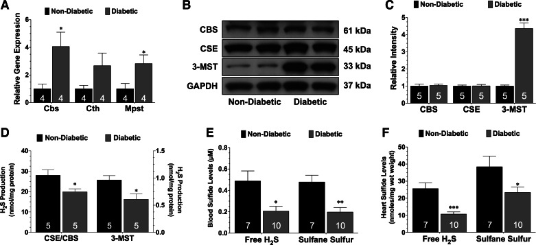
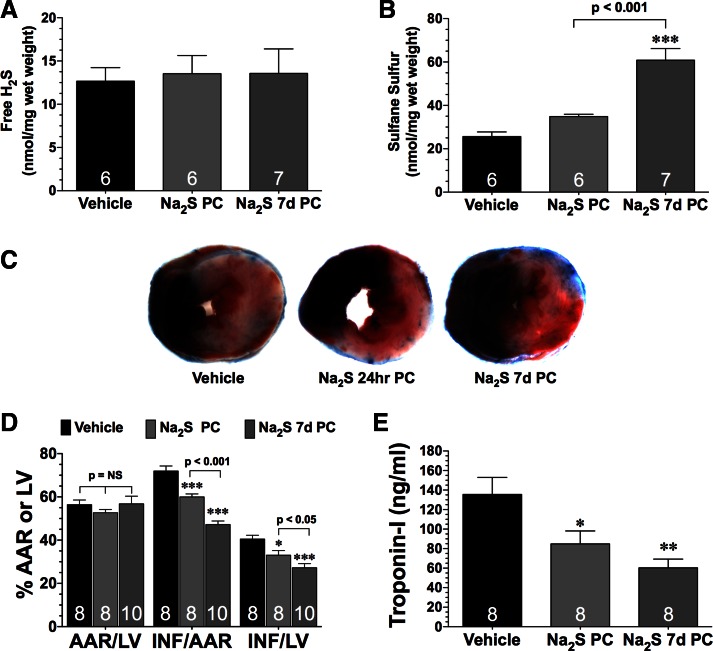
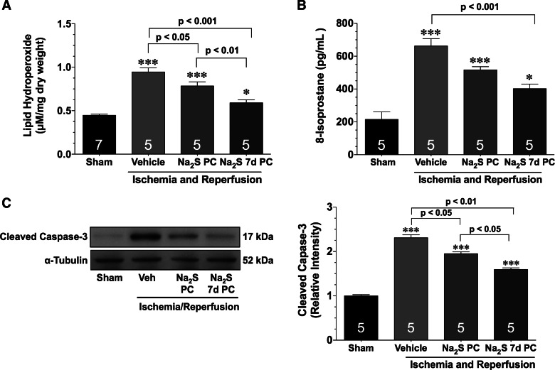
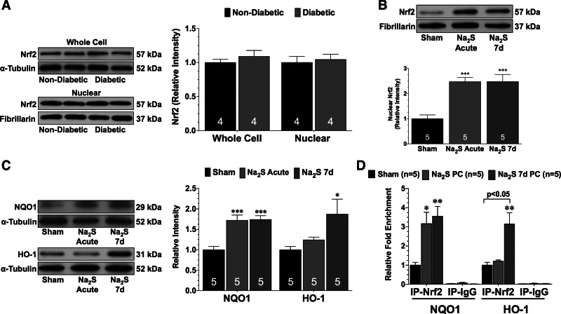
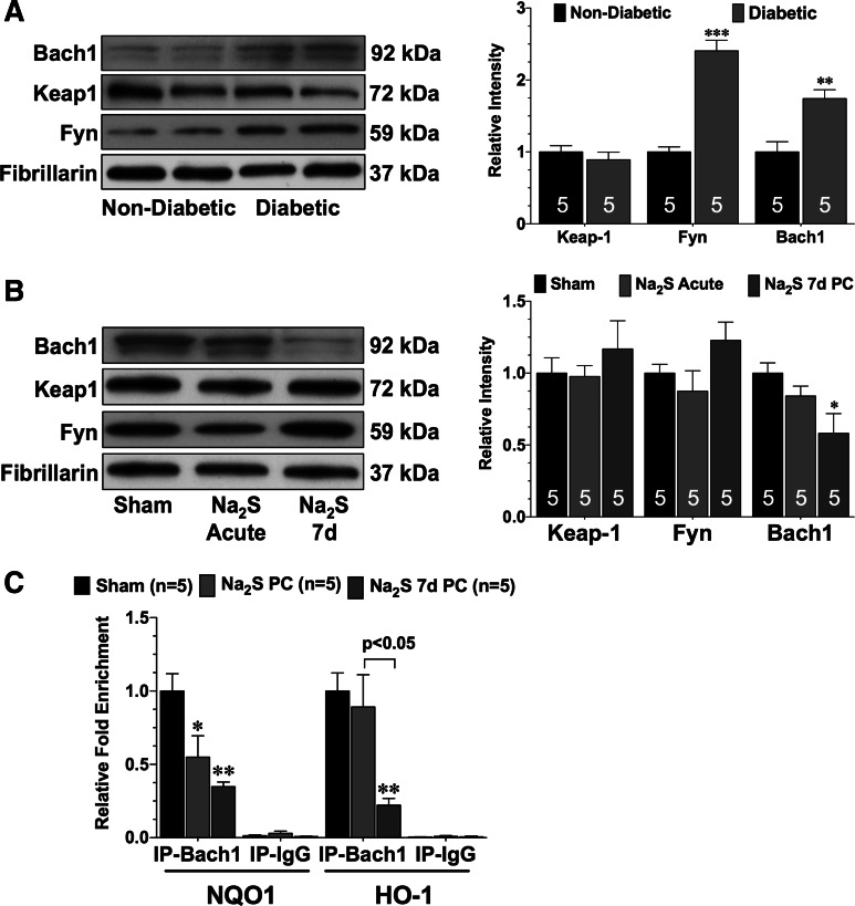
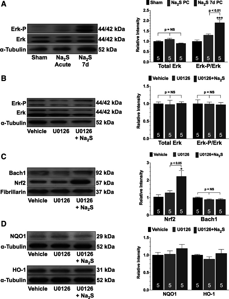
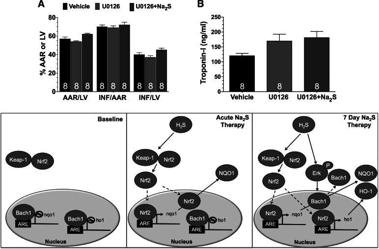
Hydrogen sulfide (H2S) therapy protects nondiabetic animals in various models of myocardial injury, including acute myocardial infarction and heart failure. Here, we sought to examine whether H2S therapy provides cardioprotection in the setting of type 2 diabetes. H2S therapy in the form of sodium sulfide (Na2S) beginning 24 h or 7 days before myocardial ischemia significantly decreased myocardial injury in db/db diabetic mice (12 wk of age). In an effort to evaluate the signaling mechanism responsible for the observed cardioprotection, we focused on the role of nuclear factor E2-related factor (Nrf2) signaling. Our results indicate that diabetes does not alter the ability of H2S to increase the nuclear localization of Nrf2, but does impair aspects of Nrf2 signaling. Specifically, the expression of NADPH quinine oxidoreductase 1 was increased after the acute treatment, whereas the expression of heme-oxygenase-1 (HO-1) was only increased after 7 days of treatment. This discrepancy was found to be the result of an increased nuclear expression of Bach1, a known repressor of HO-1 transcription, which blocked the binding of Nrf2 to the HO-1 promoter. Further analysis revealed that 7 days of Na2S treatment overcame this impairment by removing Bach1 from the nucleus in an Erk1/2-dependent manner. Our findings demonstrate for the first time that exogenous administration of Na2S attenuates myocardial ischemia-reperfusion injury in db/db mice, suggesting the potential therapeutic effects of H2S in treating a heart attack in the setting of type 2 diabetes.

Keywords: hydrogen sulfide; myocardial infarction; nuclear factor E2-related factor; type 2 diabetes.


Figures

Fig. 1.
Fig. 1.
Diabetes reduces sulfide levels. A: mRNA expression of the genes that encode for cystathionine-β-synthase (Cbs), cystathionine-γ-lyase (Cth), and 3-mercaptopyruvate sulfutransferase (Mpst) in the hearts of nondiabetic and diabetic mice. B and C: representative immunoblots and densitometric analysis of cystathionine-γ-lyase (CSE), CBS, and 3-mercaptopyruvate sulfurtransferase (3-MST) proteins in the hearts of nondiabetic and diabetic mice. D: biosynthesis of hydrogen sulfide (H2S) from the pyridoxal-5′-phosphate (PLP)-dependent enzymes (CBS and CSE) and from 3-MST. E: circulating and myocardial free (F) H2S and sulfane sulfur levels in nondiabetic and diabetic mice. Values are means ± SE. Numbers inside of the bars indicate the number of animals that were investigated in each group. Data were compared with a Student's t-test. *P < 0.05, **P < 0.01, and ***P < 0.001 vs. nondiabetic.
Fig. 2.
Fig. 2.
H2S therapy in the form of sodium sulfide (Na2S) pretreatment reduces the extent of myocardial injury in diabetic mice after ischemia-reperfusion. Myocardial free H2S (A) and sulfane sulfur (B) levels were from diabetic mice treated with vehicle or a single tail vein injection of Na2S the day before experimentation (Na2S PC; 0.1 mg/kg) or Na2S for 7 days before experimentation (Na2S 7d PC). C: representative midventricular photomicrographs of hearts, myocardial infarct size relative to the area-at-risk (INF/AAR) and INF relative to the left ventricle (INF/LV) (D), and circulating Troponin I levels (E) from the experimental groups after 30 min of ischemia and 2 h of reperfusion. Values are means ± SE. Data were compared through the use of a 1-way ANOVA with a Tukey test as the post hoc analysis. *P < 0.05, **P < 0.01, and ***P < 0.001 vs. vehicle.
Fig. 3.
Fig. 3.
Na2S pretreatment reduces oxidative stress and apoptosis after myocardial ischemia and reperfusion. Lipid hydroperoxide (A), 8-isoprostane levels (B), and representative immunoblots and densitometric analysis (C) of cleaved caspase-3 expression from hearts of sham, vehicle (Veh), Na2S PC, and Na2S 7d PC mice after 30 min of ischemia and 1 h of reperfusion were shown. Values are means ± SE. Data were compared through the use of a 1-way ANOVA with a Tukey test as the post hoc analysis. *P < 0.05 and ***P < 0.001 vs. sham.
Fig. 4.
Fig. 4.
Na2S increases the nuclear expression of nuclear factor E2-related factor (Nrf2). A: representative immunoblots and densitometric analysis of whole cell and nuclear Nrf2 from the hearts of nondiabetic and diabetic mice. B: representative immunoblots and densitometric analysis of nuclear Nrf2 in the hearts of diabetic mice treated with either a single injection of Na2S (Na2S acute) or with daily injections for 7 days (Na2S 7d). C: representative immunoblots and densitometric analysis of NADPH quinine oxidoreductase 1 (NQO1) and heme-oxygenase-1 (HO-1) in the hearts of sham, Na2S acute, and Na2S 7d mice. D: chromatin immunoprecipitation (ChIP) analysis of Nrf2 binding to the NQO1 or HO-1 promoter in the hearts of sham, Na2S acute, and Na2S 7d mice. A parallel ChIP assay was performed with IgG as a ChIP assay control. Data in A were compared with a Student's t-test. All other data were compared through the use of a 1-way ANOVA with a Tukey test as the post hoc analysis. *P < 0.05, **P < 0.01, and ***P < 0.001 vs. sham. IP, immunoprecipitation.
Fig. 5.
Fig. 5.
Na2S treatment for 7 days increases the phosphorylation of Erk and decreases the nuclear expression of Bach1. A: representative immunoblots and densitometric analysis of Bach1, kelch-like ECH-associated protein 1 (Keap1), and Fyn kinase in the nuclear fractions of hearts taken from nondiabetic and diabetic mice. B: representative immunoblots and densitometric analysis of Bach1, Keap1, and Fyn kinase in the nuclear fractions of hearts taken from sham, Na2S acute, and Na2S 7d mice. C: ChIP analysis of Bach1 binding to the NQO1 or HO-1 promoter in the hearts of sham, Na2S acute, and Na2S 7d mice. Values are means ± SE. Data in A were compared with a Student's t-test. All other data were compared through the use of a 1-way ANOVA with a Tukey test as the post hoc analysis. *P < 0.05, **P < 0.01, and ***P < 0.001 vs. sham or nondiabetic. IP, immunoprecipitation.
Fig. 6.
Fig. 6.
Na2S treatment for 7 days fails to decrease the nuclear expression of Bach1 when Erk is inhibited. A: representative immunoblots and densitometric analysis of total Erk and phosphorylated Erk in the hearts of sham, Na2S acute, and Na2S 7d mice. Representative immunoblots and densitometric analysis of total Erk and phosphorylated Erk (B), nuclear Bach1 and Nrf2 (C), and NQO1 and HO-1 (D) in the hearts of diabetic mice treated with vehicle, U0126, or U0126 and Na2S (U0126 + Na2S) for 7 days are shown. Values are means ± SE. Data were compared through the use of a 1-way ANOVA with a Tukey test as the post hoc analysis. *P < 0.05, ***P < 0.001 vs. sham or vehicle. NS, not significant.
Fig. 7.
Fig. 7.
Pretreatment with Na2S for 7 days does not provide protection against myocardial ischemia and reperfusion injury when Erk is inhibited. Myocardial INF/AAR and INF/LV (A) and circulating Troponin I levels (B) from following 30 min of ischemia and 2 h of reperfusion are shown. Mice were treated with vehicle, U0126, or U0126 and Na2S (U0126 + Na2S) for 7 days before myocardial ischemia. Bottom: schematic depicting the proposed signaling mechanism by which H2S activates Nrf2 signaling. Values are means ± SE. Data were compared through the use of a 1-way ANOVA with a Tukey test as the post hoc analysis. ARE, antioxidant response element.

References

    1. Cai H, Harrison DG. Endothelial dysfunction in cardiovascular diseases: the role of oxidant stress. Circ Res 87: 840–844, 2000 - PubMed
    1. Calvert JW, Gundewar S, Jha S, Elrod JW, Lefer DJ. Hydrogen sulfide therapy attenuates left ventricular dysfunction and reduces mortality in a murine model of heart failure. Circulation 118: S441–S331, 2008
    1. Calvert JW, Jha S, Gundewar S, Elrod JW, Ramachandran A, Pattillo CB, Kevil CG, Lefer DJ. Hydrogen sulfide mediates cardioprotection through Nrf2 signaling. Circ Res 105: 365–374, 2009 - PMC - PubMed
    1. Denizalti M, Bozkurt TE, Akpulat U, Sahin-Erdemli I, Abacioglu N. The vasorelaxant effect of hydrogen sulfide is enhanced in streptozotocin-induced diabetic rats. Naunyn Schmiedebergs Arch Pharmacol 383: 509–517, 2011 - PubMed
    1. El-Seweidy MM, Sadik NA, Shaker OG. Role of sulfurous mineral water and sodium hydrosulfide as potent inhibitors of fibrosis in the heart of diabetic rats. Arch Biochem Biophys 506: 48–57, 2011 - PubMed

Publication types

MeSH terms