. 2013 Mar;15(3):328-34.
doi: 10.1593/neo.121706.

HIF2A and IGF2 expression correlates in human neuroblastoma cells and normal immature sympathetic neuroblasts

Affiliations

HIF2A and IGF2 expression correlates in human neuroblastoma cells and normal immature sympathetic neuroblasts

Sofie Mohlin et al. Neoplasia. 2013 Mar.

Abstract

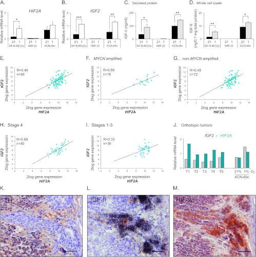
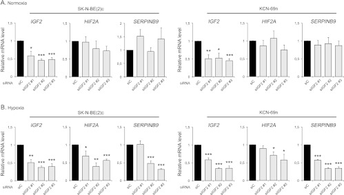
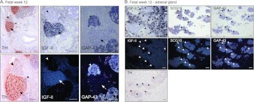
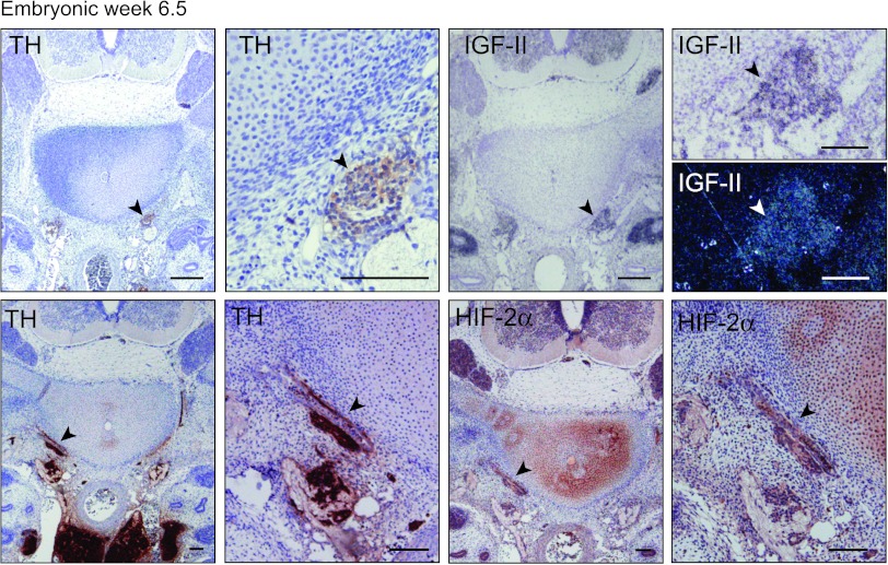
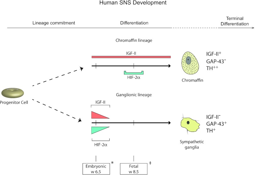
During normal sympathetic nervous system (SNS) development, cells of the ganglionic lineage can malignantly transform and develop into the childhood tumor neuroblastoma. Hypoxia-inducible transcription factors (HIFs) mediate cellular responses during normal development and are central in the adaptation to oxygen shortage. HIFs are also implicated in the progression of several cancer forms, and high HIF-2α expression correlates with disseminated disease and poor outcome in neuroblastoma. During normal SNS development, HIF2A is transiently expressed in neuroblasts and chromaffin cells. SNS cells can, during development, be distinguished by distinct gene expression patterns, and insulin-like growth factor 2 (IGF2) is a marker of sympathetic chromaffin cells, whereas sympathetic neuroblasts lack IGF2 expression. Despite the neuronal derivation of neuroblastomas, we show that neuroblastoma cell lines and specimens express IGF2 and that expression of HIF2A and IGF2 correlates, with the strongest correlation in high-stage tumors. In neuroblastoma, both IGF2 and HIF2A are hypoxia-driven and knocking down IGF2 at hypoxia resulted in downregulated HIF2A levels. HIF-2α and IGF2 were strongly expressed in subsets of immature neuroblastoma cells, suggesting that these two genes could be co-expressed also at early stages of SNS development. We show that IGF2 is indeed expressed in sympathetic chain ganglia at embryonic week 6.5, a developmental stage when HIF-2α is present. These findings provide a rationale for the unexpected IGF2 expression in neuroblastomas and might suggest that IGF2 and HIF2A positive neuroblastoma cells are arrested at an embryonic differentiation stage corresponding to the stage when sympathetic chain ganglia begins to coalesce.


Figures

Figure 1
Figure 1
HIF2A and IGF2 correlate and are co-expressed in neuroblastoma specimen. (A) Quantitative real-time-PCR (qRT-PCR) analysis of HIF2A mRNA expression in SK-N-BE(2)c, KCN-69n, and IMR-32 cells cultured for 72 hours at 21% or 1% oxygen. (B) qRT-PCR analysis of IGF2 mRNA expression in cells cultured for 72 hours at 21% or 1% oxygen. (C) Secreted IGF-II protein levels detected in conditioned medium by human IGF-II ELISA. (D) Intracellular IGF-II protein levels detected by human IGF-II ELISA. (A–D) Data are presented as means ± SEM from three independent experiments; P values were calculated using Student's t test (*P < .05, **P < .01, and ***P < .001). (E) HIF2A and IGF2 gene expression correlation in a data set containing 88 neuroblastoma tumors (Versteeg-88-MAS5.0-u133p2). (F) HIF2A and IGF2 gene expression correlation in the subgroup of MYCN-amplified tumors from the data set used in D. (G) HIF2A and IGF2 gene expression correlation in the subgroup of non-MYCN-amplified tumors from the data set used in D. (H) HIF2A and IGF2 gene expression correlation in the subgroup of International Neuroblastoma Staging System (INSS) stage 4 tumors from the data set used in D. (I) HIF2A and IGF2 gene expression correlation in the joint subgroup of INSS stage 1 to 3 tumors from the data set used in D. (E–I) R value was calculated using Pearson correlation and n is equal to the number of tumors in each subset. (J) Levels of IGF2 and HIF2A mRNA expression in orthotopic neuroblastoma tumors as measured by qRT-PCR. KCN-69n neuroblastoma cells were grown at 21% O2 for 24 hours before transplantation into the adrenal gland fat pad of NMRI-Nu/Nu mice. Tumors were excised 4 weeks after cell implantation. Gene expression levels in KCN-69n cells cultured in vitro at 21% or 1% O2 for 72 hours were used as comparison. (K–M) Immunohistochemical TH (K) and HIF-2α (M) staining and IGF-II in situ hybridization (L) of a neuroblastoma specimen. Asterisk indicates a blood vessel. Scale bars represent 50 µm.
Figure 2
Figure 2
IGF-II regulates HIF2A mRNA expression at hypoxia. (A) Expression of HIF2A and HIF-2-driven gene SERPINB9 mRNA after down-regulation of IGF2 in SK-N-BE(2)c and KCN-69n cells using three different siRNAs targeting the IGF2 gene at normoxia (21% O2), as measured by qRT-PCR. (B) Expression of HIF2A and SERPINB9 mRNA after down-regulation of IGF2 in SK-N-BE(2)c and KCN-69n cells using three different siRNAs targeting the IGF2 gene at hypoxia (1% O2), as measured by qRT-PCR. Data are presented as means ± SEM from three independent experiments, and mRNA expression after IGF2 knockdown is normalized against siC within each experiment. Statistical significance is calculated compared to siC using Student's t test (*P < .05, **P < .01, and ***P < .001).
Figure 3
Figure 3
IGF-II expression is absent in human fetal sympathetic ganglia. (A) Immunohistochemical staining of TH and in situ hybridization of IGF-II and GAP43 (bright field and dark field) in human fetus at week 12. Arrowheads indicate sympathetic ganglia; arrows indicate paraganglia. *Area of magnification. Scale bars represent 200 µm. (B) IGF-II, SCG10, and GAP43 in situ hybridization (bright field and dark field) and TH immunostaining in a 12-week-old fetal adrenal gland. Arrowheads indicate sympathetic ganglia. Scale bars represent 100 µm.
Figure 4
Figure 4
Embryonic neuroblasts express HIF-2α and IGF-II. Immunohistochemical staining of TH and HIF-2α and in situ hybridization of IGF-II (bright field and dark field) in a week 6.5 human embryo at different magnifications. Arrowheads indicate sympathetic ganglia. Scale bars represent 100 µm.
Figure 5
Figure 5
HIF-2α and IGF-II are co-expressed in sympathetic neuroblasts during early human development. Sympathoadrenal progenitor cells commit to either of three lineages, the sympathetic ganglionic, chromaffin, or SIF (not shown) cell lineages. IGF-II is expressed in both chromaffin and sympathetic ganglionic cells during late embryogenesis, but expression becomes restricted to cells of the chromaffin lineage during fetal development. HIF-2α can be detected in sympathetic neuroblasts of the ganglionic lineage only during late embryo-genesis, whereas it is transiently expressed around fetal week 8.5 in chromaffin cells. Hence, IGF-II and HIF-2α are co-expressed at early developmental stages before sympathetic ganglionic cells lose expression of both proteins. Marker gene expression for chromaffin cells (IGF-II+, GAP-43-,TH++) and sympathetic ganglionic cells (IGF-II-, GAP-43+,TH+) as depicted. Two distinct time points are shown: *embryonic week 6.5 and fetal week 8.5.

References

    1. Anderson DJ. Molecular control of cell fate in the neural crest: the sympathoadrenal lineage. Annu Rev Neurosci. 1993;16:129–158. - PubMed
    1. Hoehner JC, Gestblom C, Hedborg F, Sandstedt B, Olsen L, Påhlman S. A developmental model of neuroblastoma: differentiating stroma-poor tumors' progress along an extra-adrenal chromaffin lineage. Lab Invest. 1996;75:659–675. - PubMed
    1. Fredlund E, Ringner M, Maris JM, Påhlman S. High Myc pathway activity and low stage of neuronal differentiation associate with poor outcome in neuroblastoma. Proc Natl Acad Sci USA. 2008;105:14094–14099. - PMC - PubMed
    1. Jögi A, Øra I, Nilsson H, Lindeheim A, Makino Y, Poellinger L, Axelson H, Påhlman S. Hypoxia alters gene expression in human neuroblastoma cells toward an immature and neural crest-like phenotype. Proc Natl Acad Sci USA. 2002;99:7021–7026. - PMC - PubMed
    1. Vaupel P, Kallinowski F, Okunieff P. Blood flow, oxygen and nutrient supply, and metabolic microenvironment of human tumors: a review. Cancer Res. 1989;49:6449–6465. - PubMed

Publication types

MeSH terms

Substances

LinkOut - more resources