. 2013 Apr 19;288(16):11304-11.
doi: 10.1074/jbc.M112.444919. Epub 2013 Mar 11.

An electron-bifurcating caffeyl-CoA reductase

Affiliations

An electron-bifurcating caffeyl-CoA reductase

Johannes Bertsch et al. J Biol Chem. .

Abstract

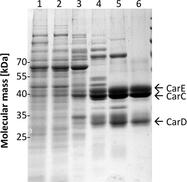
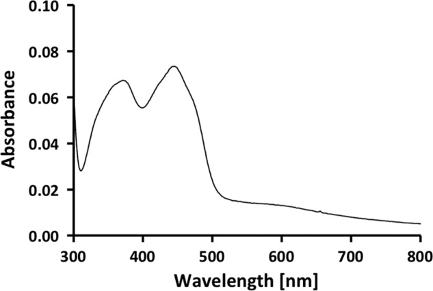
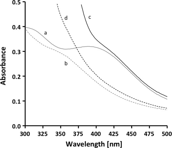
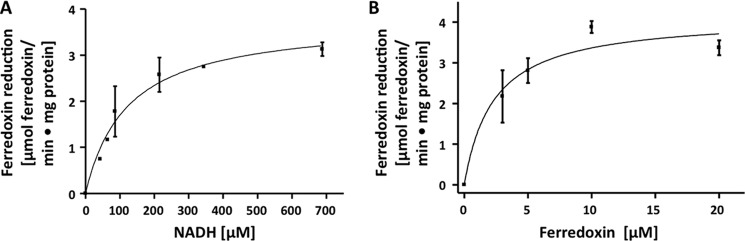
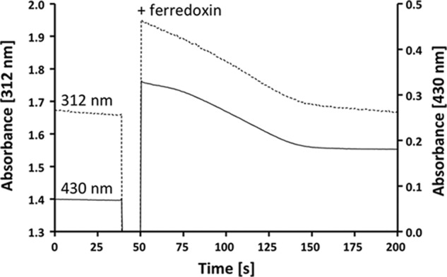
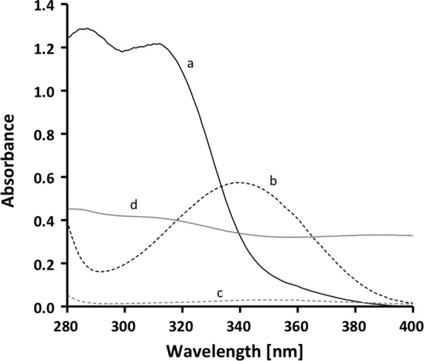
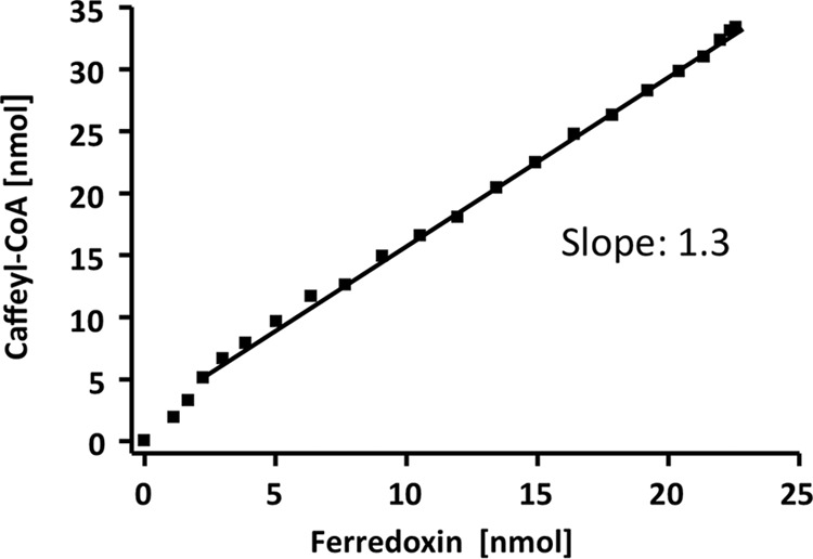
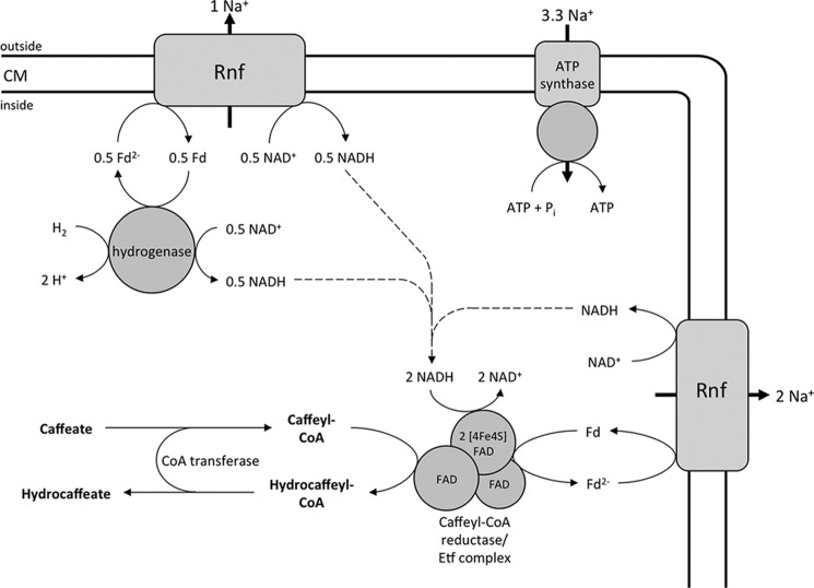
A low potential electron carrier ferredoxin (E0' ≈ -500 mV) is used to fuel the only bioenergetic coupling site, a sodium-motive ferredoxin:NAD(+) oxidoreductase (Rnf) in the acetogenic bacterium Acetobacterium woodii. Because ferredoxin reduction with physiological electron donors is highly endergonic, it must be coupled to an exergonic reaction. One candidate is NADH-dependent caffeyl-CoA reduction. We have purified a complex from A. woodii that contains a caffeyl-CoA reductase and an electron transfer flavoprotein. The enzyme contains three subunits encoded by the carCDE genes and is predicted to have, in addition to FAD, two [4Fe-4S] clusters as cofactor, which is consistent with the experimental determination of 4 mol of FAD, 9 mol of iron, and 9 mol of acid-labile sulfur. The enzyme complex catalyzed caffeyl-CoA-dependent oxidation of reduced methyl viologen. With NADH as donor, it catalyzed caffeyl-CoA reduction, but this reaction was highly stimulated by the addition of ferredoxin. Spectroscopic analyses revealed that ferredoxin and caffeyl-CoA were reduced simultaneously, and a stoichiometry of 1.3:1 was determined. Apparently, the caffeyl-CoA reductase-Etf complex of A. woodii uses the novel mechanism of flavin-dependent electron bifurcation to drive the endergonic ferredoxin reduction with NADH as reductant by coupling it to the exergonic NADH-dependent reduction of caffeyl-CoA.


Figures

FIGURE 1.
FIGURE 1.
Purification of the caffeyl-CoA reductase-Etf complex. Samples from the different purification steps were separated by SDS-PAGE, and proteins were stained with Coomassie Brilliant Blue. Lane 1, cell extract; lane 2, cytoplasm; lane 3, pooled fractions from Q Sepharose; lane 4, pooled fractions from phenyl-Sepharose; lane 5, pooled fractions from Blue Sepharose; lane 6, pooled fractions from Sephacryl S-300. 10 μg of protein was applied to each lane.
FIGURE 2.
FIGURE 2.
UV-visible spectrum of the purified caffeyl-CoA reductase-Etf complex. The spectrum of the enzyme (0.57 mg/ml) was recorded in buffer B.
FIGURE 3.
FIGURE 3.
Caffeyl-CoA-dependent ferredoxin reduction. The assay buffer (buffer B) contained 0.2 mm NADH, 20 μm ferredoxin, 5 μm caffeyl-CoA, the caffeyl-CoA regeneration system (200 units of the CoA transferase CarA and 0.2 mm caffeate), and 8 μg of the purified CarCDE complex. Trace a, ferredoxin; trace b, ferredoxin reduced with CO dehydrogenase purified from A. woodii and 100% CO in the gas phase; trace c, ferredoxin, NADH, CarCDE, and the caffeyl-CoA regeneration system (with no caffeyl-CoA present); trace d, same as trace c after the addition of caffeyl-CoA and incubation of 5 min at 30 °C.
FIGURE 4.
FIGURE 4.
Dependence of ferredoxin reduction on NADH (A) and ferredoxin (B). Enzyme activity was measured in assay buffer (buffer B) containing the caffeyl-CoA regeneration system (200 units of the CoA transferase CarA and 0.2 mm caffeate), 8 μg of protein, and 5 μm caffeyl-CoA. All assays were done at 30 °C under an atmosphere of 100% nitrogen. For NADH dependence (A), NADH was added, and the reaction was started by the addition of 20 μm ferredoxin. For ferredoxin dependence (B), 0.5 mm NADH was added, and the reaction was started by the addition of ferredoxin.
FIGURE 5.
FIGURE 5.
The purified CarCDE complex catalyzes reduction of ferredoxin and caffeyl-CoA simultaneously. The assay buffer (buffer B) contained 0.2 mm NADH, 5 μm caffeyl-CoA, 8 μg of the purified CarCDE complex, and the caffeyl-CoA regeneration system (200 units of the CoA transferase CarA and 0.2 mm caffeate). The reaction was started by the addition of 20 μm ferredoxin. Reduction of caffeyl-CoA (dashed line; monitored at 312 nm via decline of caffeate) and reduction of ferredoxin (solid line; monitored at 430 nm) were monitored simultaneously.
FIGURE 6.
FIGURE 6.
Spectral properties of components used in the enzyme assay. Trace a, 0.2 mm caffeate; trace b, 0.2 mm NADH; trace c, 5 μm caffeyl-CoA; trace d, 20 μm ferredoxin (measured in buffer B).
FIGURE 7.
FIGURE 7.
The purified CarCDE complex reduces ferredoxin and caffeyl-CoA in stoichiometric amounts. The reduction of caffeyl-CoA and ferredoxin was monitored simultaneously (see Fig. 5). Data points from 5-s intervals were taken to calculate the amount of reduced electron carrier from the absorbance difference and the molar extinction coefficient. The amount of reduced caffeyl-CoA is plotted against the amount of reduced ferredoxin.
FIGURE 8.
FIGURE 8.
Enzymology and bioenergetics of hydrogen-dependent caffeate respiration in A. woodii. The electron-bifurcating hydrogenase reduces ferredoxin (Fd) and NAD+ simultaneously. The Rnf complex couples the exergonic electron transfer from reduced ferredoxin to NAD+ with the translocation of Na+ ions. The electron acceptor caffeyl-CoA is reduced by the caffeyl-CoA reductase-Etf complex with NADH, which is coupled to the endergonic reduction of ferredoxin with NADH. The reduced ferredoxin is re-oxidized at the Rnf complex. The electrochemical Na+ gradient drives ATP synthesis by the Na+ F1F0-ATP synthase. The hydrocaffeyl-CoA:caffeate-CoA transferase CarA uses hydrocaffeyl-CoA to activate caffeate to caffeyl-CoA; this energy-saving CoA loop provides the regeneration of the acceptor under steady-state conditions. Initially, caffeate is activated by the caffeyl-CoA synthetase CarB at the expense of ATP (not shown). CM, cytoplasmic membrane.

References

    1. Müller V. (2008) Bacterial fermentation. in Encyclopedia of Life Sciences, John Wiley & Sons Ltd., Chichester, United Kingdom, DOI: 10.1002/9780470015902.a0001415.pub2 - DOI
    1. Schmitz R. A., Daniel R., Deppenmeier U., Gottschalk G. (2006) The anaerobic way of life. in The Prokaryotes (Dworkin M., Falkow S., Rosenberg E., Schleifer K. H., Stackebrandt E., eds) Vol. 2, pp. 86–101, Springer-Verlag, New York
    1. Dimroth P., von Ballmoos C. (2008) ATP synthesis by decarboxylation phosphorylation. Results Probl. Cell Differ. 45, 153–184 - PubMed
    1. Gottschalk G., Thauer R. K. (2001) The Na+-translocating methyltransferase complex from methanogenic archaea. Biochim. Biophys. Acta 1505, 28–36 - PubMed
    1. Buckel W. (2001) Sodium ion-translocating decarboxylases. Biochim. Biophys. Acta 1505, 15–27 - PubMed

Publication types

MeSH terms

LinkOut - more resources