. 2013 Apr 19;288(16):11436-47.
doi: 10.1074/jbc.M112.437103. Epub 2013 Mar 11.

Deletion of autophagy-related 5 (Atg5) and Pik3c3 genes in the lens causes cataract independent of programmed organelle degradation

Affiliations

Deletion of autophagy-related 5 (Atg5) and Pik3c3 genes in the lens causes cataract independent of programmed organelle degradation

Hideaki Morishita et al. J Biol Chem. .

Abstract

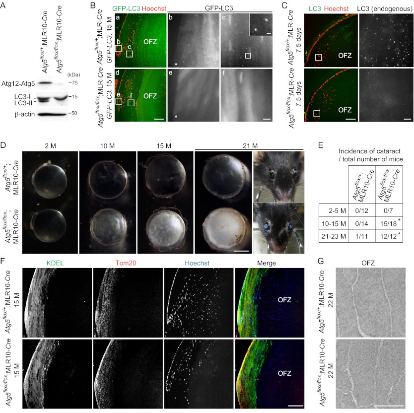
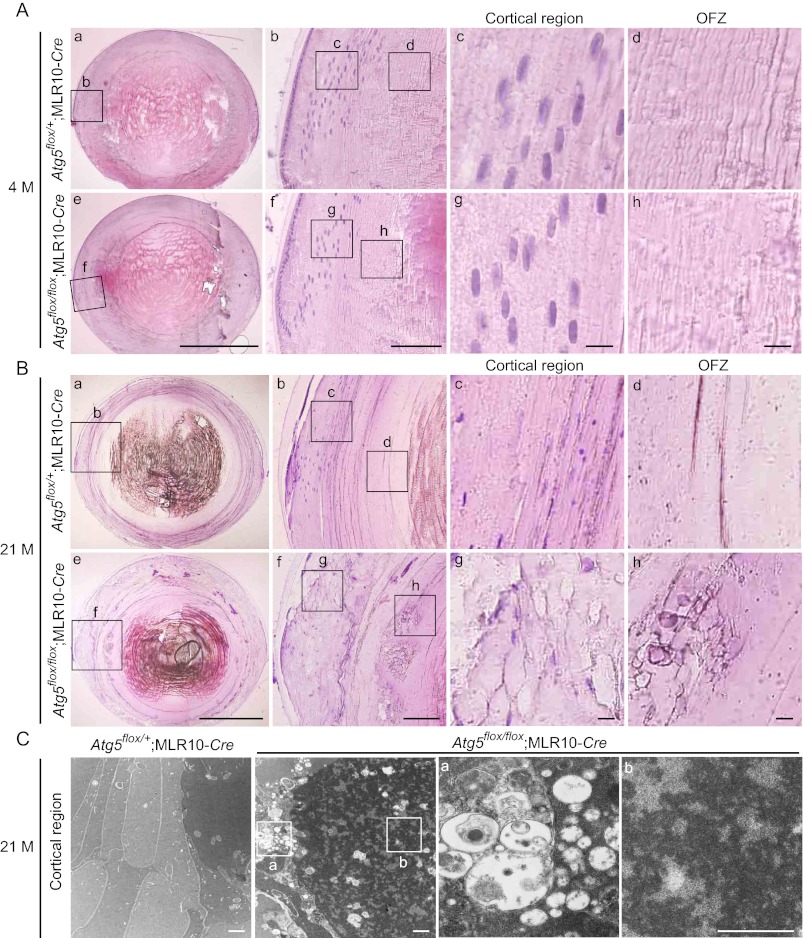
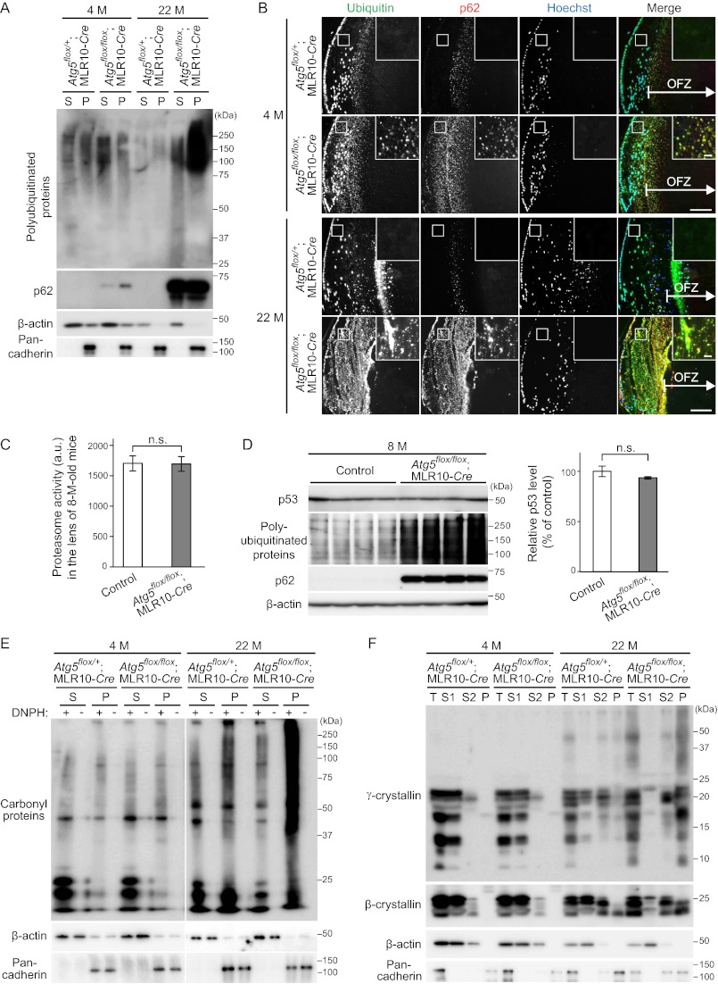
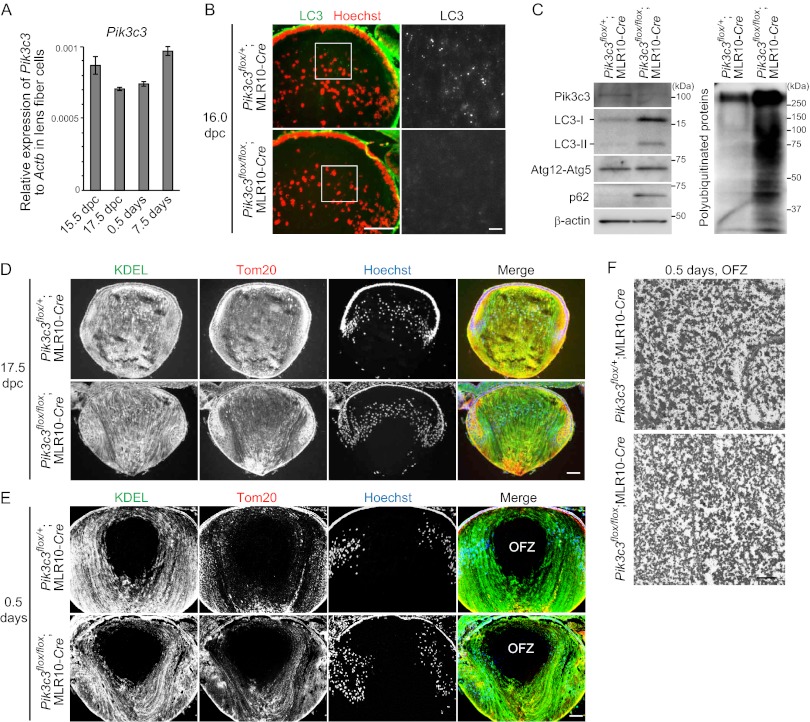
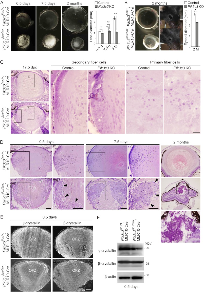
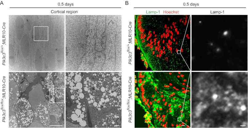
The lens of the eye is composed of fiber cells, which differentiate from epithelial cells and undergo programmed organelle degradation during terminal differentiation. Although autophagy, a major intracellular degradation system, is constitutively active in these cells, its physiological role has remained unclear. We have previously shown that Atg5-dependent macroautophagy is not necessary for lens organelle degradation, at least during the embryonic period. Here, we generated lens-specific Atg5 knock-out mice and showed that Atg5 is not required for lens organelle degradation at any period of life. However, deletion of Atg5 in the lens results in age-related cataract, which is accompanied by accumulation of polyubiquitinated and oxidized proteins, p62, and insoluble crystallins, suggesting a defect in intracellular quality control. We also produced lens-specific Pik3c3 knock-out mice to elucidate the possible involvement of Atg5-independent alternative autophagy, which is proposed to be dependent on Pik3c3 (also known as Vps34), in lens organelle degradation. Deletion of Pik3c3 in the lens does not affect lens organelle degradation, but it leads to congenital cataract and a defect in lens development after birth likely due to an impairment of the endocytic pathway. Taken together, these results suggest that clearance of lens organelles is independent of macroautophagy. These findings also clarify the physiological role of Atg5 and Pik3c3 in quality control and development of the lens, respectively.


Figures

FIGURE 1.
FIGURE 1.
Atg5 is required for suppression of age-related cataract but not for programmed organelle degradation in the lens. A, expression of Atg5 and LC3B in the lens of 15-month (M)-old Atg5flox/+;MLR10-Cre and Atg5flox/flox;MLR10-Cre mice. Triton X-100-soluble fractions of lens homogenates were prepared and subjected to immunoblot analysis using antibodies against Atg5 and LC3. β-Actin was used as a loading control. The positions of the Atg12-Atg5 conjugate, LC3-I and LC3-II (LC3-PE conjugate), are indicated. B, GFP-LC3 puncta formation in the lens of 15-month-old Atg5flox/+;MLR10-Cre;GFP-LC3 (panels a–c) and Atg5flox/flox;MLR10-Cre;GFP-LC3 (panels d–f) mice. The nuclei were stained with Hoechst 33342. GFP signals in the indicated regions in panels a and d are magnified and shown in panels b, c, e, and f. The GFP-LC3 puncta in panel c are further magnified and shown in the inset. Asterisks indicate lens epithelial cells. OFZ, organelle-free zone. Scale bars, 100 μm (panels a and d), 10 μm (panels b, c, e, and f), and 1 μm in inset (panel c). C, immunohistochemical analysis of the lens of 7.5-day-old Atg5flox/+;MLR10-Cre and Atg5flox/flox;MLR10-Cre mice. Slices were stained with anti-LC3 antibody and Hoechst 33342 (left). Endogenous LC3 signals in the indicated regions were magnified and are shown in the right panels. Scale bars, 100 μm (left) and 10 μm (right). D, representative dark field images of the lens of 2-, 10-, 15-, and 21-month-old mice. The appearance of a 21-month-old mouse is also shown. Scale bar, 1 mm. E, incidence of cataract at indicated ages. Cataract was defined as visible opacity by gross examination and microscopy. p < 0.0001, χ2 test comparing the two genotypes. F, immunohistochemical staining of the lens of 15-month-old mice using anti-KDEL (marker of the ER) and anti-Tom20 (mitochondrial marker) antibodies and Hoechst 33342. Data are representatives of four independent experiments. Scale bar, 100 μm. G, electron micrographs of fiber cells in the OFZ of 22-month-old mice. Scale bar, 1 μm.
FIGURE 2.
FIGURE 2.
Fiber cells are disorganized in the cortical region of the lens in aged lens-specific Atg5-deficient mice. A and B, hematoxylin and eosin staining of the lens of 4-month (M)-old (A) and 21-month-old (B) Atg5flox/+;MLR10-Cre (panels a–d) and Atg5flox/flox;MLR10-Cre (panels e–h) mice. Magnified images of the indicated cortical region (panels c and g) and OFZ (panels d and h) are shown. Scale bars, 1 mm (panels a and e), 100 μm (panels b and f), and 10 μm (panels c, d, g, and h). C, electron micrographs of fiber cells in the cortical region of the lens of 21-month-old mice. Magnified images of the indicated regions are shown in the right panels (panels a and b). Scale bars, 1 μm.
FIGURE 3.
FIGURE 3.
Atg5-dependent autophagy is required for quality control in lens cells. A, immunoblot analysis of polyubiquitinated proteins and p62 of the lens of 4-month (M)-old and 22-month-old mice. Lens homogenates separated into 1% Triton X-100-soluble (S) or -insoluble (P) fractions were analyzed by immunoblotting using anti-ubiquitin and anti-p62 antibodies. β-Actin and pan-cadherin were used as loading controls for the soluble and insoluble fractions, respectively. B, immunohistochemical analysis of ubiquitinated proteins and p62. Lenses were stained using anti-ubiquitin and anti-p62 antibodies and Hoechst 33342. The indicated regions are shown and magnified in the insets. Scale bars, 100 μm and 10 μm in inset. C, chymotryptic activities of the proteasome in the lens of 8-month (M)-old control (Atg5flox/flox and Atg5flox/+;MLR10-Cre) and Atg5flox/flox;MLR10-Cre mice. Lens homogenates were mixed with fluorogenic peptide substrate succinyl-Leu-Leu-Val-Tyr-7-amino-4-methylcoumarin in the presence or absence of MG132. MG132-sensitive activities were quantified (mean ± S.E.). n.s., not significant, unpaired Student's t test; n = 6. D, immunoblot analysis of p53, a proteasome substrate, in the lens of 8-month-old control and Atg5flox/flox;MLR10-Cre mice. Lens homogenates were analyzed by immunoblotting using anti-p53, anti-ubiquitin, and anti-p62 antibodies. β-Actin was used as a loading control. Relative protein levels of p53 to β-actin were quantified by densitometric analysis (mean ± S.E.). n.s., not significant, unpaired Student's t test; n = 4. E, immunoblot analysis of oxidized (carbonylated) proteins in the lens. Triton X-100-soluble and -insoluble fractions were treated with or without 2,4-dinitrophenylhydrazine to derivatize carbonyl groups in oxidized proteins. These samples were subjected to immunoblot analysis using anti-2,4-dinitrophenol (DNP) antibody. F, immunoblot analysis of crystallins using anti-γ- and anti-β-crystallin antibodies. Total lens homogenates (T) were first separated into water-soluble (S1) and -insoluble fractions. The latter was treated with NaOH and separated into NaOH-soluble (S2) and -insoluble (P) fractions. β-Actin and pan-cadherin were used as loading controls. Data are representatives of two (A, C, D, E, and F) or three (B) independent experiments.
FIGURE 4.
FIGURE 4.
Pik3c3-dependent autophagy is not required for lens organelle degradation. A, quantitative RT-PCR analysis of Pik3c3 in lens fiber cells of embryos at 15.5 and 17.5 dpc and neonates at 0.5 and 7.5 days old. Relative mRNA expression of Pik3c3 to Actb is shown. B, immunohistochemical analysis of endogenous LC3 in the lens of Pik3c3flox/+;MLR10-Cre and Pik3c3flox/flox;MLR10-Cre embryos at 16.0 dpc. Sections were stained with anti-LC3 antibody and Hoechst 33342 (left panel). Magnified images of LC3 staining are shown in the right panels. Scale bars, 100 μm (left panel) and 10 μm (right panel). C, immunoblot analysis of Pik3c3, LC3, Atg5, p62, and polyubiquitinated proteins in the lens of 0.5-day-old neonates. Triton X-100-soluble fractions of lens homogenates were prepared and subjected to immunoblotting using the indicated antibodies. β-Actin was used as a loading control. D and E, immunohistochemical analysis of the lens of mice at 17.5 dpc (D) and 0.5 days after birth (E) using anti-KDEL (marker of the ER) and anti-Tom20 (mitochondrial marker) antibodies and Hoechst 33342. Data are representatives of seven independent experiments. Scale bar, 100 μm. F, electron micrographs of the OFZ in the lens of 0.5-day-old neonates. Scale bar, 1 μm.
FIGURE 5.
FIGURE 5.
Pik3c3 is required for secondary fiber cell differentiation and suppression of congenital cataract and microphthalmia. A, representative dark field images of dissected lens of Pik3c3flox/+;MLR10-Cre (control) and Pik3c3flox/flox;MLR10-Cre mice (Pik3c3 KO) at 0.5 and 7.5 days and 2 months. Scale bar, 1 mm. The equatorial diameter of the lens was quantified (mean ± S.E.). *, p < 0.05; **, p < 0.01, unpaired Student's t test; n = 6 (0.5 days), 6 (7.5 days), and 4 (2 months). B, representative images of eyeball and appearance of a 2-month (M)-old mouse. Scale bar, 1 mm. The equatorial diameter of the eyeball at 2 months was quantified (mean ± S.E.). *, p < 0.05, unpaired Student's t test comparing indicated pairs; n = 3. C, hematoxylin and eosin staining of the lens of 17.5-dpc Pik3c3flox/+;MLR10-Cre (control, panels a–c) and Pik3c3flox/flox;MLR10-Cre (Pik3c3 KO, panels d–f) embryos. Magnified images of secondary fiber (panels b and e) and primary fiber cells (panels c and f) in the indicated regions are shown in the right panels. Scale bars, 0.1 mm (panels a and d) and 20 μm (panels b, c, e, and f). D, hematoxylin and eosin staining of the lens from mice at 0.5 and 7.5 days and 2 months. Magnified images in the indicated regions are shown in the right (0.5 and 7.5 days) or lower (2 months) panels. Arrowheads indicate cellular aggregates. Scale bars, 0.1 mm. E, immunohistochemical analysis of crystallins using anti-β- and anti-γ-crystallin antibodies in the lens of 0.5-day-old neonates. Scale bar, 100 μm. F, immunoblot analysis of γ- and β-crystallins in the lens of 0.5-day-old neonates. Triton X-100-soluble fractions of lens homogenates were prepared and subjected to immunoblotting using the indicated antibodies. β-Actin was used as a loading control.
FIGURE 6.
FIGURE 6.
Accumulation of vacuoles in Pik3c3-deficient secondary lens fiber cells. A, electron micrographs of fiber cells in the cortical region of the lens in 0.5-day-old mice. Magnified images in the indicated regions are shown in the right panels. Scale bars, 10 μm (left) and 1 μm (right). B, immunohistochemical analysis of Lamp-1 in 0.5-day-old mice. Lenses were stained using anti-Lamp-1 antibody and Hoechst 33342 (left). Magnified images in the indicated regions are shown in the right panels. Scale bars, 10 μm (left) and 1 μm (right).

References

    1. Piatigorsky J. (1981) Lens differentiation in vertebrates. A review of cellular and molecular features. Differentiation 19, 134–153 - PubMed
    1. McAvoy J. W., Chamberlain C. G., de Iongh R. U., Hales A. M., Lovicu F. J. (1999) Lens development. Eye 13, 425–437 - PubMed
    1. Kuwabara T., Imaizumi M. (1974) Denucleation process of the lens. Invest. Ophthalmol. 13, 973–981 - PubMed
    1. Bassnett S., Shi Y., Vrensen G. F. (2011) Biological glass: structural determinants of eye lens transparency. Philos. Trans. R. Soc. Lond. B Biol. Sci. 366, 1250–1264 - PMC - PubMed
    1. Wride M. A. (2011) Lens fibre cell differentiation and organelle loss: many paths lead to clarity. Philos. Trans. R. Soc. Lond. B Biol. Sci. 366, 1219–1233 - PMC - PubMed

Publication types

MeSH terms

Substances

LinkOut - more resources