. 2013 Mar 18;200(6):709-20.
doi: 10.1083/jcb.201208175. Epub 2013 Mar 11.

Rer1p maintains ciliary length and signaling by regulating γ-secretase activity and Foxj1a levels

Affiliations

Rer1p maintains ciliary length and signaling by regulating γ-secretase activity and Foxj1a levels

Nathalie Jurisch-Yaksi et al. J Cell Biol. .

Abstract

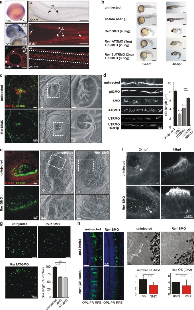
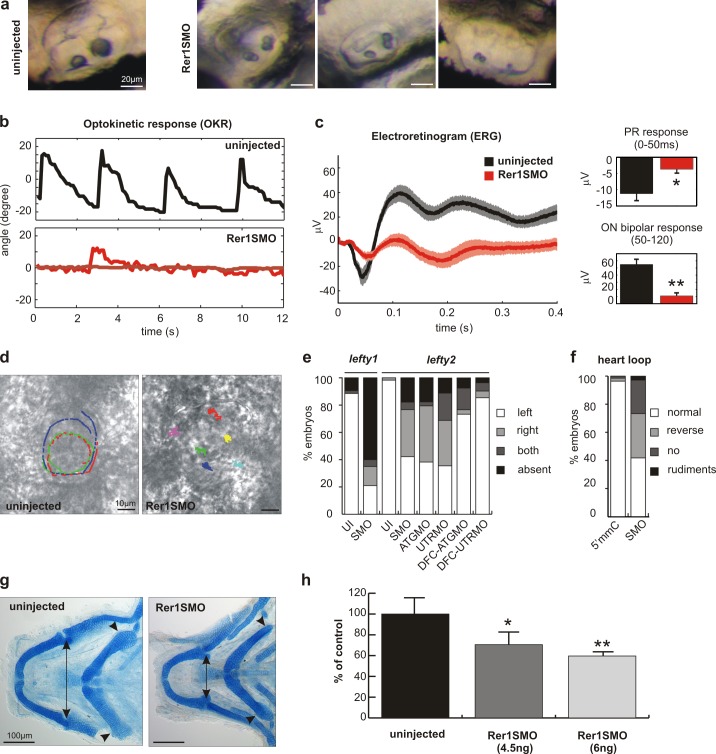
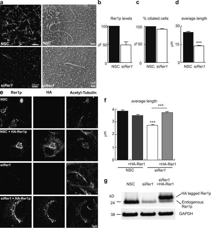
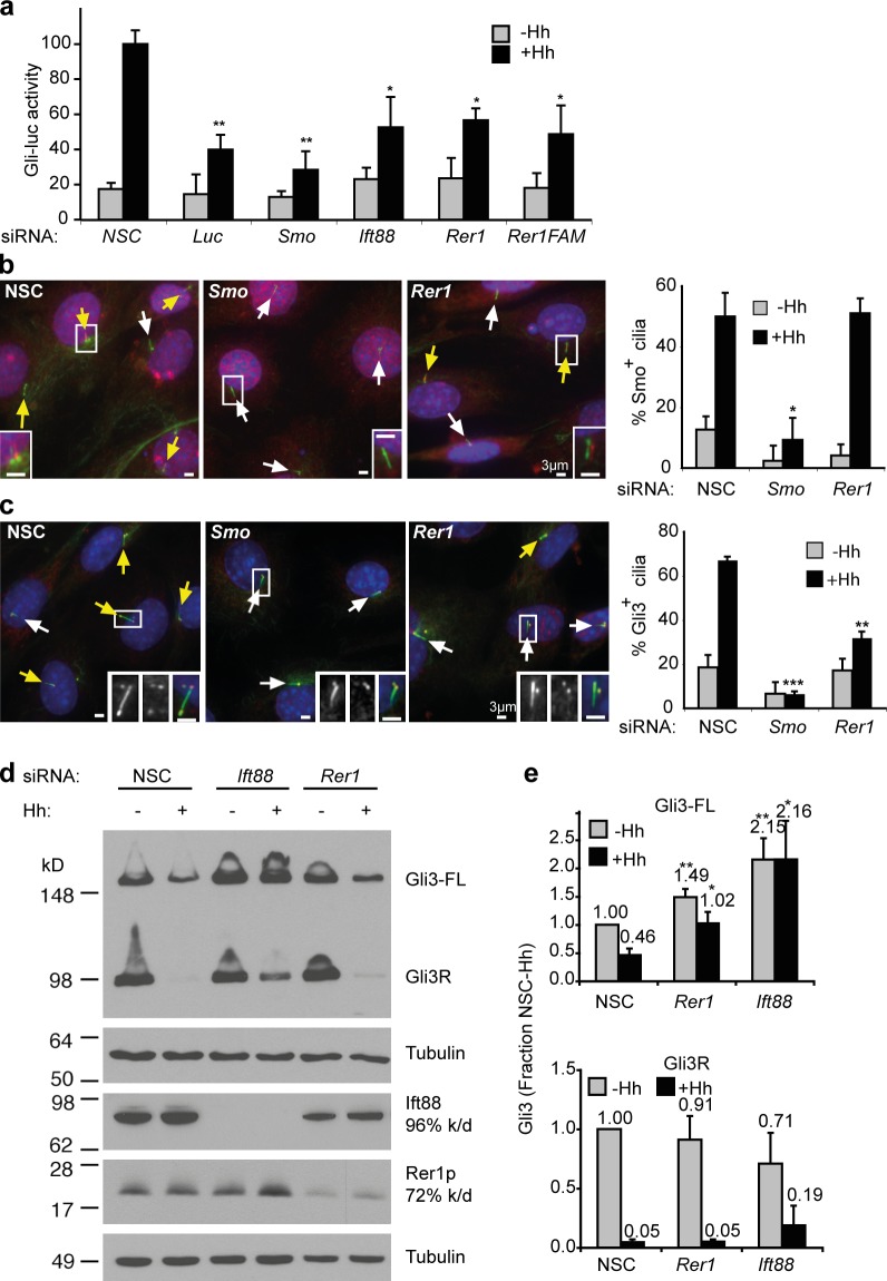
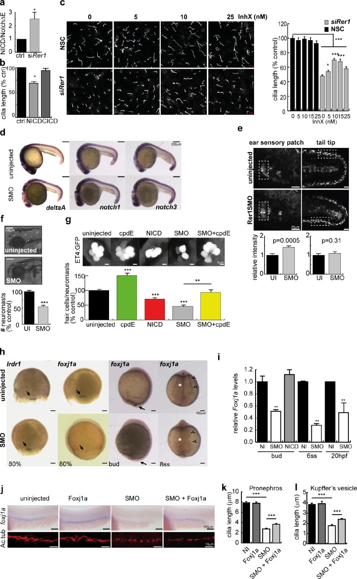
Cilia project from the surface of most vertebrate cells and are important for several physiological and developmental processes. Ciliary defects are linked to a variety of human diseases, named ciliopathies, underscoring the importance of understanding signaling pathways involved in cilia formation and maintenance. In this paper, we identified Rer1p as the first endoplasmic reticulum/cis-Golgi-localized membrane protein involved in ciliogenesis. Rer1p, a protein quality control receptor, was highly expressed in zebrafish ciliated organs and regulated ciliary structure and function. Both in zebrafish and mammalian cells, loss of Rer1p resulted in the shortening of cilium and impairment of its motile or sensory function, which was reflected by hearing, vision, and left-right asymmetry defects as well as decreased Hedgehog signaling. We further demonstrate that Rer1p depletion reduced ciliary length and function by increasing γ-secretase complex assembly and activity and, consequently, enhancing Notch signaling as well as reducing Foxj1a expression.


Figures

Figure 1.
Figure 1.
Rer1p is highly expressed in ciliated organs and affects ciliogenesis in zebrafish. (a) WISH (iiii) and immunostaining (iv–vi) show that rer1 is highly expressed in ciliated organs: KV (delineated with a dashed box in i), lateral views of the otic vesicle (OV; different hybridization probe in inset; ii), ALL and PLL neuromasts (iiv), and pronephros (delineated with dashed lines in vi). (b) Rer1p knockdown results in a “curly tail down” phenotype after 24 hpf. (c) Ciliary defects in SMO ALL neuromast cells at 48 hpf (confocal) and 72 hpf (scanning EM; zoomed area of boxes in right images). Rer1p staining is absent in SMO. (d) Pronephric cilia are shortened in Rer1p morphants (SMO, ATGMO, and UTRMO) compared with control (uninjected and p53MO) and rescued upon reexpression of Rer1p (n > 9; three experiments). (e) Ciliary defects of olfactory pits in SMO at 48 hpf (confocal; scanning EM). Boxed areas are enlarged in the bottom images. (f) Short cilia in the inner ear sensory patches of SMO at 24 hpf (indicated with arrows) and 48 hpf. (g) Reduced cilia length in the KV (10-somite stage [ss]) of SMO/ATGMO (n = 10). (h, left) Impaired photoreceptor outer segments (OS) in 4-dpf SMO. OPL, outer plexiform layer; PR, photoreceptor; RPE, retinal pigment epithelium. (right) EM of OS of 4-dpf larvae. Decreased number and mean size of the outer segments in SMO (n = 4 embryos). uninj., uninjected. In all panels except h, cilia are visualized using anti–acetylated tubulin (ac.tub) staining. Means ± SD (h) or SEM (d and g); ***, P < 0.001, Wilcoxon rank sum test (h) or Student’s t test (d and g). Bars: (a) 100 µm; (b) 500 µm.
Figure 2.
Figure 2.
Functional impairment of the ciliated organs in Rer1p morphant embryos. (a) Defective otolith formation in the inner ear of SMO. (b) Reduced or absent optokinetic response of 4.5-dpf SMO. The relative angle of eye rotation is shown for a control and two SMO larvae (n > 10). (c) Impaired retinal activity as measured by ERG in 4.5-dpf SMO; mean ± SEM is represented by shading. Decreased photoreceptor (PR) response and concomitant ON bipolar response (n ≥ 16). (d) Directional fluid flow in the KV is absent in the SMO as shown by fluorescent bead tracking (see Videos 3 and 4). Colored tracks show individual bead movements. (e) Randomization or loss of expression of Nodal antagonists lefty1 and lefty2 in SMO, ATGMO, and UTRMO as well as in DFC-specific injection of ATGMO and UTRMO at 20 hpf. Bars indicate percentages of embryos with expression on either side, both sides, or absent (n ≥ 90). UI, uninjected. (f) Reversal or absence of heart looping in SMO at 48 hpf. Bars indicate percentages of normal, reverse, or no heart looping. (g) Dorsal view of Alcian-stained cartilage of zebrafish heads. Arrowheads point to changes in pharyngeal cartilage chondrocyte number/arrangement; morphometric differences indicated by arrows are quantified in h. Means ± SEM (c) or SD (h); *, P < 0.05; **, P < 0.01, Wilcoxon rank sum test (c) or Student’s t test (h).
Figure 3.
Figure 3.
The regulatory role of Rer1p in cilia formation is conserved in mammalian cells. (a) Anti–acetylated tubulin staining and scanning EM demonstrates shorter cilia after rer1 knockdown in CL4 cells. (b) Efficiency of rer1 knockdown by RNAi (siRer1) in CL4 cells compared with nonspecific control (NSC) using at least three Western blots (normalized to GAPDH). (c) Unaffected number of ciliated cells upon rer1 knockdown in CL4. (d) Twofold decrease in mean cilia length upon rer1 knockdown in CL4 (n > 684). (e) Shorter cilia after rer1 down-regulation in RPE cells are rescued upon reexpression of HA-tagged Rer1p. Western blot is shown in g. Quantification (n ≥ 95) shown in f. Means ± SEM (d and f) or SD (b). ***, P < 0.001, Wilcoxon rank sum test.
Figure 4.
Figure 4.
Rer1p knockdown in S12 cells decreases Hh signaling. (a) Gli luciferase reporter assay reveals that Rer1p depletion inhibits Hh signaling similarly to Ift88 knockdown but less than depletion of Smoothened (Smo) or luciferase (Luc); Rer1F is a FAM-labeled Rer1 siRNA. NSC, nonspecific control. (b, left) Smoothened is transported normally to the cilia of Rer1p-deficient cells after 21-h Hh stimulation. (insets) Magnification of boxed areas with the red Smoothened channel displaced six pixels to the right. Arrows point to tips of cilia: white for Smoothened and yellow for Smoothened+ cilia. (right) Quantification of Smoothened translocation to cilia. (c, left) Rer1p inhibits ciliary accumulation of Gli3 (red). Arrows point to cilia tips: white for Gli3 and yellow for Gli3+ cilia. (right) Quantification of Gli3 accumulation at cilia tips. (b and c, insets) Bars, 3 µm. (d) Hh-dependent Gli3 degradation, but not processing, is inhibited by Rer1 depletion. (e) Representative Western blot and quantification (n = 4 independent experiments) of Gli3FL and Gli3R normalized to tubulin. Acetylated and γ-tubulin are shown in green, and DAPI is shown in blue (b and c). Represented are means ± SD of more than three experiments. *, P ≤ 0.05; **, P ≤ 0.01 by Student’s t test.
Figure 5.
Figure 5.
Reduced cilium length is caused by enhanced γ-secretase activity and diminished Foxj1a levels. (a) 2.5-fold increased γ-secretase activity in Rer1p-depleted CL4 cells as indicated by increased NICD production (n = 7). ctrl, control. (b) Overexpression of NICD but not of Cadherin ICD (CICD) reduces cilium length in CL4 cells. (c) Nanomolar concentrations of γ-secretase inhibition (InhX) rescue cilia length in Rer1p-depleted CL4 cells without affecting cilia length in control cells. Acetylated tubulin staining (n = 150; three experiments). NSC, nonspecific control. (d) Reduced deltaA but unaffected notch1 and notch3 levels in 24-hpf SMO. (e) Enhanced Notch signaling in the ear sensory patch, but not in the tail, indicated by increased fluorescence of the Notch reporter line Tp1:hmgb1-mCherry. Quantification is shown of highlighted regions (n > 15). UI, uninjected. (f) FM1-43 dye labeling shows a twofold reduction in the number of neuromasts in 3-dpf SMO. (g) Twofold reduction in the number of hair cells per neuromast (PLL) in 2-dpf SMO-injected ET4 transgenic embryos. Increased Notch signaling (100 pg NICD mRNA) results in less hair cells per neuromast (71% of control), whereas inhibiting Notch signaling (25 µM compound E [cpdE]) increases hair cell number (150% of control) and rescues the effect of SMO from 46 to 93.6% (n > 12). (h) WISH shows normal differentiation of the DFC/KV lineage (lrdr1) but reduced foxj1a levels in DFC (80% epiboly, arrows), KV (bud, arrows), pronephros (arrowheads), and notochord (asterisks). ss, somite stage. (i) Quantification of foxj1a levels by quantitative PCR in SMO. NICD does not affect foxj1a levels in the bud. NI, not injected. (j–l) Partial rescue of pronephros (j and k) and KV (l) cilia length upon coinjection of Foxj1a (n ≥ 11 embryos; three experiments). Ac.tub, acetylated tubulin. Means ± SEM; *, P < 0.05; **, P < 0.01; ***, P < 0.001, Wilcoxon rank sum test (b and c) or Student’s t test (a, e, g, i, k, and l).

Similar articles

Cited by

References

    1. Daudet N., Lewis J. 2005. Two contrasting roles for Notch activity in chick inner ear development: specification of prosensory patches and lateral inhibition of hair-cell differentiation. Development. 132:541–551 10.1242/dev.01589 - DOI - PubMed
    1. De Strooper B., Annaert W. 2010. Novel research horizons for presenilins and γ-secretases in cell biology and disease. Annu. Rev. Cell Dev. Biol. 26:235–260 10.1146/annurev-cellbio-100109-104117 - DOI - PubMed
    1. De Strooper B., Annaert W., Cupers P., Saftig P., Craessaerts K., Mumm J.S., Schroeter E.H., Schrijvers V., Wolfe M.S., Ray W.J., et al. 1999. A presenilin-1-dependent gamma-secretase-like protease mediates release of Notch intracellular domain. Nature. 398:518–522 10.1038/19083 - DOI - PubMed
    1. Eggenschwiler J.T., Anderson K.V. 2007. Cilia and developmental signaling. Annu. Rev. Cell Dev. Biol. 23:345–373 10.1146/annurev.cellbio.23.090506.123249 - DOI - PMC - PubMed
    1. Ellgaard L., Helenius A. 2003. Quality control in the endoplasmic reticulum. Nat. Rev. Mol. Cell Biol. 4:181–191 10.1038/nrm1052 - DOI - PubMed

Publication types

MeSH terms