. 2013 May 15;4(5):761-71.
doi: 10.1021/cn400031v. Epub 2013 Mar 26.

Controlled iontophoresis coupled with fast-scan cyclic voltammetry/electrophysiology in awake, freely moving animals

Affiliations

Controlled iontophoresis coupled with fast-scan cyclic voltammetry/electrophysiology in awake, freely moving animals

Anna M Belle et al. ACS Chem Neurosci. .

Abstract

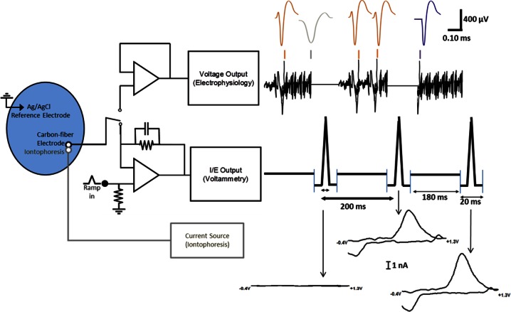
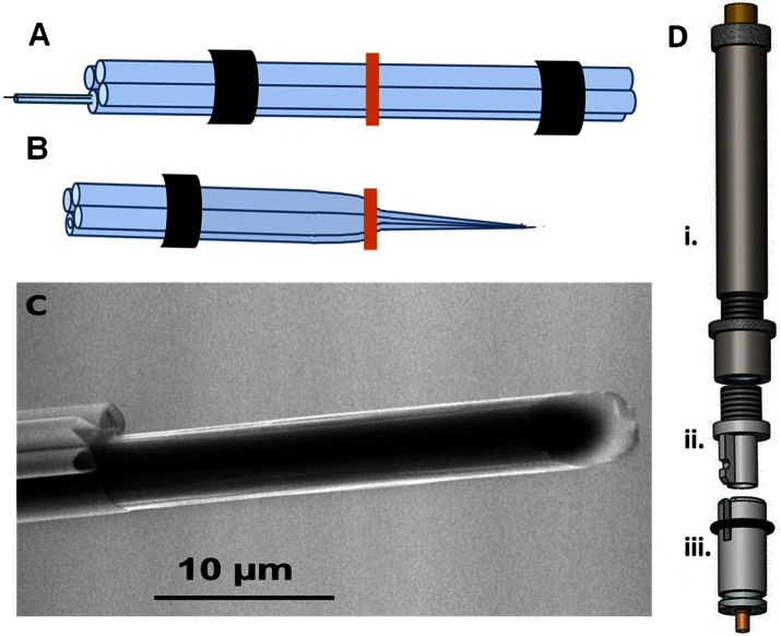
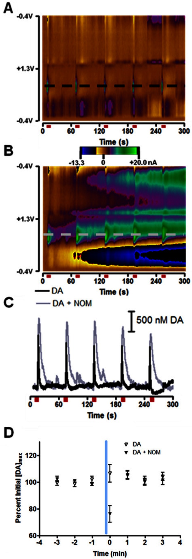
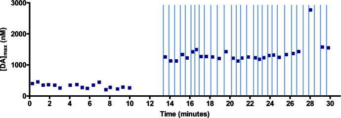
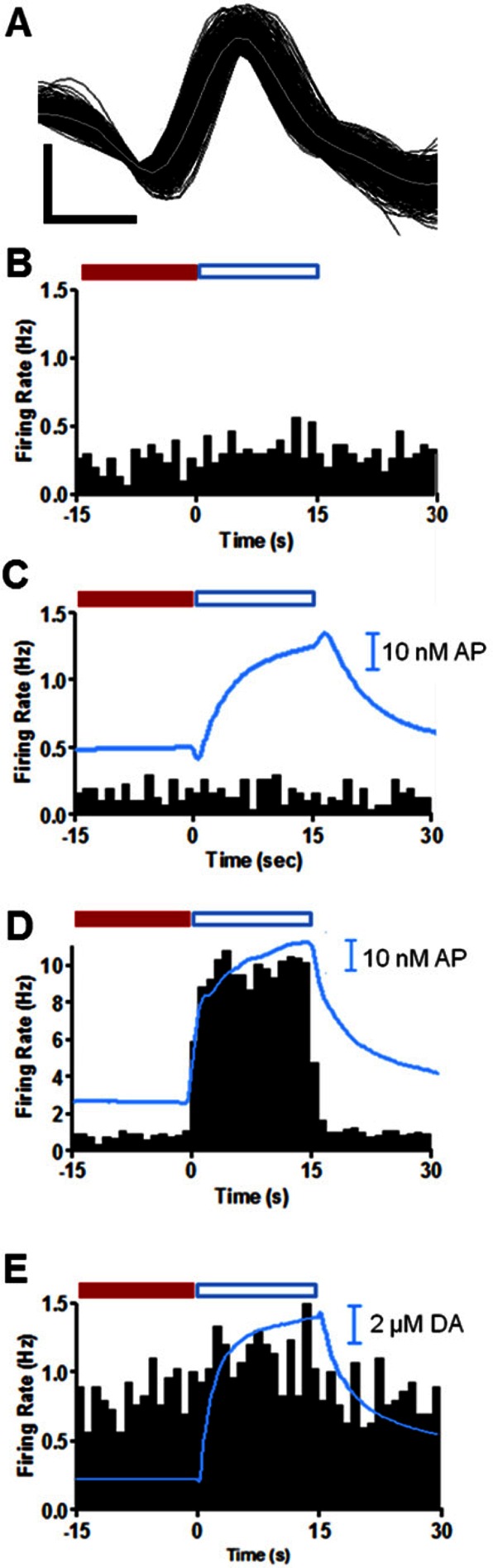
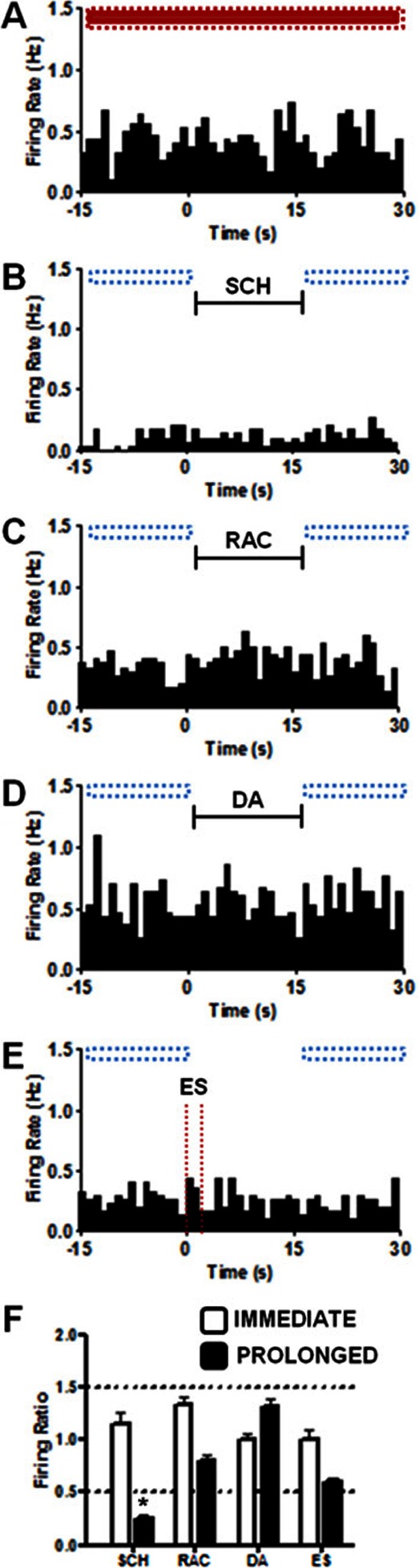
Simultaneous electrochemical and electrophysiological data were recorded to evaluate the effects of controlled local application of dopaminergic agonists and antagonists in awake rats. Measurements were made with a probe consisting of a carbon-fiber microelectrode fused to three iontophoretic barrels used to introduce the drugs of interest. The probe and the manipulator used to position it in the brain of behaving animals were optimized to improve their performance. The effect of the dopamine autoreceptor on electrically stimulated release was demonstrated. Dopamine inhibited the release of endogenous dopamine whereas raclopride, a D2 antagonist, enhanced it, with similar responses in anesthetized and awake animals. We also examined changes in the firing rate of nucleus accumbens (NAc) neurons in awake animals during and after brief (15 s) iontophoretic ejections of SCH 23390 (D1 receptor antagonist) or raclopride. Changes in response to these antagonists were seen both immediately and on a prolonged time scale. Application of raclopride increased the firing rate in 40% of medium spiny neurons (MSNs), of which half responded immediately. Decreases in firing rate were observed in 46% of MSNs after SCH 23390 application. Only 11% of MSNs responded to both antagonists and one MSN (3%) showed no response to either drug. The same prolonged response in firing rate was seen for electrically stimulated and locally applied dopamine in 75% of MSNs. These results are in agreement with previously reported distributions for dopamine receptor subtypes on MSNs and probe the effects of dopamine on these cell populations.


Figures

Figure 1
Figure 1
Diagram of the electronics and outputs for combined fast-scan cyclic voltammetry/electrophysiology. The same carbon-fiber electrode and reference electrode were used for both circuits and the reference electrode also served as ground for the iontophoresis circuit. The output of the carbon-fiber electrode was connected to a voltage follower to monitor cell firing (upper circuitry). Every 200 ms, the carbon-fiber microelectrode was switched from the electrophysiology circuit to the lower voltammetry circuitry for 20 ms. In this position, the amplifier controlled the electrode potential and the current was monitored. The synchronized outputs for both circuits are shown to the right of the circuitry. When the lower circuit was completed, the electrode was held at −0.4 V then over 8.5 ms scanned from to +1.3 V and back to −0.4 V. The changes in current during each scan are indicated with arrows. During this time, there were no voltage changes seen in the electrophysiology (when circuit was open but program still recorded data). With the upper circuit connected, changes in voltage were monitored. The colored marks above the voltage read out indicate regions of the 180 ms that have been expanded above. Orange spikes are firing of the MSN of interest; purple spikes are firing that looks similar to the MSN of interest but are not included due to the horizontal portion at the start of the spike. The gray spike is an example of an excluded event.
Figure 2
Figure 2
Modifications to probe construction and hardware for iontophoresis in freely moving animals. (A) Four-barrel prefused pipet ready for pulling with heat shrink on either end where the glass will contact the chucks of the puller. A smaller capillary containing a T-650 carbon fiber is loaded into one barrel. (B) One of the two electrodes created from the two-pull electrode making technique. The vertical bar indicates the midpoint of the pipet before pulling. (C) Environmental scanning electron micrograph of the probe tip showing the glass iontophoresis barrels and carbon fiber. (D) Hardware used to attach and lower iontophoresis probe into the head of a freely moving animal. (i) Commercially available Biela manipulator from Crist Instrument Co. (Part# 3-MMB-3D). (ii) Custom machined adapter that allows use of Biela manipulator with a guide cannula from Bioanalytical Systems, Inc. fabricated by UNC Physics Machine Shop (Chapel Hill, NC). (iii) Commercially available guide cannula from Bioanalytical Systems.
Figure 3
Figure 3
Effect of dopamine transporter on exogenous dopamine diffusion. (A) Two-dimensional color plot where current is shown in false color on the potential vs time axes for current changes in the nucleus accumbens (NAc). The positive (green) currents are indicative of dopamine release. Anesthetized rats received a 24 pulse, 125 μA, 60 Hz electrical stimulation every 60 s, and maximum dopamine concentration ([DA]max) evoked with each stimulation recorded. Stimulations are indicated by red bars below the color plot. (B) In the same animal, the same experiment was carried out as in (A) but 54 ± 2 μM nomifensine (NOM) was constantly iontophoresed during the stimulations. (C) Dopamine (DA) oxidation currents as a function of time. Traces were taken from the potentials marked with dashed lines in panels (A) and (B). (D) 15 s ejection of 16 ± 1 μM dopamine occurred at the vertical blue line and ended 10 s before electrical stimulation at t = 0 s. Open triangles indicate dopamine ejections, while black triangles indicate dopamine ejections made during constant application of nomifensine. [DA]max evoked was greater in the presence of nomifensine, but all data is presented as percent of pre-ejection [DA]max for each data set.
Figure 4
Figure 4
Consistent modulation of electrically evoked dopamine release with raclopride antagonism of the presynaptic D2 receptor during ICSS in a behaving rat. Stimulated release of dopamine occurred each time the animal pressed a lever, and the maximum amplitude was recorded as the maximum dopamine concentration ([DA]max). The lever was made available to the animal once every 18–27 s. Raclopride iontophoretic ejections (15 s, 100 nM) began at 13 min. The ejections terminated 3 s before the lever became available (indicated by vertical lines).
Figure 5
Figure 5
Immediate response of a single NAc MSN to iontophoretic application of dopamine receptor antagonists, SCH 23390 and raclopride, and dopamine in an awake animal. (A) Sample waveforms (150) shown in black with the average of the waveforms in gray for a NAc MSN collected in these experiments. The horizontal scale bar represents 0.3 ms, and vertical scale bar is 300 μV. (B) MSN firing with no iontophoretic ejections. (C) MSN firing during iontophoresis of 23 nM SCH 23390 for 15 s starting at t = 0 s. (D) MSN firing with 40 nM raclopride iontophoresed for 15 s starting at t = 0 s. (E) MSN firing with 12 μM of dopamine iontophoresed for 15 s starting at t = 0 s. Histograms are the average of 30 consecutive trials (60 s duration) and each bar is the average frequency recorded during the indicated 1 s bin. All histograms were collected in the same location in the NAc of the same animal. The blue traces superimposed over histograms are the average concentration of acetaminophen (AP) (C, D) or dopamine (DA) (E) ejected during the 45 s window shown for all 30 trials. In panels (C)–(E), iontophoretic drug delivery started at t = 0 s and ended at t = 15 s. Immediate changes in firing ratio were determined by comparison of cell firing during a baseline period (−15 to 0 s, solid red bar) to the average firing rate during the ejection (blue boxed region, 0 to 15 s). A quotient that differed from the control by ±0.5 was taken as an immediate change in cell firing rate.
Figure 6
Figure 6
Firing rates measured at another MSN indicating the prolonged analysis method. Each histogram was constructed from the results of 30 trials for each condition, (A) Control where no event occurs (B) 15 s SCH 23390 (SCH) ejection, indicated by back bar (C) 15 s raclopride (RAC) ejection, indicated by back bar (D) 15s dopamine (DA) ejection, indicated by back bar (E) 60 Hz, 24 p electrical stimulation (ES), indicated with dashed vertical lines. The horizontal boxes above each histogram indicate where comparisons were made to determine prolonged changes in firing rate. Red indicates baseline (−15 to 30 s in panel A) and blue outlined boxes indicate regions compared to baseline (−15 to 0 s and 15 to 30 s). (F) Summary graph of immediate and prolonged changes in firing ratio from the single unit in panels (A)–(E). Prolonged firing ratios were determined by dividing the average firing rate during periods with blue boxes over them in a panel by the red boxed region in panel (A). Immediate firing ratios were determined as described in Figure 5 and the text. A quotient greater than 1.5 or less than 0.5 qualified as a change in frequency of cell firing. Horizontal dashed lines enclose the region where changes in firing ratio are less than 50% of the original signal. The only effect seen at this cell was a prolonged depression to SCH23390 as indicated by the asterisk (*) in panel (F).

References

    1. Beyene M.; Carelli R. M.; Wightman R. M. (2010) Cue-evoked dopamine release in the nucleus accumbens shell tracks reinforcer magnitude during intracranial self-stimulation. Neuroscience 169, 1682–1688. - PMC - PubMed
    1. Day J. J.; Jones J. L.; Wightman R. M.; Carelli R. M. (2010) Phasic nucleus accumbens dopamine release encodes effort- and delay-related costs. Biol. Psychiatry 68, 306–309. - PMC - PubMed
    1. Owesson-White C. A.; Ariansen J.; Stuber G. D.; Cleaveland N. A.; Cheer J. F.; Wightman R. M.; Carelli R. M. (2009) Neural encoding of cocaine-seeking behavior is coincident with phasic dopamine release in the accumbens core and shell. Eur. J. Neurosci. 30, 1117–1127. - PMC - PubMed
    1. Phillips P. E.; Stuber G. D.; Heien M. L.; Wightman R. M.; Carelli R. M. (2003) Subsecond dopamine release promotes cocaine seeking. Nature 422, 614–618. - PubMed
    1. Robinson D. L., and Wightman R. M. (2007) Rapid dopamine release in freely moving rats. In Electrochemical Methods for Neuroscience (Michael A. C., and Borland L. M., Eds.), pp 17–36, CRC Press, Boca Raton, FL. - PubMed

Publication types