Review
. 2013 Jan-Feb;50(1):23-36.
doi: 10.3109/10408363.2013.764490.

Biomarkers of sepsis

Affiliations
Review

Biomarkers of sepsis

James D Faix. Crit Rev Clin Lab Sci. 2013 Jan-Feb.

Abstract

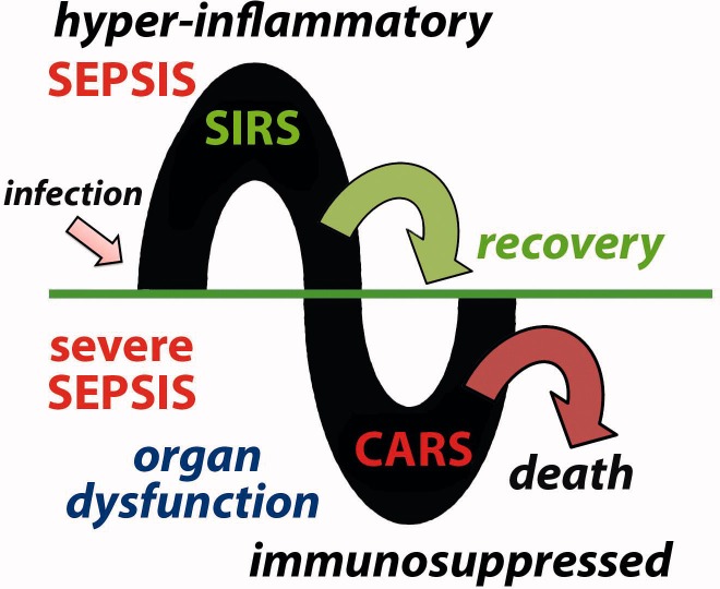
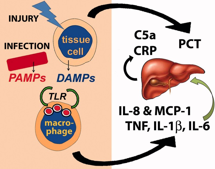
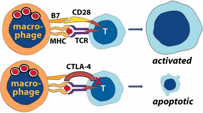
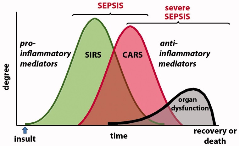
Sepsis is an unusual systemic reaction to what is sometimes an otherwise ordinary infection, and it probably represents a pattern of response by the immune system to injury. A hyper-inflammatory response is followed by an immunosuppressive phase during which multiple organ dysfunction is present and the patient is susceptible to nosocomial infection. Biomarkers to diagnose sepsis may allow early intervention which, although primarily supportive, can reduce the risk of death. Although lactate is currently the most commonly used biomarker to identify sepsis, other biomarkers may help to enhance lactate's effectiveness; these include markers of the hyper-inflammatory phase of sepsis, such as pro-inflammatory cytokines and chemokines; proteins such as C-reactive protein and procalcitonin which are synthesized in response to infection and inflammation; and markers of neutrophil and monocyte activation. Recently, markers of the immunosuppressive phase of sepsis, such as anti-inflammatory cytokines, and alterations of the cell surface markers of monocytes and lymphocytes have been examined. Combinations of pro- and anti-inflammatory biomarkers in a multi-marker panel may help identify patients who are developing severe sepsis before organ dysfunction has advanced too far. Combined with innovative approaches to treatment that target the immunosuppressive phase, these biomarkers may help to reduce the mortality rate associated with severe sepsis which, despite advances in supportive measures, remains high.


Figures

Figure 1.
Figure 1.
Sepsis may be divided into two phases. Following infection, a hyper-inflammatory phase is characterized by SIRS. This may resolve or the patient may progress to what is called severe sepsis. During this phase, there is evidence of CARS with immunosuppression and multiple organ dysfunction. This may also resolve, especially with appropriate support, but it often leads to death.
Figure 2.
Figure 2.
Sepsis begins with either infection or tissue injury. PAMPs from invading organisms or DAMPs from injured tissue cells (or both) are recognized by macrophage receptors such as the TLRs. This results in the production of pro-inflammatory cytokines such as TNF, IL-1β and IL-6 and chemokines such as IL-8 and MCP-1. IL-6 stimulates the liver to produce CRP and complement proteins. Many cells in the body also produce PCT in response to both infection and injury.
Figure 3.
Figure 3.
Activated inflammatory cells up-regulate a number of proteins which may be detected as biomarkers of sepsis, either on the cell surface or as soluble forms in plasma. (a) An unstimulated PMN; (b) a stimulated PMN with darker (“toxic”) granules and a Dohle body (arrow); (c) frequently utilized biomarkers of sepsis related to PMNs include CD64, the soluble forms of TREM-1 and CD11b, and HBP. (d) Frequently utilized biomarkers of sepsis related to macrophages or monocytes include the soluble forms of CD14 (which facilitates recognition of bacterial lipopolysaccharides) and the receptor for RAGE.
Figure 4.
Figure 4.
There is significant evidence that patients with severe sepsis have defective adaptive immunity. Macrophages (or monocytes) may lose expression of the Class II MHC proteins which display foreign peptide to the TCR. However, more importantly, T-cells upregulate expression of CTLA-4, an alternative ligand for the co-stimulator B7 on the antigen-presenting cell. Instead of providing co-stimulation and activation of the T-cell, which would occur if B7 interacted with CD28, interaction with CTLA-4 results in T-cell unresponsiveness and, eventually, death by apoptosis.
Figure 5.
Figure 5.
An alternative model for the progression of sepsis to severe sepsis proposes that the CARS begins while the pro-inflammatory SIRS is still present. Understanding the interplay of these opposing features may help investigators discover the pathogenesis of the organ dysfunction that occurs in patients who develop severe sepsis (and die).

References

    1. Levy MM, Dellinger RP, Townsend SR, et al. The Surviving Sepsis Campaign: results of an international guideline-based performance improvement program targeting severe sepsis. Intensive Care Med. 2010;36:222–31. - PMC - PubMed
    1. Hall MJ, Williams SN, DeFrances CJ, Golosinsky A. Inpatient care for septicemia or sepsis: a challenge for patients and hospitals. National Center for Health Statistics Data Brief No. 62. U.S. Department of Health and Human Services, Centers for Disease Control and Prevention, Jun. 2011. http://www.cdc.gov/nchs/data/databriefs/db62.htm Available from: [last accessed 2 Jan 2013]
    1. Wang HE, Shapiro NI, Angus DC, Yealy DM. National estimates of severe sepsis in United States emergency departments. Crit Care Med. 2007;35:1928–36. - PubMed
    1. Ulevitch RJ, Tobias PS. Recognition of gram-negative bacteria and endotoxin by the innate immune system. Curr Opin Immunol. 1999;11:19–22. - PubMed
    1. Kumar H, Kawai T, Akira S. Pathogen recognition by the innate immune system. Int Rev Immunol. 2011;30:16–34. - PubMed