

Molecular and cellular pathology of very-long-chain acyl-CoA dehydrogenase deficiency
- PMID: 23480858
- PMCID: PMC3628282
- DOI: 10.1016/j.ymgme.2013.02.002
Molecular and cellular pathology of very-long-chain acyl-CoA dehydrogenase deficiency
Abstract
Background: Very-long-chain acyl-CoA dehydrogenase (VLCAD) deficiency (VLCADD) is diagnosed in the US through newborn screening (NBS). NBS often unequivocally identifies affected individuals, but a growing number of variant patterns can represent mild disease or heterozygous carriers.
Aims: To evaluate the validity of standard diagnostic procedures for VLCADD by using functional in vitro tools.
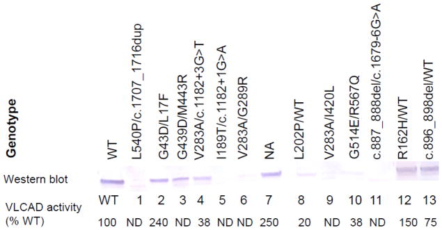
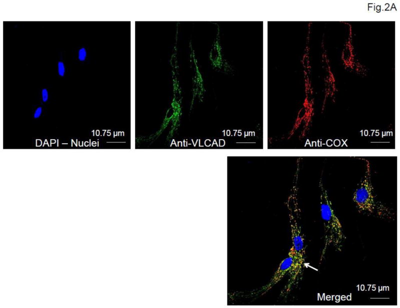
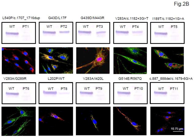
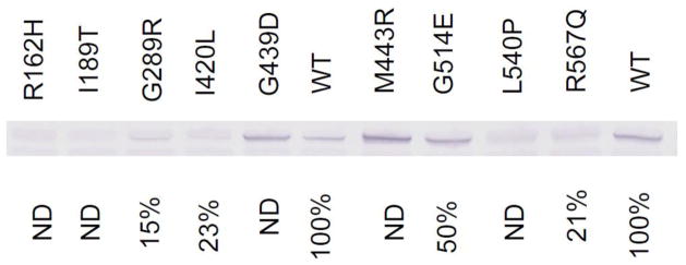
Methods: We retrospectively investigated 13 patient samples referred to our laboratory because of a suspicion of VLCADD but with some uncertainty to the diagnosis. All 13 patients were suspected of having VLCADD either because of abnormal NBS or suggestive clinical findings. ACADVL genomic DNA sequencing data were available for twelve of them. Ten of the patients had an abnormal NBS suggestive of VLCADD, with three samples showing equivocal results. Three exhibited suggestive clinical findings and blood acylcarnitine profile (two of them had a normal NBS and the third one was unscreened). Assay of VLCAD activity and immunoblotting or immunohistologic staining for VLCAD were performed on fibroblasts. Prokaryotic mutagenesis and expression studies were performed for nine uncharacterized ACADVL missense mutations.
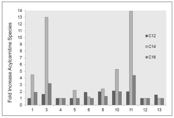
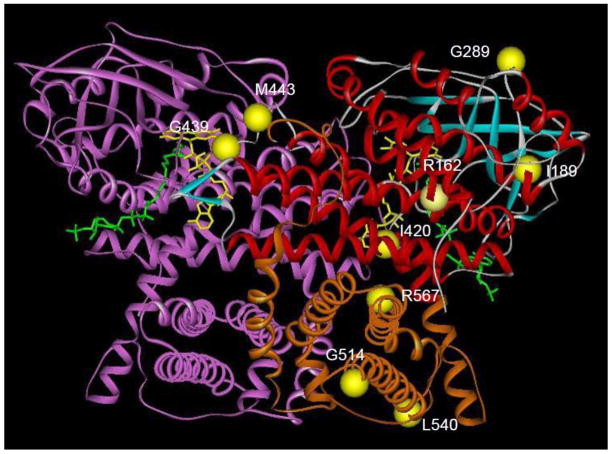
Results: VLCAD activity was abnormal in fibroblast cells from 9 patients (8 identified through abnormal NBS, 1 through clinical symptoms). For these 9 patients, immunoblotting/staining showed the variable presence of VLCAD; all but one had two mutated alleles. Two patients with equivocal NBS results (and a heterozygous genotype) and the two patients with normal NBS exhibited normal VLCAD activity and normal VLCAD protein on immunoblotting/staining thus ruling out VLCAD deficiency. Nine pathogenic missense mutations were characterized with prokaryotic expression studies and showed a decrease in enzyme activity and variable stability of VLCAD antigen.
Conclusions: These results emphasize the importance of functional investigation of abnormal NBS or clinical testing suggestive but not diagnostic of VLCADD. A larger prospective study is necessary to better define the clinical and metabolic ramifications of the defects identified in such patients.
Copyright © 2013 Elsevier Inc. All rights reserved.
Figures
Similar articles
-
Recurrent ACADVL molecular findings in individuals with a positive newborn screen for very long chain acyl-coA dehydrogenase (VLCAD) deficiency in the United States.Mol Genet Metab. 2015 Nov;116(3):139-45. doi: 10.1016/j.ymgme.2015.08.011. Epub 2015 Sep 2. Mol Genet Metab. 2015. PMID: 26385305 Free PMC article.
-
Impact of newborn screening for very-long-chain acyl-CoA dehydrogenase deficiency on genetic, enzymatic, and clinical outcomes.J Inherit Metab Dis. 2019 May;42(3):414-423. doi: 10.1002/jimd.12075. Epub 2019 Apr 8. J Inherit Metab Dis. 2019. PMID: 30761551
-
Characterization of exonic variants of uncertain significance in very long-chain acyl-CoA dehydrogenase identified through newborn screening.J Inherit Metab Dis. 2022 May;45(3):529-540. doi: 10.1002/jimd.12492. Epub 2022 Mar 11. J Inherit Metab Dis. 2022. PMID: 35218577 Free PMC article.
-
The Pathogenesis of Very Long-Chain Acyl-CoA Dehydrogenase Deficiency.Biomolecules. 2025 Mar 14;15(3):416. doi: 10.3390/biom15030416. Biomolecules. 2025. PMID: 40149952 Free PMC article. Review.
-
Nutrition management guideline for very-long chain acyl-CoA dehydrogenase deficiency (VLCAD): An evidence- and consensus-based approach.Mol Genet Metab. 2020 Sep-Oct;131(1-2):23-37. doi: 10.1016/j.ymgme.2020.10.001. Epub 2020 Oct 6. Mol Genet Metab. 2020. PMID: 33093005 Review.
Cited by
-
Metabolic Outcomes of Anaplerotic Dodecanedioic Acid Supplementation in Very Long Chain Acyl-CoA Dehydrogenase (VLCAD) Deficient Fibroblasts.Metabolites. 2021 Aug 13;11(8):538. doi: 10.3390/metabo11080538. Metabolites. 2021. PMID: 34436479 Free PMC article.
-
Four Years' Experience in the Diagnosis of Very Long-Chain Acyl-CoA Dehydrogenase Deficiency in Infants Detected in Three Spanish Newborn Screening Centers.JIMD Rep. 2018;39:63-74. doi: 10.1007/8904_2017_40. Epub 2017 Jul 29. JIMD Rep. 2018. PMID: 28755359 Free PMC article.
-
ITCH deficiency clinical phenotype expansion and mitochondrial dysfunction.Mol Genet Metab Rep. 2022 Oct 29;33:100932. doi: 10.1016/j.ymgmr.2022.100932. eCollection 2022 Dec. Mol Genet Metab Rep. 2022. PMID: 36338154 Free PMC article.
-
Sturge-Weber syndrome coexisting with episodes of rhabdomyolysis.BMC Neurol. 2013 Nov 11;13:169. doi: 10.1186/1471-2377-13-169. BMC Neurol. 2013. PMID: 24207015 Free PMC article.
-
Acylcarnitines--old actors auditioning for new roles in metabolic physiology.Nat Rev Endocrinol. 2015 Oct;11(10):617-25. doi: 10.1038/nrendo.2015.129. Epub 2015 Aug 25. Nat Rev Endocrinol. 2015. PMID: 26303601 Free PMC article. Review.
References
-
- Vockley J, Whiteman DA. Defects of mitochondrial beta-oxidation: a growing group of disorders. Neuromuscul Disord. 2002;12:235–246. - PubMed
-
- McHugh DM, Cameron CA, Abdenur JE, Abdulrahman M, Adair O, Al Nuaimi SA, Ahlman H, Allen JJ, Antonozzi I, Archer S, Au S, Auray-Blais C, Baker M, Bamforth F, Beckmann K, Pino GB, Berberich SL, Binard R, Boemer F, Bonham J, Breen NN, Bryant SC, Caggana M, Caldwell SG, Camilot M, Campbell C, Carducci C, Cariappa R, Carlisle C, Caruso U, Cassanello M, Castilla AM, Ramos DE, Chakraborty P, Chandrasekar R, Ramos AC, Cheillan D, Chien YH, Childs TA, Chrastina P, Sica YC, de Juan JA, Colandre ME, Espinoza VC, Corso G, Currier R, Cyr D, Czuczy N, D’Apolito O, Davis T, de Sain-Van der Velden MG, Delgado Pecellin C, Di Gangi IM, Di Stefano CM, Dotsikas Y, Downing M, Downs SM, Dy B, Dymerski M, Rueda I, Elvers B, Eaton R, Eckerd BM, El Mougy F, Eroh S, Espada M, Evans C, Fawbush S, Fisher L, Franzson L, Frazier DM, Garcia LR, Bermejo MS, Gavrilov D, Gerace R, Giordano G, Irazabal YG, Greed LC, Grier R, Grycki E, Gu X, Gulamali-Majid F, Hagar AF, Han L, Hannon WH, Haslip C, Hassan FA, He M, Hietala A, Himstedt L, Hoffman GL, Hoffman W, Hoggatt P, Hopkins PV, Hougaard DM, Hughes K, Hunt PR, Hwu WL, Hynes J, Ibarra-Gonzalez I, Ingham CA, Ivanova M, Jacox WB, John C, Johnson JP, Jonsson JJ, Karg E, Kasper D, Klopper B, Katakouzinos D, Khneisser I, Knoll D, Kobayashi H, Koneski R, Kozich V, Kouapei R, Kohlmueller D, Kremensky I, la Marca G, Lavochkin M, Lee SY, Lehotay DC, Lemes A, Lepage J, Lesko B, Lewis B, Lim C, Linard S, Lindner M, Lloyd-Puryear MA, Lorey F, Loukas YL, Luedtke J, Maffitt N, Magee JF, Manning A, Manos S, Marie S, Hadachi SM, Marquardt G, Martin SJ, Matern D, Mayfield Gibson SK, Mayne P, McCallister TD, McCann M, McClure J, McGill JJ, McKeever CD, McNeilly B, Morrissey MA, Moutsatsou P, Mulcahy EA, Nikoloudis D, Norgaard-Pedersen B, Oglesbee D, Oltarzewski M, Ombrone D, Ojodu J, Papakonstantinou V, Reoyo SP, Park HD, Pasquali M, Pasquini E, Patel P, Pass KA, Peterson C, Pettersen RD, Pitt JJ, Poh S, Pollak A, Porter C, Poston PA, Price RW, Queijo C, Quesada J, Randell E, Ranieri E, Raymond K, Reddic JE, Reuben A, Ricciardi C, Rinaldo P, Rivera JD, Roberts A, Rocha H, Roche G, Greenberg CR, Mellado JM, Juan-Fita MJ, Ruiz C, Ruoppolo M, Rutledge SL, Ryu E, Saban C, Sahai I, Garcia-Blanco MI, Santiago-Borrero P, Schenone A, Schoos R, Schweitzer B, Scott P, Seashore MR, Seeterlin MA, Sesser DE, Sevier DW, Shone SM, Sinclair G, Skrinska VA, Stanley EL, Strovel ET, Jones AL, Sunny S, Takats Z, Tanyalcin T, Teofoli F, Thompson JR, Tomashitis K, Domingos MT, Torres J, Torres R, Tortorelli S, Turi S, Turner K, Tzanakos N, Valiente AG, Vallance H, Vela-Amieva M, Vilarinho L, von Dobeln U, Vincent MF, Vorster BC, Watson MS, Webster D, Weiss S, Wilcken B, Wiley V, Williams SK, Willis SA, Woontner M, Wright K, Yahyaoui R, Yamaguchi S, Yssel M, Zakowicz WM. Clinical validation of cutoff target ranges in newborn screening of metabolic disorders by tandem mass spectrometry: a worldwide collaborative project. Genet Med. 2011;13:230–254. - PubMed
-
- Andresen BS, Bross P, Vianey-Saban C, Divry P, Zabot MT, Roe CR, Nada MA, Byskov A, Kruse TA, Neve S, Kristiansen K, Knudsen I, Corydon MJ, Gregersen N. Cloning and characterization of human very-long-chain acyl-CoA dehydrogenase cDNA, chromosomal assignment of the gene and identification in four patients of nine different mutations within the VLCAD gene. Hum Mol Genet. 1996;5:461–472. - PubMed
-
- Andresen BS, Olpin S, Poorthuis BJ, Scholte HR, Vianey-Saban C, Wanders R, Ijlst L, Morris A, Pourfarzam M, Bartlett K, Baumgartner ER, deKlerk JB, Schroeder LD, Corydon TJ, Lund H, Winter V, Bross P, Bolund L, Gregersen N. Clear correlation of genotype with disease phenotype in very-long-chain acyl-CoA dehydrogenase deficiency. Am J Hum Genet. 1999;64:479–494. - PMC - PubMed
Publication types
MeSH terms
Substances
Supplementary concepts
Grants and funding
LinkOut - more resources
Full Text Sources
Other Literature Sources
Medical
Research Materials
Miscellaneous
