Review
. 2013:116:95-118.
doi: 10.1016/B978-0-12-394311-8.00005-4.

Structure, function, and regulation of desmosomes

Affiliations
Review

Structure, function, and regulation of desmosomes

Andrew P Kowalczyk et al. Prog Mol Biol Transl Sci. 2013.

Abstract

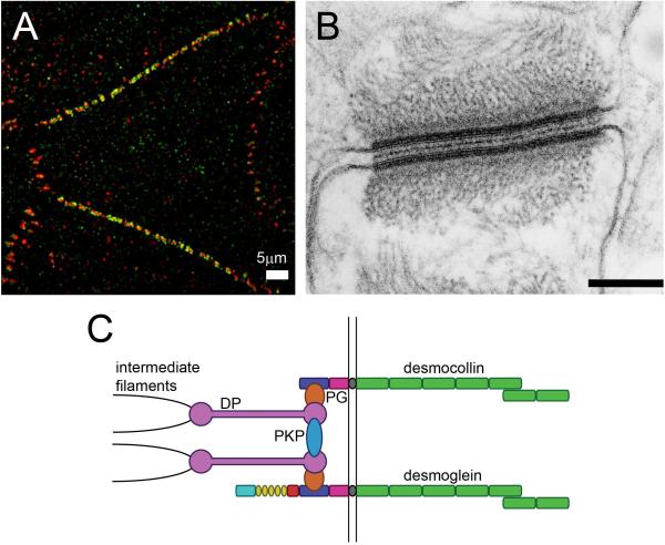
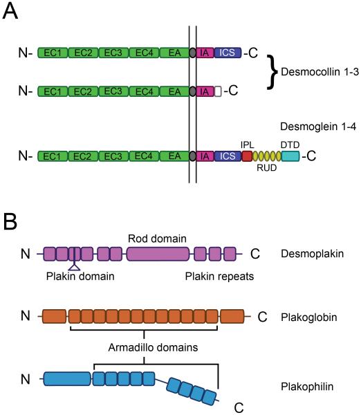
Desmosomes are adhesive intercellular junctions that mechanically integrate adjacent cells by coupling adhesive interactions mediated by desmosomal cadherins to the intermediate filament cytoskeletal network. Desmosomal cadherins are connected to intermediate filaments by densely clustered cytoplasmic plaque proteins comprising members of the armadillo gene family, including plakoglobin and plakophilins, and members of the plakin family of cytolinkers, such as desmoplakin. The importance of desmosomes in tissue integrity is highlighted by human diseases caused by mutations in desmosomal genes, autoantibody attack of desmosomal cadherins, and bacterial toxins that selectively target desmosomal cadherins. In addition to reviewing the well-known roles of desmosomal proteins in tissue integrity, this chapter also highlights the growing appreciation for how desmosomal proteins are integrated with cell signaling pathways to contribute to vertebrate tissue organization and differentiation.


Figures

Figure 1
Figure 1. Desmosome architecture and molecular composition
A. Super-resolution immunofluorescence localization of desmoglein-3 (red) and desmoplakin (green) in cultured keratinocytes obtained using structured illumination microscopy (Nikon N-SIM) B. Electron micrograph of a desmosome from bovine tongue epithelium. C. Molecular components and model of desmosomal protein organization (see Fig. 2 for domain annotations of desmosomal cadherins).Scale bar= 0.25 μm.
Figure 2
Figure 2. Domain structure of major desmosomal proteins
A. Desmosomal cadherins comprise five extracellular cadherin repeats (ECs), a single pass transmembrane domain, and an intracellular domain that associates with desmosomal plaque proteins (EA, extracellular anchor; IA, intracellular anchor; ICS, intracellular cadherin-like sequence; IPL, intracellular proline-rich linker; RUD, repeat unit domain; DTD, desmoglein terminal domain). B. Desmoplakin is a prototypical member of the plakin family with amino and carboxyl terminal globular domains joined by a central alpha-helical coiled-coil rod domain. C. Plakoglobin comprises amino and carboxyl terminal domains flanking 12 central armadillo repeat (Arm) domains. D. Plakophilins are also armadillo family proteins, but contain nine Arm repeats as well as amino and carboxyl terminal domains which can vary between isoforms as a result of alternative RNA splicing. Please refer to text for details of domain organization and function.

References

    1. Thomason HA, Scothern A, McHarg S, Garrod DR. Desmosomes: adhesive strength and signalling in health and disease. Biochemical Journal. 2010;429:419–433. - PubMed
    1. Delva E, Tucker DK, Kowalczyk AP. The desmosome. Cold Spring Harb Perspect Biol. 2009;1:a002543. - PMC - PubMed
    1. Saito MT, Kholhorst D, Niessen CM, Kowalczyk AP. Classical and Desmosomal Cadherins at a Glance. J Cell Science. 2012;125:2547–2552. D.K. - PMC - PubMed
    1. Amagai M, Stanley JR. Desmoglein as a target in skin disease and beyond. J Invest Dermatol. 2012;132:776–784. - PMC - PubMed
    1. Petrof G, Mellerio JE, McGrath JA. Desmosomal genodermatoses. Br J Dermatol. 2012;166:36–45. - PubMed

Publication types

Substances