. 2013 Apr 3;32(7):970-81.
doi: 10.1038/emboj.2013.50. Epub 2013 Mar 12.

p57 controls adult neural stem cell quiescence and modulates the pace of lifelong neurogenesis

Affiliations

p57 controls adult neural stem cell quiescence and modulates the pace of lifelong neurogenesis

Shohei Furutachi et al. EMBO J. .

Abstract

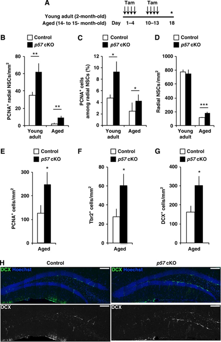
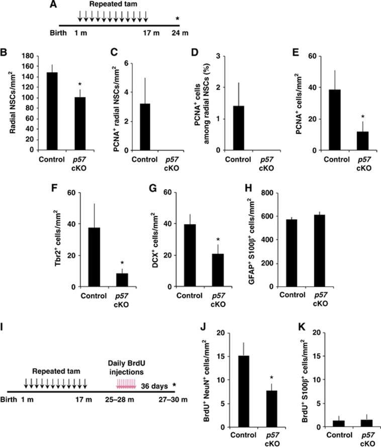
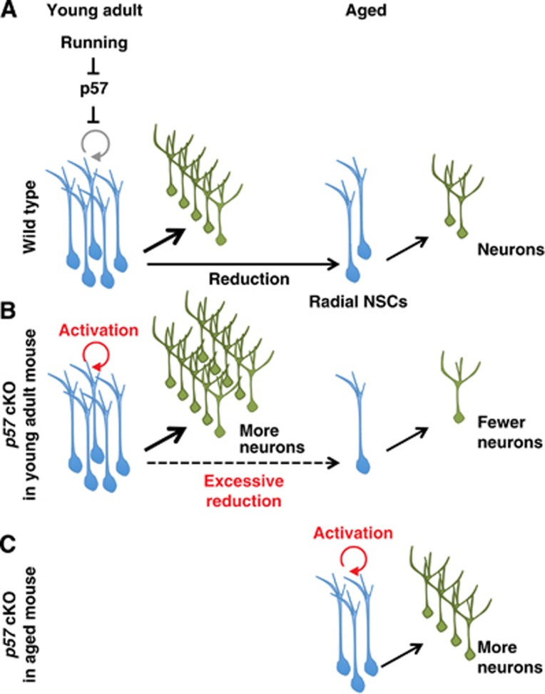
Throughout life, neural stem cells (NSCs) in the adult hippocampus persistently generate new neurons that modify the neural circuitry. Adult NSCs constitute a relatively quiescent cell population but can be activated by extrinsic neurogenic stimuli. However, the molecular mechanism that controls such reversible quiescence and its physiological significance have remained unknown. Here, we show that the cyclin-dependent kinase inhibitor p57(kip2) (p57) is required for NSC quiescence. In addition, our results suggest that reduction of p57 protein in NSCs contributes to the abrogation of NSC quiescence triggered by extrinsic neurogenic stimuli such as running. Moreover, deletion of p57 in NSCs initially resulted in increased neurogenesis in young adult and aged mice. Long-term p57 deletion, on the contrary, led to NSC exhaustion and impaired neurogenesis in aged mice. The regulation of NSC quiescence by p57 might thus have important implications for the short-term (extrinsic stimuli-dependent) and long-term (age-related) modulation of neurogenesis.


Conflict of interest statement

The authors declare that they have no conflict of interest.

Figures

Figure 1
Figure 1
Radial NSCs in the adult hippocampus express p57. (A) p57 is expressed in adult hippocampal radial NSCs. Immunohistochemistry on brain sections of 3-month-old mice showing GFAP (blue), Nestin (red), and p57 (green) triple-labelled cells projecting radially from the SGZ of the DG (arrowhead). Nuclei were counterstained with Hoechst. Scale bar, 5 μm. (BF) Expression of p57 in differentiated cell populations in the SGZ/GCL. Strong p57 staining was detected in DCX+ cells (D) and some S100β+ cells (F), but not in the majority of Ascl1+ IPCs (B), BrdU+ proliferating cells (2 h pulse, C) and NeuN+ mature neurons (E). Arrowheads indicate differentiated cell populations. Nuclei were counterstained with Hoechst (blue). Scale bar, 10 μm.
Figure 2
Figure 2
Conditional deletion of p57 promotes radial NSC proliferation. (A) Experimental design employed for the selective deletion of p57. A p57-floxed mouse and a Nestin-CreERT2 mouse were crossed to conditionally knockout p57 in the adult hippocampal NSCs. Two-month-old mice were administered with tamoxifen for 4 days and then sacrificed at day 5. p57 f/f; Nestin-CreERT2 mice and/or p57 +/f; Nestin-CreERT2 mice were used for p57 deletion. (BD) Acute activation of hippocampal radial NSCs after p57 conditional deletion. The numbers of PCNA+ radial NSCs (B), the proportion of PCNA+ cells among radial NSCs (C), and the PCNA+ cells in the SGZ/GCL (D) are shown. n=5 and 4 animals for control and p57 cKO, respectively. (E) Experimental design for quantification of the PCNA+ cells in the SGZ/GCL at 17 days after p57 deletion. (F) The number of PCNA+ cells in the SGZ/GCL was increased in p57 cKO mice at day 18 (n=5 and 4 animals for control and p57 cKO, respectively). (G) Experimental design for the quantification of quiescent radial NSCs. (H) Quantification of remaining radial NSCs immediately after AraC infusion. Four control and three p57 cKO mice were infused with AraC. As a control, three control and three p57 cKO mice were infused with PBS. (I) Representative images used in visualization and quantification of remaining radial NSCs after AraC treatment in (H) are shown. Scale bar, 25 μm. (J) Experimental design for the quantification of infrequently dividing radial NSCs. BrdU was injected 10 days after the administration of tamoxifen. Animals were sacrificed 36 days later. (K) Quantification of label-retaining radial NSCs. The number of BrdU+ radial NSCs (BrdU+ GFAP+ Nestin+ Radial fibre+) from SGZ in control (n=3) and p57 cKO mice (n=4) is shown. (L) Representative images used in visualization and quantification of label-retaining radial NSCs in (K) are shown. Arrowheads indicate BrdU+ radial NSCs. Close-ups are shown in the bottom. Asterisk indicates non-specific signal caused by cross-reaction of secondary antibody. Scale bar, 50 μm. (M) Experimental design for the quantification of the size of radial NSC pool at various time points after p57 deletion. (N) Transient expansion of radial NSCs after p57 deletion. The number of radial NSCs at various time points after p57 deletion is shown. Note that p57 deletion increased radial NSCs (left), but this expansion was transient (middle and right). Values represent mean±s.e.m. ***P<0.001; **P<0.01; *P<0.05; Student’s t-test.
Figure 3
Figure 3
Reduction of p57 contributes to radial NSC activation induced by running. (A) Two-month-old female mice initially housed in small cages without a running wheel were exposed to spacious housing with running wheels (run) or small housing (control) for 12 days and then sacrificed. (B) The staining intensity of p57 was measured in the nucleus of radial NSCs identified by their radial morphology and expression of GFAP and Nestin. The intensity of each nucleus was plotted for comparison. Red horizontal bar represents average staining intensity. n=3 for both control and run. (C) Two-month-old female mice initially housed in small cages without a running wheel were administered with tamoxifen and then exposed to spacious housing with running wheels (run) or small housing (control) for 12 days and then sacrificed. (D) Quantification of PCNA+ radial NSCs in the SGZ. Three control and p57 cKO mice were exposed to small housing and four control and three p57 cKO mice were exposed to spacious housing with running wheels. Values represent mean±s.e.m. ***P<0.001; *P<0.05; Student’s t-test.
Figure 4
Figure 4
Enhanced hippocampal neurogenesis by conditional deletion of p57. (A) Experimental design for the quantification of IPCs and neuroblasts/immature neurons after p57 deletion. Asterisk indicates time point of sacrifice. (BE) Quantification of number of Tbr2+ IPCs (B) and DCX+ neuroblasts/immature neurons (D) in the SGZ/GCL of control (n=5) and p57 cKO mice (n=3). The proportion of Ki67+ cells among Tbr2+ cells (n=3, 3, C) and PCNA+ cells among DCX+ cells (n=5, 3, E) are shown. (F) Representative images of DCX+ cells in p57 cKO and control mice. Scale bar, 100 μm. (G) Experimental design for the quantification of newly generated mature neurons and astrocytes after p57 deletion. BrdU was injected 10 days after the administration of tamoxifen. Animals were sacrificed 36 days later. (H, I) Quantification of the number of BrdU+ cells expressing NeuN (H) and S100β (I) in the SGZ/GCL of control (n=3) and p57 cKO mice (n=4). Values represent mean±s.e.m. ***P<0.001; **P<0.01; Student’s t-test. See also Supplementary Figure S5.
Figure 5
Figure 5
Reactivation of radial NSC proliferation and hippocampal neurogenesis by conditional deletion of p57 in aged mice. (A) Tamoxifen was administered to young adult (2-month-old) and aged (14- to 15-month-old) mice. Animals were sacrificed 17 days after the first tamoxifen administration. (BD) Quantification of the number of PCNA+ radial NSCs (B), the proportion of PCNA+ radial NSCs (C), and the number of radial NSCs (D) in the DG of young adult (left) and aged mice (right). n=5 and 3 for young adult control and p57 cKO, respectively, and n=4 and 4 for aged control and p57 cKO, respectively. (EG) Quantification of PCNA+ cells (E), Tbr2+ IPCs (F), and DCX+ neuroblasts/immature neurons (G) in the SGZ/GCL of aged mice. (H) Representative images of DCX+ cells in aged control and p57 cKO mice. Scale bar, 100 μm. Values represent mean±s.e.m. *P<0.05; Mann–Whitney U-test in (C) and (E). Others, ***P<0.001; **P<0.01; *P<0.05; Student’s t-test.
Figure 6
Figure 6
Excessive loss of radial NSCs by long-term p57 deletion. (A) Experimental design for the quantification of the number and the proliferation of radial NSCs and the generation of new neurons after long-term deletion of p57. Tamoxifen was administered repeatedly from 1 month to 17 months after birth, and then mice were sacrificed at 24 months after birth. (BH) Quantification of the number of radial NSCs (B), the number of PCNA+ radial NSCs (C), the proportion of PCNA+ among radial NSCs (D), the number of PCNA+ cells (E), the number of Tbr2+ IPCs (F), and the number of DCX+ neuroblasts/immature neurons (G), and GFAP and S100β double-positive cells (H) in the SGZ/GCL of control (n=4) and p57 cKO (n=4). *P<0.05; Student’s t-test in (B). *P<0.05; Mann–Whitney test in (EG). In (C, D), P=0.093; Mann–Whitney test. (I) Experimental design for the quantification of newly generated mature neurons and astrocytes after long-term p57 deletion. Tamoxifen was administered repeatedly from 1 month to 17 months after birth, and then animals were sacrificed at 27–30 months after birth (36 days after 10 days of BrdU injections). (J, K) Quantification of the number of BrdU+ cells expressing NeuN (J) and S100β (K) in the SGZ/GCL of control (n=7) and p57 cKO mice (n=4). *P<0.05; Student’s t-test. Values represent mean±s.e.m.
Figure 7
Figure 7
A model for the regulation and significance of hippocampal NSC quiescence. (A) In wild-type mice, p57 restricts the proliferation of hippocampal radial NSCs. Reduction of p57 protein in NSCs contributes to the abrogation of NSC quiescence triggered by various extrinsic neurogenic stimuli. (B) Selective deletion of p57 in NSCs initially enhances NSC proliferation and neurogenesis in young adult mouse, but later leads to the NSC exhaustion and excessive reduction of neurogenesis in aged mice, suggesting that regulation of NSC quiescence by p57 is significant in modulating the pace of lifelong neurogenesis. (C) Acute p57 deletion in aged mice reactivates hippocampal NSC proliferation and neurogenesis.

References

    1. Ahn S, Joyner AL (2005) In vivo analysis of quiescent adult neural stem cells responding to Sonic hedgehog. Nature 437: 894–897 - PubMed
    1. Artegiani B, Calegari F (2012) Age-related cognitive decline: can neural stem cells help us? Aging (Albany NY) 4: 176–186 - PMC - PubMed
    1. Ben Abdallah NM, Slomianka L, Vyssotski AL, Lipp HP (2010) Early age-related changes in adult hippocampal neurogenesis in C57 mice. Neurobiol Aging 31: 151–161 - PubMed
    1. Bonaguidi MA, Wheeler MA, Shapiro JS, Stadel RP, Sun GJ, Ming GL, Song H (2011) In vivo clonal analysis reveals self-renewing and multipotent adult neural stem cell characteristics. Cell 145: 1142–1155 - PMC - PubMed
    1. Bondolfi L, Ermini F, Long JM, Ingram DK, Jucker M (2004) Impact of age and caloric restriction on neurogenesis in the dentate gyrus of C57BL/6 mice. Neurobiol Aging 25: 333–340 - PubMed

Publication types

Substances