Case Reports
. 2013 May 2;32(9):1225-37.
doi: 10.1038/emboj.2013.51. Epub 2013 Mar 12.

Deficiency of terminal ADP-ribose protein glycohydrolase TARG1/C6orf130 in neurodegenerative disease

Affiliations
Case Reports

Deficiency of terminal ADP-ribose protein glycohydrolase TARG1/C6orf130 in neurodegenerative disease

Reza Sharifi et al. EMBO J. .

Abstract

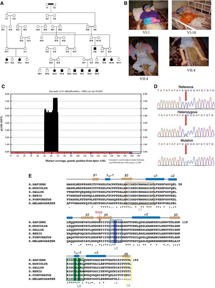
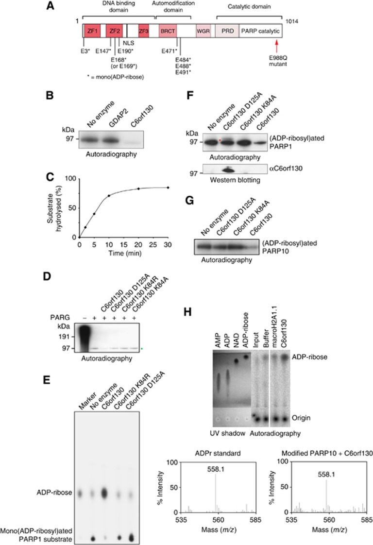
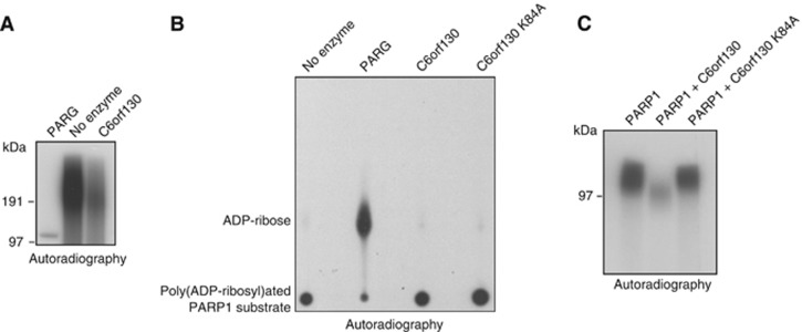
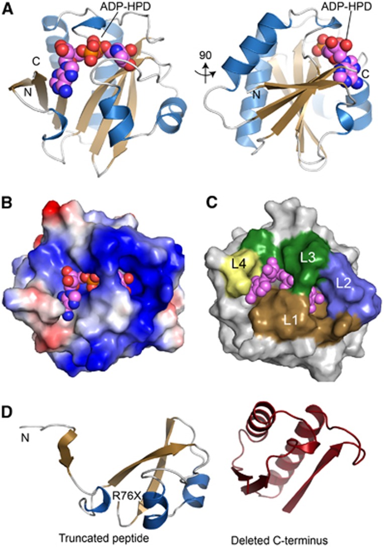
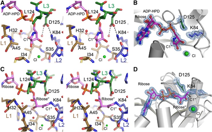
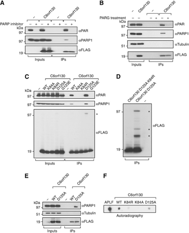
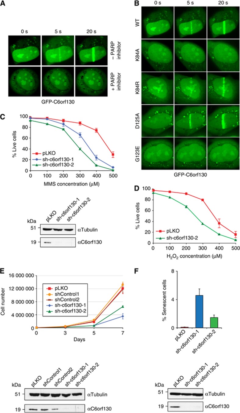
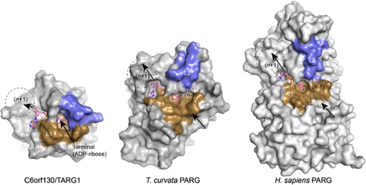
Adenosine diphosphate (ADP)-ribosylation is a post-translational protein modification implicated in the regulation of a range of cellular processes. A family of proteins that catalyse ADP-ribosylation reactions are the poly(ADP-ribose) (PAR) polymerases (PARPs). PARPs covalently attach an ADP-ribose nucleotide to target proteins and some PARP family members can subsequently add additional ADP-ribose units to generate a PAR chain. The hydrolysis of PAR chains is catalysed by PAR glycohydrolase (PARG). PARG is unable to cleave the mono(ADP-ribose) unit directly linked to the protein and although the enzymatic activity that catalyses this reaction has been detected in mammalian cell extracts, the protein(s) responsible remain unknown. Here, we report the homozygous mutation of the c6orf130 gene in patients with severe neurodegeneration, and identify C6orf130 as a PARP-interacting protein that removes mono(ADP-ribosyl)ation on glutamate amino acid residues in PARP-modified proteins. X-ray structures and biochemical analysis of C6orf130 suggest a mechanism of catalytic reversal involving a transient C6orf130 lysyl-(ADP-ribose) intermediate. Furthermore, depletion of C6orf130 protein in cells leads to proliferation and DNA repair defects. Collectively, our data suggest that C6orf130 enzymatic activity has a role in the turnover and recycling of protein ADP-ribosylation, and we have implicated the importance of this protein in supporting normal cellular function in humans.


Conflict of interest statement

The authors declare that they have no conflict of interest.

Figures

Figure 1
Figure 1
The genetic and clinical data. (A) Pedigree diagram of family. (B) Photographs of individuals VI:1,VI:10, VII:4 affected by neurodegeneration. (C) EasyLinkage Plus v.5.08 output of parametric analysis of chromosome 6 under an autosomal recessive model by Simwalk2.91. (D) Electropherograms showing the identified mutation in c6orf130 gene (NC_000006.11:g.41037831G>A; NM_145063.2:c.227C>T; NP_659500.1:p.R76X). (E) Structure-based sequence alignment of C6orf30 homologues. The conserved residues K84 and G123/D125 are highlighted in blue and green, respectively. The predicted C6orf130 protein truncation in the patients analysed in this study is marked with red arrow.
Figure 2
Figure 2
C6orf130 protein is a mono(ADP-ribosyl) protein hydrolase. (A) Schematic representation of PARP1 E988Q mutant protein. Residues found to be mono(ADP-ribosyl)ated by mass spectrometry analysis of automodified recombinant PARP1 E988Q protein are shown and marked with an asterisk. (B) SDS–PAGE based assay showing that purified recombinant human C6orf130 de(ADP-ribosyl)ates 32P-labelled mono(ADP-ribosyl)ated PARP1 E988Q protein. Macrodomain-containing protein GDAP2 served as a negative control. (C) The time curve showing the activity of C6orf130 on ADP-ribosylated PARP1 E988Q protein. (D) Hydrolytic activity of C6orf130 wild-type and mutated proteins on mono(ADP-ribosyl) PARP1 peptide derived from the activity of PARG protein on [32P]-labelled poly(ADP-ribosyl)ated wt PARP1. Mono(ADP-ribosyl)ated PARP1 species is indicated by green asterisk. (E) Analysis of the product of the C6orf130 reaction on mono(ADP-ribosyl)ated PARP E988Q by thin-layer chromatography (TLC). (F) Catalytic activity of C6orf130 proteins carrying point mutations in the indicated residues (top panel). The band appearing above 97 kDa in the SDS–PAGE in the C6orf130 D125A mutant lane (top panel, red asterisk) contains a crosslinked C6orf130 protein as revealed by western blotting (bottom panel). (G) De(ADP-ribosyl)ation of the PARP10 substrate by C6orf130. (H) Analysis of the reaction products of C6orf130 on ADP-ribosylated PARP10 substrate by TLC (top panel) and mass spectrometry (bottom panel).
Figure 3
Figure 3
Activities of C6orf130 on poly(ADP-ribosyl)ated PARP1 substrates. (A) Hydrolysis of PAR by C6orf130 and PARG analysed by SDS–PAGE and (B) by thin-layer chromatography-based assays. (C) C6orf130 inhibits automodification activity of PARP1 in vitro.
Figure 4
Figure 4
Crystal structure of the C6orf130 ADP-HPD complex. (A) Orthogonal views of C6orf130 (blue and tan) bound to ADP-HPD (spheres). (B) Surface charge representation of C6orf130 (blue, positive; red, negative; grey, neutral or hydrophobic) showing a positively charged binding site for ADP-HPD. (C) ADP-HPD binding site is composed of four sequence motifs, L1–L4. (D) The C6orf130 protein truncation (R76X) in the patients analysed in this study.
Figure 5
Figure 5
C6orf130 active site architecture and ligand interactions. (A) Stereo view of the active site of C6orf130. A chloride ion (green) interacts with the 2′′- and 3′′-hydroxyls of the ADP-HPD. (B) Final 1.25 Å sigma-A weighted 2Fo-Fc electron density map (contoured at 1.0σ) showing ADP-HPD bound in the C6orf130 active site. Lys84 and Asp125 and a bound chloride ion are in close proximity to the pyrrolidine ring of ADP-HPD. (C) Stereo view of the active site of C6orf130 (ADP-ribose) complex. Molecule D (Supplementary Figure 5) is displayed. (D) Final 1.55 Å sigma-A weighted 2Fo-Fc electron density map (contoured at 1.0σ) showing the covalent lysyl-ADP-ribose adduct of chain D.
Figure 6
Figure 6
C6orf130 is a PARP1 interacting factor. (A) Interactions of C6orf130 with PARP1 and poly(ADP-ribose) in 293T human cells are diminished by blocking the active poly(ADP-ribose) synthesis using the PARP inhibitor olaparib. (B) Interactions of C6orf130 with PAR and PARP1 are abolished when the complexes are treated with purified, recombinant human PARG protein. (C) Effects of C6orf130 point mutations on its interactions with PARP1 and poly(ADP-ribose). (D) The crosslinking of C6orf130 to proteins is dependent on K84 residue. The crosslinked protein species in C6orf130 Asp125Ala immunoprecipitates in (C, D) are indicated by asterisks. (E) Covalent crosslink between the D125A mutant and PARP1 is retained at high salt concentrations. (F) Binding of C6orf130 and the indicated mutants to free poly(ADP-ribose) in vitro revealed by dot blot assay using radioactively labelled PAR.
Figure 7
Figure 7
C6orf130 depletion affects DNA repair, cell proliferation, and senescence. (A) PARP-dependent localization of GFP-tagged C6orf130 to the sites of laser-induced microirradiation. The recruitment is abolished by pre-treatment of cells using the PARP inhibitor olaparib. (B) Recruitment of GFP-tagged C6orf130 point mutants to the sites of laser-induced damage. (C) Sensitivity of C6orf130 knockdown 293T cells to the DNA-damaging agent, methyl methanosulphonate (MMS) and (D) hydrogen peroxide, and the knockdown efficiencies of C6orf130 shRNAs in analysed cells assessed by western blotting (bottom panel). (E) Reduction in cell proliferation observed under stable C6orf130 knockdown in 293T cells and knockdown efficiencies of C6orf130 shRNAs in 293T cells assessed by western blotting (bottom panel). (F) Induction of senescence in c6orf130 knockdown U2OS cells as judged by β-galactosidase staining and knockdown efficiencies of C6orf130 shRNAs in U2OS cells (bottom panel).
Figure 8
Figure 8
Structural comparisons of TARG1/C6orf130 and PARG enzymes. L1 and L2 substrate binding regions of TARG1 encircle the terminal ADP-ribose. Conversely, exposed surfaces of PARG homologues are well suited for PAR binding and endoglycosidic catalysis.

Comment in

References

    1. Adams PD, Afonine PV, Bunkóczi G, Chen VB, Davis IW, Echols N, Headd JJ, Hung LW, Kapral GJ, Grosse-Kunstleve RW, McCoy AJ, Moriarty NW, Oeffner R, Read RJ, Richardson DC, Richardson JS, Terwilliger TC, Zwart PH (2010) PHENIX: a comprehensive Python-based system for macromolecular structure solution. Acta Crystallogr D Biol Crystallogr 66: 213–221 - PMC - PubMed
    1. Ahel D, Horejsí Z, Wiechens N, Polo SE, Garcia-Wilson E, Ahel I, Flynn H, Skehel M, West SC, Jackson SP, Owen-Hughes T, Boulton SJ (2009) Poly(ADP-ribose)-dependent regulation of DNA repair by the chromatin remodeling enzyme ALC1. Science 325: 1240–1243 - PMC - PubMed
    1. Ahel I, Ahel D, Matsusaka T, Clark AJ, Pines J, Boulton SJ, West SC (2008) Poly(ADP-ribose)-binding zinc finger motifs in DNA repair/checkpoint proteins. Nature 451: 81–85 - PubMed
    1. Andrabi SA, Kim NS, Yu SW, Wang H, Koh DW, Sasaki M, Klaus JA, Otsuka T, Zhang Z, Koehler RC, Hurn PD, Poirier GG, Dawson VL, Dawson TM (2006) Poly(ADP-ribose) (PAR) polymer is a death signal. Proc Natl Acad Sci USA 103: 18308–18313 - PMC - PubMed
    1. Berthold CL, Wang H, Nordlund S, Hogbom M (2009) Mechanism of ADP-ribosylation removal revealed by the structure and ligand complexes of the dimanganese mono-ADP-ribosylhydrolase DraG. Proc Natl Acad USA 106: 14247–14252 - PMC - PubMed

Publication types

MeSH terms