. 2013 May;21(5):986-94.
doi: 10.1038/mt.2013.35. Epub 2013 Mar 12.

Targeting miR-21 for the therapy of pancreatic cancer

Affiliations

Targeting miR-21 for the therapy of pancreatic cancer

Flavie Sicard et al. Mol Ther. 2013 May.

Abstract

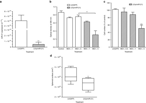
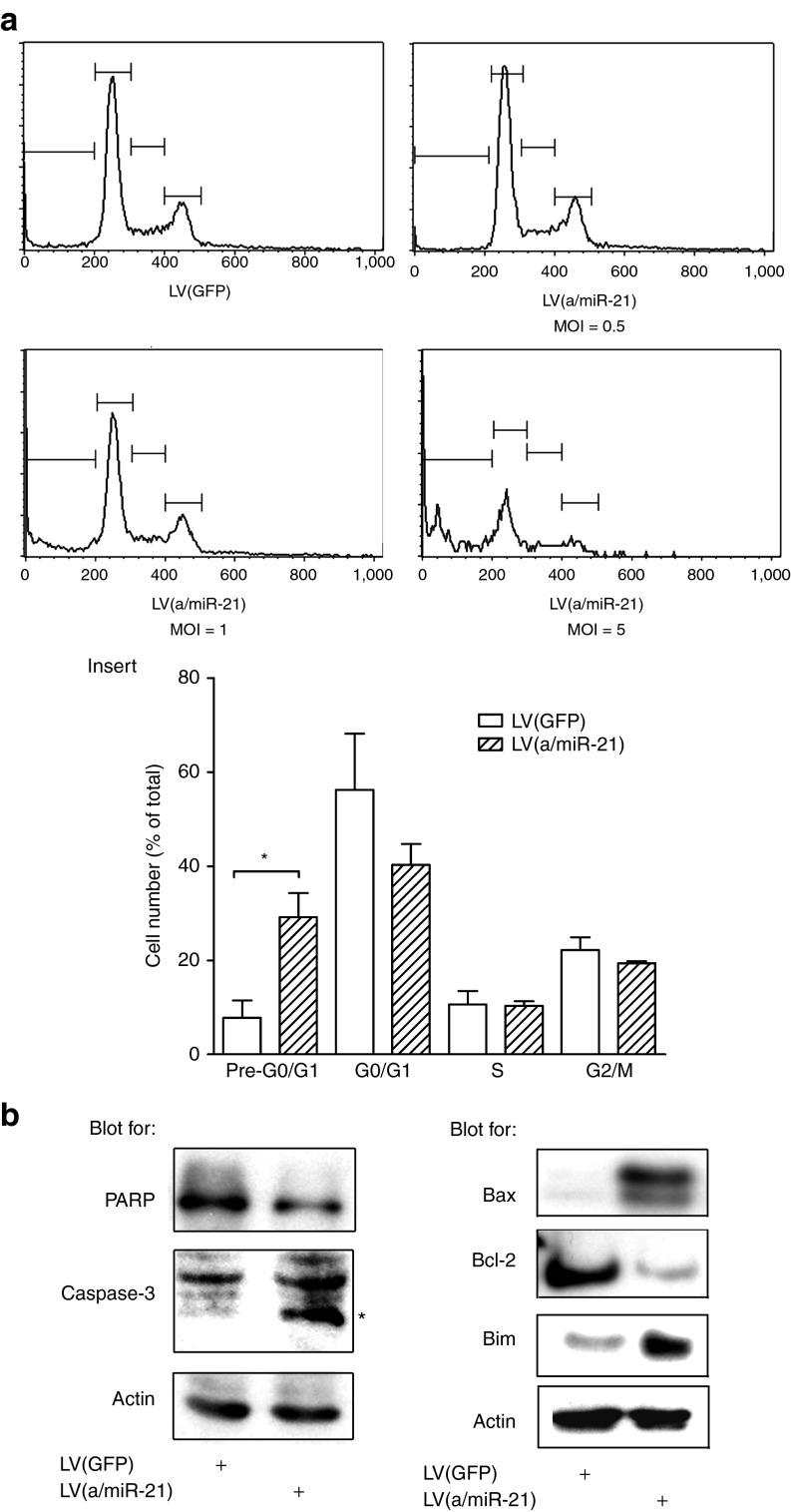
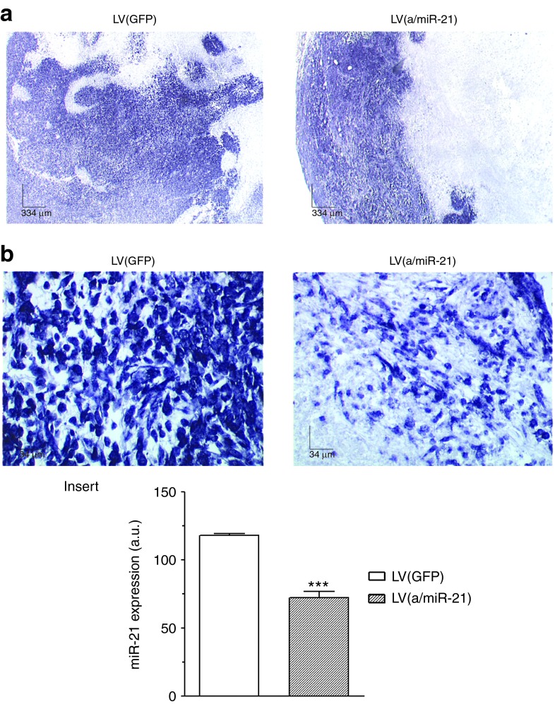
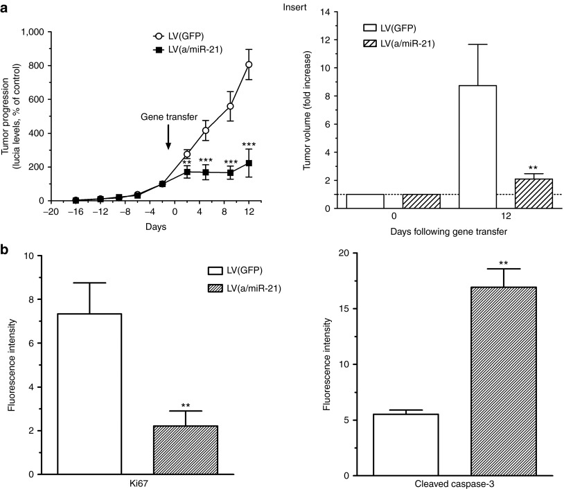
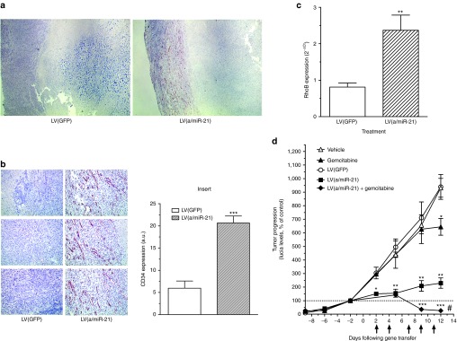
Despite tremendous efforts worldwide from clinicians and cancer scientists, pancreatic ductal adenocarcinoma (PDA) remains a deadly disease for which no cure is available. Recently, microRNAs (miRNAs) have emerged as key actors in carcinogenesis and we demonstrated that microRNA-21 (miR-21), oncomiR is expressed early during PDA. In the present study, we asked whether targeting miR-21 in human PDA-derived cell lines using lentiviral vectors (LVs) may impede tumor growth. We demonstrated that LVs-transduced human PDA efficiently downregulated miR-21 expression, both in vitro and in vivo. Consequently, cell proliferation was strongly inhibited and PDA-derived cell lines died by apoptosis through the mitochondrial pathway. In vivo, miR-21 depletion stopped the progression of a very aggressive model of PDA, to induce cell death by apoptosis; furthermore, combining miR-21 targeting and chemotherapeutic treatment provoked tumor regression. We demonstrate herein for the first time that targeting oncogenic miRNA strongly inhibit pancreatic cancer tumor growth both in vitro and in vivo. Because miR-21 is overexpressed in most human tumors; therapeutic delivery of miR-21 antagonists may still be beneficial for a large number of cancers for which no cure is available.


Figures

Figure 1
Figure 1
In vitro targeting of miR-21 using lentiviral vectors decreases miR-21 cellular content and strongly inhibits PDA cell proliferation and viability. Mia PaCa-2 cells were transduced by LV(a/miR-21) encoding for miR-21 antisenses at the indicated MOI. Control cells were transduced with GFP-encoding lentiviral vectors, or left untransduced. (a) miR-21 expression was determined by qRT-PCR. Results, expressed as 2−ΔCt, are mean ± SEM of five independent experiments performed in duplicate. (b) Cell viability and (c) cell proliferation were determined 72 hours following transduction. Results are mean ± SEM of five independent experiments performed in triplicate. (d) Capan-2 cells were stably transduced with LV(a/miR-21). Control cells were stably transduced with LV(GFP). After 2 weeks in culture, spheroids surface was measured using ImageJ software. Results are mean ± SEM of three independent experiments of eight replicates. *P < 0.05; **P < 0.01. GFP, green fluorescent protein; LV, lentiviral vector; MOI, multiplicity of infection; PDA, pancreatic ductal adenocarcinoma; qRT-PCR, quantitative reverse transcriptase-PCR.
Figure 2
Figure 2
miR-21 inhibition induces human PDA-derived cells' death by apoptosis via the mitochondrial pathway. Mia PaCa-2 cells were transduced by LV(a/miR-21) encoding for miR-21 antisenses at the indicated MOI. Control cells were transduced with GFP-encoding lentiviral vectors. (a) Flow cytometric analysis of propidium iodide-stained cells. Histograms are representative of three independent experiments. Insert: quantification of the DNA content distribution in each phase of the cell cycle. Results are mean ± SEM of three independent experiments. *P < 0.05. (b) Western blot analysis of PARP and caspase-3 cleavage, and of Bax, Bcl-2, and Bim expression in the transduced cells. Results are representative of three independent experiments. GFP, green fluorescent protein; LV, lentiviral vector; MOI, multiplicity of infection; PDA, pancreatic ductal adenocarcinoma.
Figure 3
Figure 3
Efficient knockdown of miR-21 in PDA tumors using lentiviral vectors. Pancreatic tissue was harvested 12 days following gene transfer for analysis of miR-21 expression by in situ hybridization. Results are representative of five different (a) low (×4) or (b) high (×40) power fields from three different tumors for each group. Insert: quantification of miR-21 expression using ImageJ software. Results are mean ± SEM of 10 different fields from three different tumors for each group. ***P < 0.005. a.u., arbitrary unit; GFP, green fluorescent protein; LV, lentiviral vector; PDA, pancreatic ductal adenocarcinoma.
Figure 4
Figure 4
Targeting miR-21 inhibits PDA tumor growth. (a) Noninvasive monitoring of PDA tumor growth was performed for 34 days before and after intratumoral gene transfer. Results are mean ± SEM of Lucia levels, expressed as % of day 0, in the serum of 12 animals per group. Insert: tumor volume was measured at day 0 and day 12 following intratumoral gene transfer using a caliper. Results are mean ± SEM of tumor volume progression in 12 animals per group. **P < 0.01; ***P < 0.005. (b) Pancreatic tissue was harvested for the analysis of Ki67 (left) or cleaved caspase-3 (right) expression in the tumors by immunofluorescence. Results are mean ± SEM of 15 different fields from three different tumors for each group. **P < 0.01. GFP, green fluorescent protein; LV, lentiviral vector; PDA, pancreatic ductal adenocarcinoma.
Figure 5
Figure 5
Targeting miR-21 induces tumor angiogenesis and potentiate gemcitabine antitumoral effect. Pancreatic tissue was harvested 12 days following gene transfer for analysis of CD34 expression by immunohistochemistry. Results are representative of (a) 5 low and (b) 15 high power fields from three different tumors per group. Insert: quantification of CD34 expression using Image J software. Results are mean ± SEM of 15 different low power fields from three different tumors for each group. ***P < 0.005. (c) Total RNA was extracted from tumors and analyzed for RhoB expression by qRT-PCR. Results, expressed as 2−ΔCt, are mean ± SEM of three different tumors for each group. (d) Lucia was quantified in mice serum every 2–3 days during the course of the experiment. Arrows indicate the intraperitoneal injection of gemcitabine. Results are mean ± SEM of Lucia levels, expressed as % of day 0, in the serum of 5–10 animals per group. *P < 0.05; **P < 0.01; ***P < 0.005, #P < 0.005 comparing gemcitabine versus gemcitabine + LV(a/miR-21) or LV(a/miR-21) versus gemcitabine + LV(a/miR-21). a.u., arbitrary unit; GFP, green fluorescent protein; LV, lentiviral vector; qRT-PCR, quantitative reverse transcriptase-PCR.

References

    1. Jemal A, Bray F, Center MM, Ferlay J, Ward E., and, Forman D. Global cancer statistics. CA Cancer J Clin. 2011;61:69–90. - PubMed
    1. Torrisani J, Bournet B, Cordelier P., and, Buscail L. [New molecular targets in pancreatic cancer] Bull Cancer. 2008;95:503–512. - PubMed
    1. Conroy T, Desseigne F, Ychou M, Bouché O, Guimbaud R, Bécouarn Y, et al. Groupe Tumeurs Digestives of Unicancer; PRODIGE Intergroup. (2011FOLFIRINOX versus gemcitabine for metastatic pancreatic cancer N Engl J Med 3641817–1825. - PubMed
    1. Delpu Y, Hanoun N, Lulka H, Sicard F, Selves J, Buscail L.et al. (2011Genetic and epigenetic alterations in pancreatic carcinogenesis Curr Genomics 1215–24. - PMC - PubMed
    1. Iorio MV., and, Croce CM. MicroRNA dysregulation in cancer: diagnostics, monitoring and therapeutics. A comprehensive review. EMBO Mol Med. 2012;4:143–159. - PMC - PubMed

Publication types

MeSH terms