. 2013:4:1590.
doi: 10.1038/ncomms2594.

Engineering the type III secretion system in non-replicating bacterial minicells for antigen delivery

Affiliations

Engineering the type III secretion system in non-replicating bacterial minicells for antigen delivery

Heather A Carleton et al. Nat Commun. 2013.

Abstract

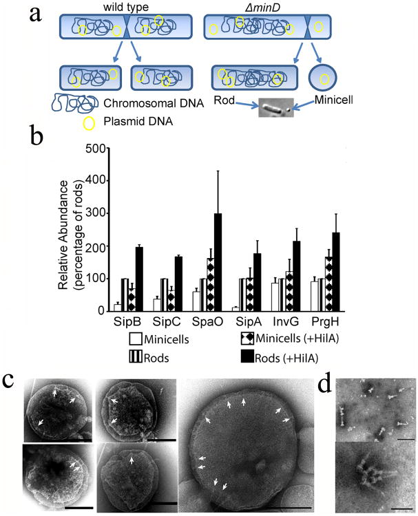
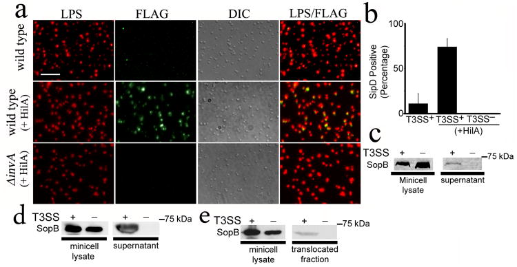
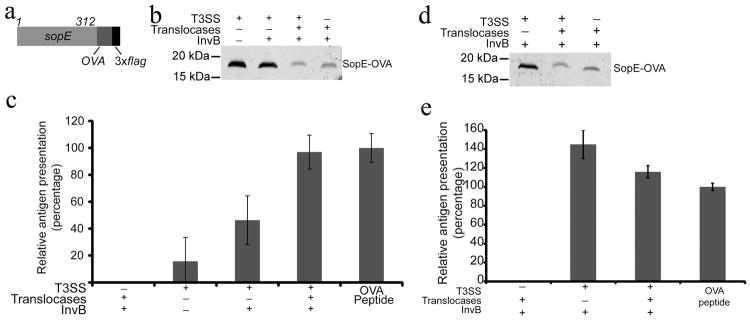
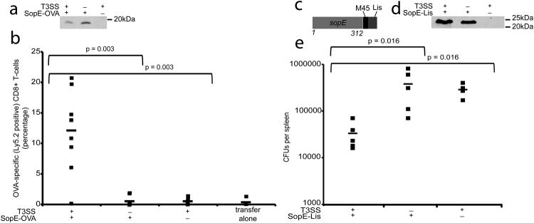
Type III protein secretion systems are being considered for vaccine development as virtually any protein antigen can be engineered for delivery by these nanomachines into the class I antigen presentation pathway to stimulate antigen-specific CD8(+) T cells. A limitation in the use of this system is that it requires live virulence-attenuated bacteria, which may preclude its use in certain populations such as children and the immunocompromised. Here we report the engineering of the Salmonella Typhimurium type III secretion system in achromosomal, non-replicating nanoparticles derived from bacterial minicells. The engineered system is shown to be functional and capable of delivering heterologous antigens to the class I antigen presentation pathway stimulating immune responses both in vitro and in vivo. This antigen delivery platform offers a novel approach for vaccine development and cellular immunotherapy.


Conflict of interest statement

Competing interests: The authors declare no competing interests

Figures

Fig. 1
Fig. 1. Bacterial minicells assemble a type III secretion system
(a) Schematic of minicell generation (see also Supplementary Fig. S1). (b) Relative abundance of selected T3SS proteins analyzed by immunoblotting. Values are standardized compared to those obtained from wild-type S. Typhimurium rods and normalized according to the total protein concentration of each sample. Values are the mean ± standard deviation of 3 independent measurements. See additional information in Materials and Methods and Supplementary Fig. S2 and S3. (c) Electron micrographs of negatively-stained osmotically-shocked minicells. Arrows indicate T3SS needle complexes on the minicell envelope. Scale bars: 200 nm. (d) Electron micrographs of negatively-stained needle complexes isolated from purified minicells. Scale bars: 100 nm (top) and 50 nm (bottom).
Fig. 2
Fig. 2. The type III secretion system in minicells is functional
(a and b) Detection of the needle complex tip protein SipD on the surface of purified minicells. Minicells were isolated from wild-type or T3SS-defective (ΔinvA) S. Typhimurium strains expressing SipD-FLAG and carrying a plasmid expressing the SPI-1 T3SS positive transcriptional regulator HilA. Minicells were stained with an antibody to LPS (red), an antibody against the FLAG tag (green), and examined by immunofluorescence and DIC microscopy. Scale bar: 2.5 μm (a). The % of minicells showing surface SipD stain is shown (b). Values represent the mean ± standard deviation of three independent experiments in which a minimum of 4,000 cells per strain were counted. (c) and (d) Secretion of de novo synthesized effector proteins by purified minicells through their SPI-1 T3SS. Minicells were isolated from wild type or T3SS-defective (ΔinvA) S. Typhimurium strains carrying a plasmid encoding the SPI-1 T3SS effector SopB expressed under the control of an arabinose-inducible promoter. Isolated minicells were incubated for 3 hs in the presence of arabinose and the presence of SopB in minicell lysates and supernatants were analyzed by western blot (c). Alternatively, minicells were exposed to cultured Henle-407 cells and the presence of SopB in supernatants examined as described above (d). (e) Minicell-mediated, type III secretion dependent protein translocation into cultured epithelial cells. Henle-407 cells were treated with minicells isolated from type III secretion competent or type III secretion-defective (ΔinvA) as described above and added to Henle-407 cells for 2.5hs. The presence of the effector protein SopB in minicell lysates and the translocated fraction was assayed by Western blot.
Fig. 3
Fig. 3. T3SS-dependent antigen delivery by minicellsin-vitro
(a) Schematic of the SopE-OVA construct used in these studies. (b) Western blot analysis of minicells obtained from wild type or T3SS-defective (ΔinvA) S. Typhimurium strains expressing the SopE-OVA construct and used in the experiment shown in (c). When indicated, the SopE-OVA construct was co-expressed with the SopE chaperone InvB and the T3SS protein translocases and their chaperones to improve protein secretion and/or translocation. Equal amount of total protein was loaded in each sample. (c) Analysis of antigen delivery by minicells to antigen-presenting cells. RMA cells (C57BL/6 mouse hybridomas) were pulsed for 3 hs with minicells isolated from wild type or T3SS-defective (ΔinvA) S. Typhimurium strains. After pulsing, RMA cells were fixed, and used as APCs in a B3Z T-cell activation assay as described in experimental procedures. Values represent the levels of antigen presentation based on the β-galactosidase activity detected in the B3Z-T cell hybridoma reporter and are normalized relative to the values of the OVA peptide positive control, which was considered 100 %. The values are the mean ± standard deviation of three independent experiments. (d and e) Minicells can deliver antigen to dendritic cells ex vivo. (d) Western blot analysis of minicells obtained from wild type or T3SS-defective (ΔinvA) S. Typhimurium strains expressing the SopE-OVA construct and used in the experiment shown in (e). Equal amount of total protein was loaded in each sample. (e) Bone marrow-derived dendritic cells were pulsed for 3 hs with minicells isolated from the indicated S. Typhimurium strains carrying a plasmid expressing SopE-OVA and the indicated SPI-1 T3SS-associated proteins. After pulsing, dendritic cells were fixed and used as APCs in a B3Z T-cell activation assay as described in Supplementary Materials. Values represent the levels of antigen presentation based on the β-galactosidase activity detected in the B3Z-T cell hybridoma reporter and they are normalized relative to the values of the OVA peptide positive control, which was considered 100 %. The values are the mean ± standard deviation of three independent experiments.
Fig. 4
Fig. 4. T3SS-dependent priming of protective CD8+ T-cell responses by minicells
(a) Western blot analysis of minicells obtained from wild type or T3SS-defective (ΔinvA) S. Typhimurium strains expressing the SopE-OVA construct and used in the experiment shown in (b). Equal amount of total protein was loaded in each sample. (b) Splenocytes from OT-I mice were adoptively transferred into recipient mice (C57BL/6/CD45.1), which were subsequently immunized with minicells isolated from the indicated S. Typhimurium Δasd strains expressing SopE-OVA. Three weeks after minicell immunization mice were boosted and the levels of OVA-specific CD8+ T cells were measured by flow cytometry as indicated in Materials and Methods. Values represent the percentage of OVA-specific CD8+ T-cells in each individual mouse (number of mice used in each category: T3SS+/SopE-OVA+: 9; T3SS/SopE-OVA+: 7; T3SS+/SopE-OVA: 7; 4 transfer alone: 4). Data were analyzed using the Student's t test. (c) Schematic of SopE-Lis construct used in the protection experiments. (d) Western blot analysis of minicells obtained from wild type or T3SS-defective (ΔinvA) S. Typhimurium strains expressing the SopE-Lis construct and used in the experiment shown in (e). (e) BMDCs prepared from Balb/c mice were incubated with minicells isolated from the indicated bacterial strains and transferred by tail vein injection into a Balb/c mouse as indicated in Materials and Methods. Six days after transfer mice were challenged with L. monocytogenes, and 3 days after challenge the c. f. u. in spleens were determined (number of mice used in each category: T3SS+/SopE-Lys+: 5; T3SS/SopE-Lys+: 5; T3SS+/SopE-Lys: 4). Data were analyzed using the Wicoxon rank test.

References

    1. Russmann H, et al. Delivery of epitopes by the Salmonella type III secretion system for vaccine development. Science. 1998;281:565–568. - PubMed
    1. Chen LM, Briones G, Donis RO, Galan JE. Optimization of the delivery of heterologous proteins by the Salmonella enterica serovar Typhimurium type III secretion system for vaccine development. Infect Immun. 2006;74:5826–5833. doi: 10.1128/IAI.00375-06. - DOI - PMC - PubMed
    1. Amanna I, Slifka M. Contributions of humoral and cellular immunity to vaccine-induced protection in humans. Virology. 2011;411:206–215. - PMC - PubMed
    1. Curtiss Rr, et al. New technologies in using recombinant attenuated Salmonella vaccine vectors. Crit Rev Immunol. 2010;30:255–270. - PMC - PubMed
    1. Hegazy W, Hensel M. Salmonella enterica as a vaccine carrier. Future Microbiol. 2012;7:111–127. - PubMed

Publication types

MeSH terms