. 2013 Apr;72(4):272-85.
doi: 10.1097/NEN.0b013e318288a8dd.

Rapid β-amyloid deposition and cognitive impairment after cholinergic denervation in APP/PS1 mice

Affiliations

Rapid β-amyloid deposition and cognitive impairment after cholinergic denervation in APP/PS1 mice

Juan Jose Ramos-Rodriguez et al. J Neuropathol Exp Neurol. 2013 Apr.

Abstract

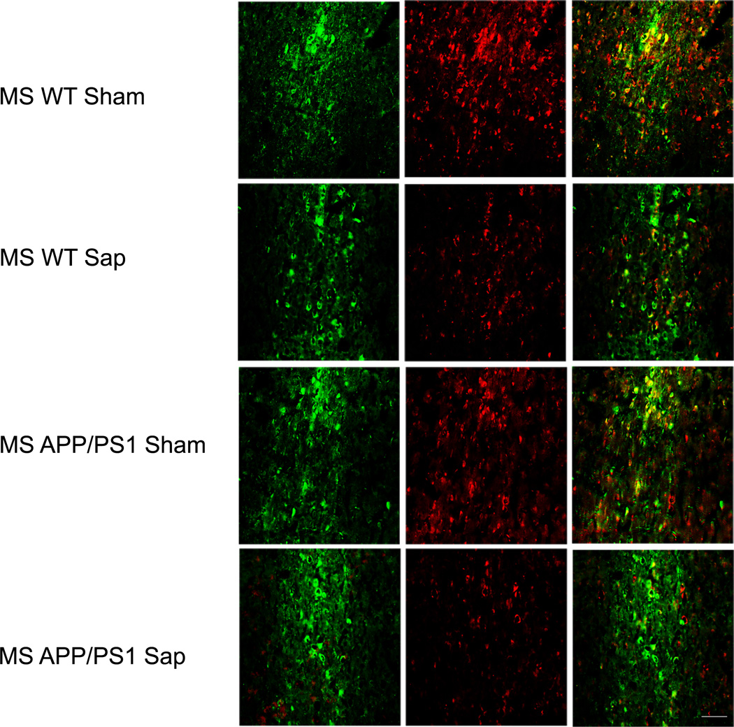
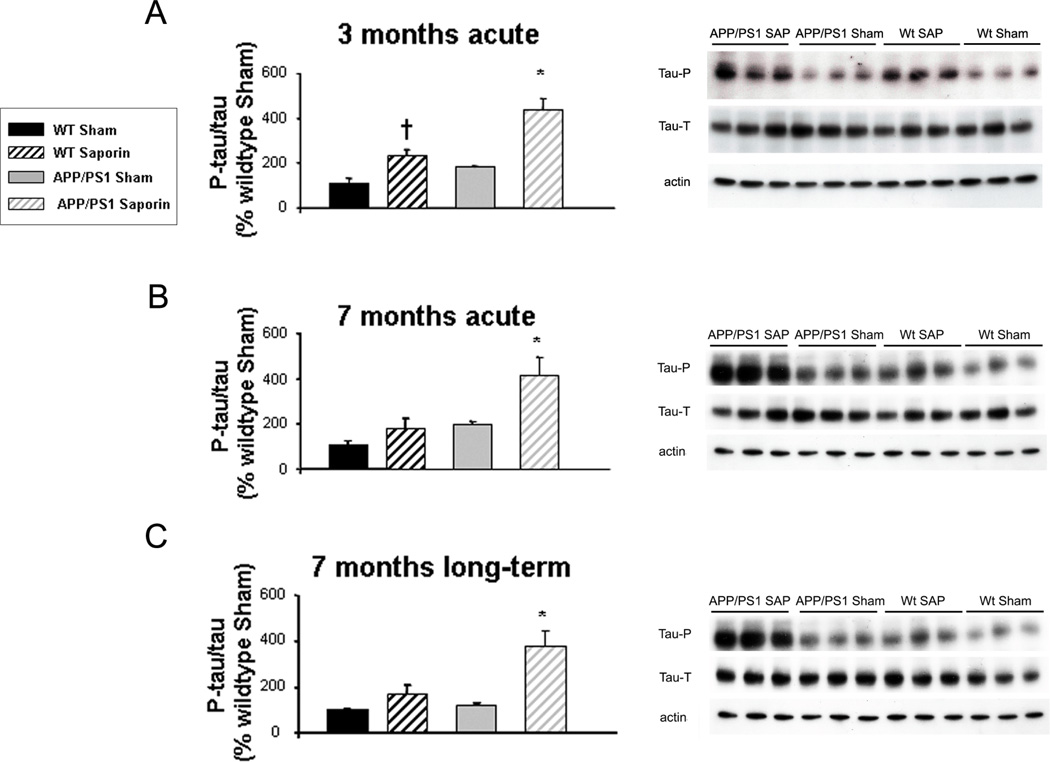
Although extensive evidence supports the role of β-amyloid (Aβ) in Alzheimer disease (AD), the neurotoxic mechanisms underlying AD pathogenesis are not understood. On the other hand, neuronal loss is the pathologic feature that best correlates with cognitive impairment. We hypothesized that cholinergic neurodegeneration may lead to Aβ deposition and tested this by inducing selective cholinergic lesions in APPswe/PS1dE9 mice with murine p75 saporin (mu p75-SAP). Intracerebroventricular lesions that removed approximately 50% of cholinergic innervation to the cortex and hippocampus were induced in animals with incipient (∼3 months) and marked (∼7 months of age) Aβ deposition. Cranial windows were implanted, and Aβ deposition was monitored in vivo using multiphoton microscopy. Deposition of Aβ was increased as soon as 7 days after the lesion, and this effect was maintained up to 3 months later. Postmortem studies using immunohistochemistry with an anti-Aβ antibody corroborated these findings in both cerebral cortex and hippocampus. Tau phosphorylation was also significantly increased after the lesions. Cholinergic denervation resulted in early memory impairment at 3 months of age that worsened with age (∼7 months); there was a synergistic effect between cholinergic denervation and the presence of APP/PS1 transgenes. Altogether, our data suggest that cholinergic denervation may trigger Aβ deposition and synergistically contribute to cognitive impairment in AD patients.


Figures

Figure 1
Figure 1
Cholinergic denervation after murine p75NTR saporin (Sap) lesions of the basal forebrain in wild type (WT) and APP/PS1 mice. There is reduction of choline acetyltransferase immunostaining (red) in the medial septum at 2 weeks after the lesion compared to vehicle-treated (Sham) mice; γ-aminobutyric acid–releasing (GABAergic) neurons indicated by parvalbumin immunostaining (green) appear preserved. Scale bar = 250 µm.
Figure 2
Figure 2
Effect of basal forebrain cholinergic denervation (Saporin) in APP/PS1 mice on episodic memory evaluated with the new object discrimination (NOD) test. There was an overall worsening performance with age for all paradigms under study (age X transgene effect: what: [F(2,1)=12.243, ** p < 0.01), where: [F(2,1)=13.81, * p = 0.023), when: [F(2,1)=19.710, ** p < 0.01). There was significant impairment in 3-month-old (short term) lesioned mice for "what", "where" and "when" parameters (* p < 0.05 vs. Wt [WT] Sham [vehicle-treated] and WT Saporin). There was a worsening effect of cholinergic denervation in older APP/PS1 lesioned mice (7 months old) (* p < 0.05 vs. the other groups). One-way ANOVA for independent samples followed by Tukey-b or Tamahane test as needed.
Figure 3
Figure 3
Effect of cholinergic denervation (Saporin) in APP/PS1 mice on spatial learning and memory evaluated using the Morris water maze test. Data are representative of 7 to 14 mice. (A) Young mice (3 months old) had some impairment locating the hidden platform during the 4 days of the acquisition phase; this effect was worsened in older transgenic mice (7 months), with acute and long-term cholinergic lesions. Differences were detected by two-way ANOVA (group X day) * p < 00.5. y-axis = time to locate the hidden platform. Daily analysis showed an overall delay to locate the platform for 3-month-old transgenic lesioned mice (day 1: [F(3,144)=9.79, ** p < 0.01 vs. wild-type (WT) Sham], day 2: [F(3,140)=17.910, ** p < 0.01 vs. WT Sham and WT p-75 SAP, †† p < 0.01 vs. WT Sham], day 3: [F(3,133)=17.346, ** p < 0.01 vs. other groups, †† p < 0.01 vs. WT Sham ], day 4: [F(3,134)=14.025, ** p < 0.01 vs. other groups]). In 7-month-old mice we observed cognitive alterations in animals bearing the APP/PS1 transgenes and we observed a worsening effect on those animals with acute cholinergic denervation (day 1: [F(3,272)=10.611, ** p < 0.01 vs. WT Sham], day 2: [F(3,272)=22.398, ** p < 0.01 vs. WT Sham], day 3: [F(3,262)=13.605, ** p < 0.01 vs. WT Sham and APP/PS1 Sham, †† p<0.01 vs. WT Sham], day 4: [F(3,266)=24.109, ** p < 0.01 vs. WT Sham and APP/PS1 Sham, †† p < 0.01 vs. WT Sham]). A similar profile was observed in 7-month-old mice with chronic cholinergic lesions. The presence of the APP/PS1 transgenes was enough to impair learning and memory abilities in the water maze, however this effect was worsened in the presence of the cholinergic lesion (day 1: [F(3,141)=19.203, ** p < 0.01 vs. WT Sham], day 2: [F(3,151)=9.385, ** p < 0.01 vs. other groups, †† vs. WT Sham], day 3: [F(3,143)=19.199 ** p < 0.01 vs. other groups, †† p < 0.01 vs. WT Sham], day 4: [F(3,143)=19.199, ** p < 0.01 vs. other groups, †† p < 0.01 vs. WT Sham]). (B) During the retention phase there was significant memory impairment in 7-month-old transgenic lesioned mice with acute and long-term cholinergic lesions. Differences were detected using one-way ANOVA followed by Tukey-b test.
Figure 4
Figure 4
Increased numbers of new senile plaques (SPs) in the cortex of APP/PS1 mice with and without murine p75NTR saporin (mu p75-SAP) lesions were detected using in vivo multiphoton microscopy. Medians, maximums and minimums are shown and statistical differences were detected between both groups using Mann-Whitney U test for independent samples. (A) Numbers of plaques appearing per mm3 in ~3-month-old APPswe/PS1dE9 mice 7 and 14 days after the lesions (7 days: ** p = 0.006 vs. Sham; 14 days: ** p < 0.01 vs. Sham). When animals were compared between the day of the lesion and 120 days later there was an increase in SPs in lesioned animals, although differences did not reach statistical significance (p = 0.190 vs. Sham mice). (B) The same profile was observed in ~7-month-old mice (7 days: ** p < 0.01 vs. Sham; 14 days: * p = 0.044 vs. Sham). (D–K) Examples of SP deposition in an APPswe/PS1dE9 mouse 7 days after mu p75-SAP lesion (D, day 0; E, day 7) vs. a Sham mouse (F, day 0; G, day 7). Higher deposition was observed in lesioned APPswe/PS1dE9 mice 120 days after surgery (H, day 0; I, day 120), vs. a Sham mouse (J, day 0; K, day 120). The same fields were located using Texas Red dextran 70K (blue) for the angiograms and amyloid deposits were stained with methoxy-XO4 (red). White arrows indicate new amyloid deposits. Scale bar = 85 µm.
Figure 5
Figure 5
Postmortem assessment of senile plaque (SP) deposition with anti-β-amyloid (Aβ) antibody and thioflavin S (TS) staining in cortex and hippocampus of APP/PS1 mice with or without murine p75NTR saporin (mu p75-SAP)-induced lesions. (A, B) There was a significant increase in plaque burden in all lesioned mice, although this effect was greater in 7-month-old mice, particularly in those with long-term lesions. The increase in amyloid burden, both in cortex (A) and hippocampus (B) was not due to increased plaque size but to an increase in the number of new deposited SPs. Data are representative of 3 to 6 mice per group. Statistical differences were detected by Student t-test for independent samples and Mann-Whitney U-test for independent samples in the case of number of plaques per mm2. * p < 0.05. (C–H) Examples of Aβ immunostaining in the cortex: C, Sham 3 months acute; D, Mu p75-SAP, 3 months acute; E, Sham 7 months acute; F, Mu p75-SAP, 7 months acute; G, Sham 7 months long-term; H, Mu p75-SAP 7 months long-term. Scale bar = 250 µm.
Figure 6
Figure 6
Increased tau phosphorylation in transgenic mice acutely lesioned with murine p75NTR saporin (SAP) at 3 months and 7 months of age and in long-term lesioned 7-month-old mice. Phosphorylated tau/ total tau ratios (left panels) are expressed as percentage of wild-type (WT) vehicle-treated (Sham) values. Differences were detected by one-way ANOVA followed by Tukey-b test or Tamhane test as required (* p < 0.05). (A) In 3-month acute lesions, F(3,28) = 16.455, (* p < 0.01 vs. other groups, † p = 0.001 vs. WT Sham). (B) 7-month acute lesions F(3,19) = 8.896, (* p = 0.01 vs. other groups). (C) 7-month long-term lesions, F(3,28) = 6.720, (* p = 0.001 vs. other groups). Right panels: Examples of western blot for total tau, phospho-tau and α-tubulin, including APP/PS1 mu p75-SAP, APP/PS1 Sham, WT SAP and WT Sham mice.

References

    1. Gomez-Isla T, Hollister R, West H, et al. Neuronal loss correlates with but exceeds neurofibrillary tangles in Alzheimer's disease. Ann Neurol. 1997;41:17–24. - PubMed
    1. Walsh DM, Selkoe DJ. Deciphering the molecular basis of memory failure in Alzheimer's disease. Neuron. 2004;44:181–193. - PubMed
    1. Roberson MR, Harrell LE. Cholinergic activity and amyloid precursor protein metabolism. Brain Res Brain Res Rev. 1997;25:50–69. - PubMed
    1. Meyer-Luehmann M, Spires-Jones TL, Prada C, et al. Rapid appearance and local toxicity of amyloid-beta plaques in a mouse model of Alzheimer's disease. Nature. 2008;451:720–724. - PMC - PubMed
    1. Brendza RP, Bacskai BJ, Cirrito JR, et al. Anti-Abeta antibody treatment promotes the rapid recovery of amyloid-associated neuritic dystrophy in PDAPP transgenic mice. J Clin Invest. 2005;115:428–433. - PMC - PubMed

Publication types

MeSH terms