. 2013 Mar;25(3):1093-107.
doi: 10.1105/tpc.112.108829. Epub 2013 Mar 12.

Protein S-ACYL Transferase10 is critical for development and salt tolerance in Arabidopsis

Affiliations

Protein S-ACYL Transferase10 is critical for development and salt tolerance in Arabidopsis

Liang-Zi Zhou et al. Plant Cell. 2013 Mar.

Abstract

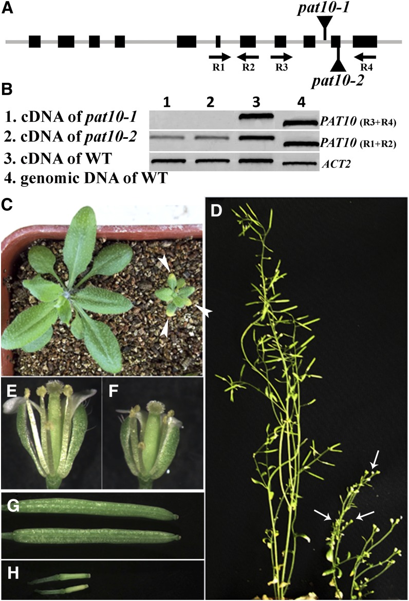
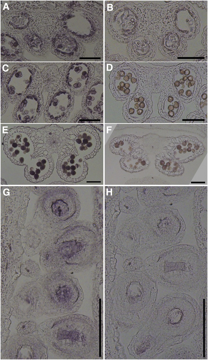
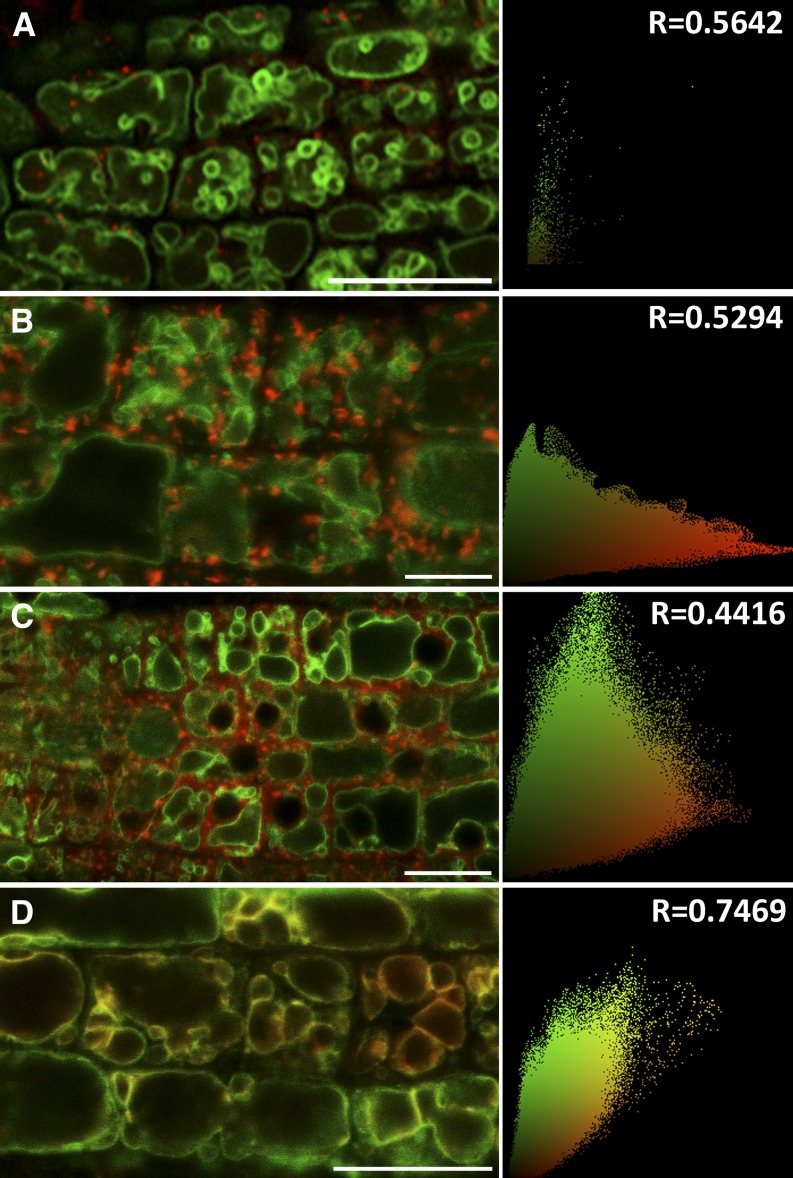
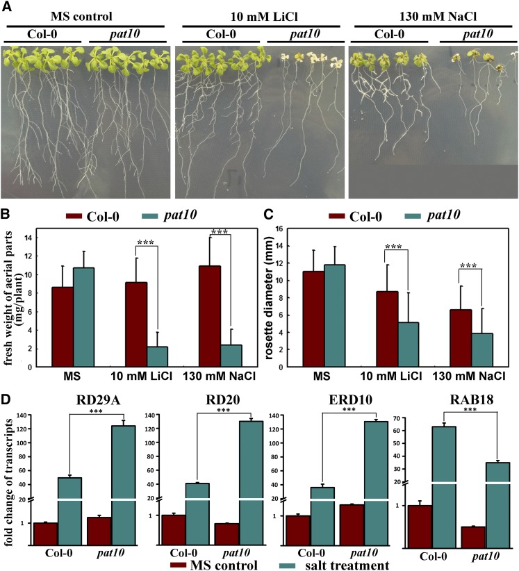
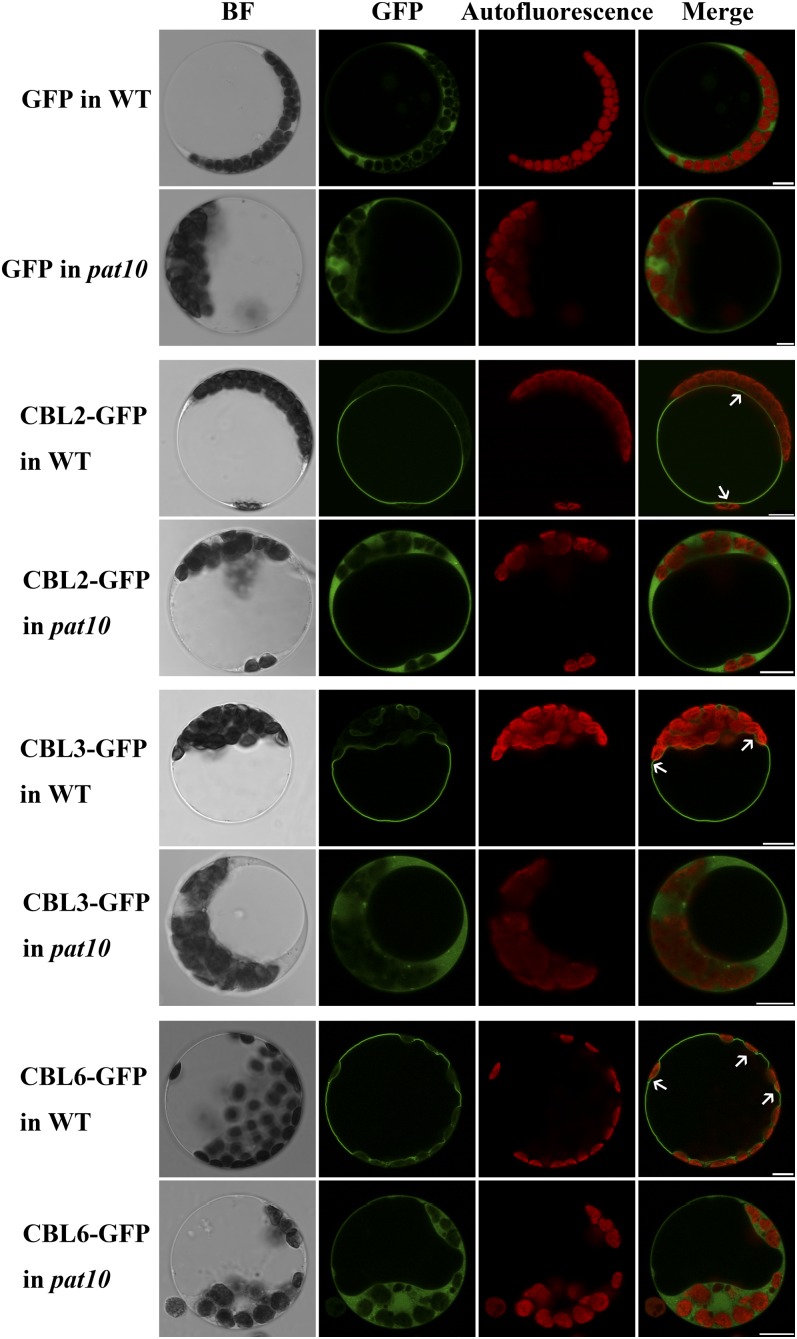
Protein S-acylation, commonly known as palmitoylation, is a reversible posttranslational modification that catalyzes the addition of a saturated lipid group, often palmitate, to the sulfhydryl group of a Cys. Palmitoylation regulates enzyme activity, protein stability, subcellular localization, and intracellular sorting. Many plant proteins are palmitoylated. However, little is known about protein S-acyl transferases (PATs), which catalyze palmitoylation. Here, we report that the tonoplast-localized PAT10 is critical for development and salt tolerance in Arabidopsis thaliana. PAT10 loss of function resulted in pleiotropic growth defects, including smaller leaves, dwarfism, and sterility. In addition, pat10 mutants are hypersensitive to salt stresses. We further show that PAT10 regulates the tonoplast localization of several calcineurin B-like proteins (CBLs), including CBL2, CBL3, and CBL6, whose membrane association also depends on palmitoylation. Introducing a C192S mutation within the highly conserved catalytic motif of PAT10 failed to complement pat10 mutants, indicating that PAT10 functions through protein palmitoylation. We propose that PAT10-mediated palmitoylation is critical for vacuolar function by regulating membrane association or the activities of tonoplast proteins.


Figures

Figure 1.
Figure 1.
Mutations at PAT10 Caused Pleiotropic Developmental Defects. (A) Schematic illustration of T-DNA insertions within the PAT10 genomic region. (B) RT-PCR analysis showing that pat10 mutants are devoid of full-length PAT10 transcripts. WT, the wild type. (C) A representative image of the wild type (left) and pat10 (right) at 20 DAG. Arrowheads indicate necrotic leaf tips in the mutant plant. (D) A representative image of the wild type (left) and pat10 (right) at 50 DAG. Arrows point to nonelongated siliques. (E) A representative image of wild-type open flowers. (F) A representative image of pat10 open flowers. (G) Fully elongated siliques in the wild type. (H) Nonelongated siliques in pat10.
Figure 2.
Figure 2.
Mutations at PAT10 Caused Pollen Developmental Defects. (A) to (C) Scanning electron micrographs (SEM) of mature pollen from the wild type (A), heterozygous pat10 mutants (B), and homozygous pat10 mutants (C). Arrowhead indicates defective pollen coat. (D) to (F) Close-ups of SEM images shown in (A) to (C), respectively. Arrowhead indicates defective pollen coat. (G) and (J) Transmission electron micrograph (TEM) of mature pollen from the wild type (G) and pat10 (J) showing pollen coat structure. Arrowheads indicate defective pollen coats. (H) and (K) TEM section of mature pollen from the wild type (H) and pat10 (K). (I) and (L) Close-ups of TEM shown in (H) and (K), respectively. Asterisks indicate lipid bodies. Bars = 5 µm in (A) to (C), 1 µm in (D) to (F), 2 µm in (H) and (K), and 200 nm in (G), (I), (J), and (L).
Figure 3.
Figure 3.
PAT10 Mutations Resulted in Defective Pollen Germination and Tube Growth. (A) In vitro germination of pollen from the wild type (WT), heterozygous pat10 mutants (heterozygous), and homozygous pat10 mutants (homozygous). (B) In vitro germination percentage of the wild type, heterozygous pat10 mutants, and homozygous pat10 mutants. Germination percentage was calculated from three independent experiments, including around 400 pollen grains in each experiment. For each experiment, samples from three genetic backgrounds were placed side by side on the same slide to reduce system variations. Results are given as means ± se. Samples with different letters (a, b, and c) are significantly different from each other by Fisher’s LSD method. (C) Tube length of in vitro grown pollen from the wild type and pat10. Data were collected from three independent experiments, each including 80 to 100 pollen tubes. For each experiment, samples from the two genetic backgrounds were placed side by side on the same slides to reduce system variations. Results are given as means ± se. Samples with different letters (a and b) are significantly different from each other by Fisher’s LSD method. (D) and (E) Aniline blue staining of emasculated wild-type pistils hand-pollinated with wild-type pollen (D) or with pat10 pollen (E) at 9 HAP. Arrows point to the growth fronts of pollen tubes. (F) and (G) Aniline blue staining of emasculated wild-type pistils hand-pollinated with wild-type pollen (F) or pat10 pollen (G) at 48 HAP. Arrowheads point to ovules fertilized by pat10 pollen. Bars = 100 µm in (A) and 200 µm in (D) to (G).
Figure 4.
Figure 4.
Female Reproductive Organs Are Defective in pat10. (A) and (B) Aniline blue staining of emasculated wild-type pistils (A) and pat10 pistils (B) hand-pollinated with wild-type pollen at 24 HAP. Arrows indicate the growth fronts of pollen tubes. (C) and (D) Scanning electron microscopy of a mature ovule from the wild type (C) and pat10 (D). (E) to (H) Optical sections of wild-type ovules from unfertilized mature pistils (E) and from pistils at 9 HAP (F), 24 HAP (G), and 48 HAP (H). Dotted lines highlight the embryo sacs. Arrows indicate the chalazal end. (I) to (L) Optical sections of pat10 ovules from unfertilized mature pistils (I) and from pistils at 9 HAP (J), 24 HAP (K), and 48 HAP (L). Dotted lines highlight the embryo sacs. Arrows indicate the chalazal end. Bars = 200 µm in (A) and (B), 20 µm in (C) and (D), 25 µm in (E) and (I), and 50 µm in (F) to (H) and (J) to (L).
Figure 5.
Figure 5.
Constitutive Expression of PAT10 by ProPAT10:GUS Reporter Analysis. (A) A mature embryo freshly dissected from rehydrated mature seeds. (B) A cotyledon showing GUS signals at vascular tissues. (C) A true leaf showing strong GUS signals at hydathodes, trichomes, and guard cells. (D) A seedling at 15 DAG showing constitutive GUS signals at both aerial parts and in roots. (E) A lateral root. (F) A transverse section of ProPAT10:GUS transgenic roots showing strong GUS signal at the phloem but not at the xylem. (G) A longitudinal section of ProPAT10:GUS transgenic roots showing strong GUS signal at the vascular bundles. (H) An open flower of a ProPAT10:GUS transgenic plant showing strong GUS signal at the style and stomata of floral organs.
Figure 6.
Figure 6.
Expression of PAT10 in Tapetum, Pollen, and Developing Ovules by RNA in Situ Hybridization Analysis. (A) and (B) A stage 6 wild-type anther labeled by the antisense probe (A) or the sense probe (B). The tapetal layer shows strong signal. (C) and (D) A stage 9 wild-type anther labeled by the antisense probe (C) or the sense probe (D). Strong signal is not only detected at the tapetal layer but also in developing pollen. (E) and (F) A stage 12 wild-type anther labeled by the antisense probe (E) or the sense probe (F). The tapetal layer is fully degenerated. Strong signals are present in mature pollen. (G) and (H) Longitudinal sections of unfertilized mature pistils from wild-type plants labeled by the antisense probe (G) or the sense probe (H). Strong signals are present at the embryo sacs and its surrounding cells. Bars = 100 µm.
Figure 7.
Figure 7.
PAT10 Localizes at Tonoplast. Confocal fluorescence micrographs of transgenic Arabidopsis roots coexpressing PAT10-GFP (green) and fluorescent markers (red) for the TGN/EE (A), Golgi apparatus (B), the prevacuolar compartments (C), and the tonoplast (D). Pearson’s correlation coefficient (R) is shown at the right side of each micrograph. Bars = 10 µm.
Figure 8.
Figure 8.
pat10 Mutants Are Hypersensitive to Salt Stresses. (A) Seedlings growing either on MS medium, on MS medium supplemented with 10 mM LiCl, or on MS medium supplemented with 130 mM NaCl. Col-0, Columbia-0. (B) and (C) Fresh weight (B) and rosette diameter (C) of seedlings (aerial parts only) grown on either MS medium, MS medium supplemented with 10 mM LiCl, or MS medium supplemented with 130 mM NaCl. All seedlings were grown on MS for 4 d before being transferred to MS medium or MS supplemented with salts for an additional 14 d. Data were collected from three independent experiments in which at least 30 seedlings were included in each experiment. Results are given as means ± se. Asterisks indicate significant differences (Student’s t test, P < 0.01). (D) Induction of salt responsive genes RD29A, RD20, ERD10, and RAB18 in the wild type or pat10. Asterisks indicate significant differences (Student’s t test, P < 0.01).
Figure 9.
Figure 9.
Localization of CBLs at the Tonoplast Depends on Functional PAT10. Representative images of each treatment (i.e., exogenous expression) are shown. In total, 60 to 80 protoplasts from three independent experiments were visualized for each treatment. BF, bright-field; WT, the wild type. Bars = 10 µm.

References

    1. Agee A.E., et al. (2010). MODIFIED VACUOLE PHENOTYPE1 is an Arabidopsis myrosinase-associated protein involved in endomembrane protein trafficking. Plant Physiol. 152: 120–132 - PMC - PubMed
    1. Apse M.P., Sottosanto J.B., Blumwald E. (2003). Vacuolar cation/H+ exchange, ion homeostasis, and leaf development are altered in a T-DNA insertional mutant of AtNHX1, the Arabidopsis vacuolar Na+/H+ antiporter. Plant J. 36: 229–239 - PubMed
    1. Baekkeskov S., Kanaani J. (2009). Palmitoylation cycles and regulation of protein function (Review). Mol. Membr. Biol. 26: 42–54 - PubMed
    1. Barragán V., Leidi E.O., Andrés Z., Rubio L., De Luca A., Fernández J.A., Cubero B., Pardo J.M. (2012). Ion exchangers NHX1 and NHX2 mediate active potassium uptake into vacuoles to regulate cell turgor and stomatal function in Arabidopsis. Plant Cell 24: 1127–1142 - PMC - PubMed
    1. Bassil E., Ohto M.A., Esumi T., Tajima H., Zhu Z., Cagnac O., Belmonte M., Peleg Z., Yamaguchi T., Blumwald E. (2011a). The Arabidopsis intracellular Na+/H+ antiporters NHX5 and NHX6 are endosome associated and necessary for plant growth and development. Plant Cell 23: 224–239 - PMC - PubMed

Publication types

MeSH terms