

Experimental models for investigating intra-stromal migration of corneal keratocytes, fibroblasts and myofibroblasts
- PMID: 23482859
- PMCID: PMC3589802
- DOI: 10.3390/jfb3010183
Experimental models for investigating intra-stromal migration of corneal keratocytes, fibroblasts and myofibroblasts
Erratum in
-
Correction: Petroll et al. Experimental Models for Investigating Intra-Stromal Migration of Corneal Keratocytes, Fibroblasts and Myofibroblasts. J. Funct. Biomater. 2012, 3, 183-198.J Funct Biomater. 2024 Jul 2;15(7):182. doi: 10.3390/jfb15070182. J Funct Biomater. 2024. PMID: 39057324 Free PMC article.
Abstract
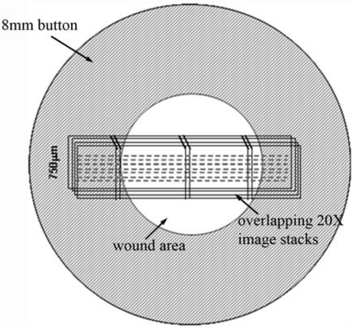
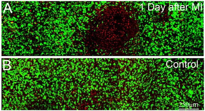
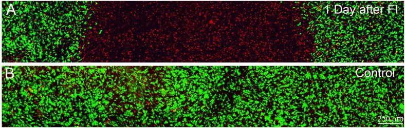
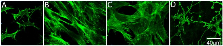
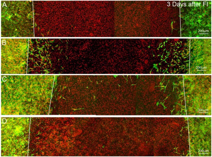
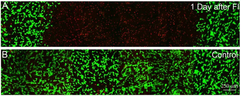
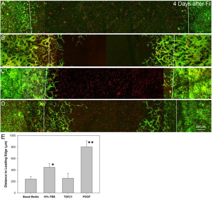
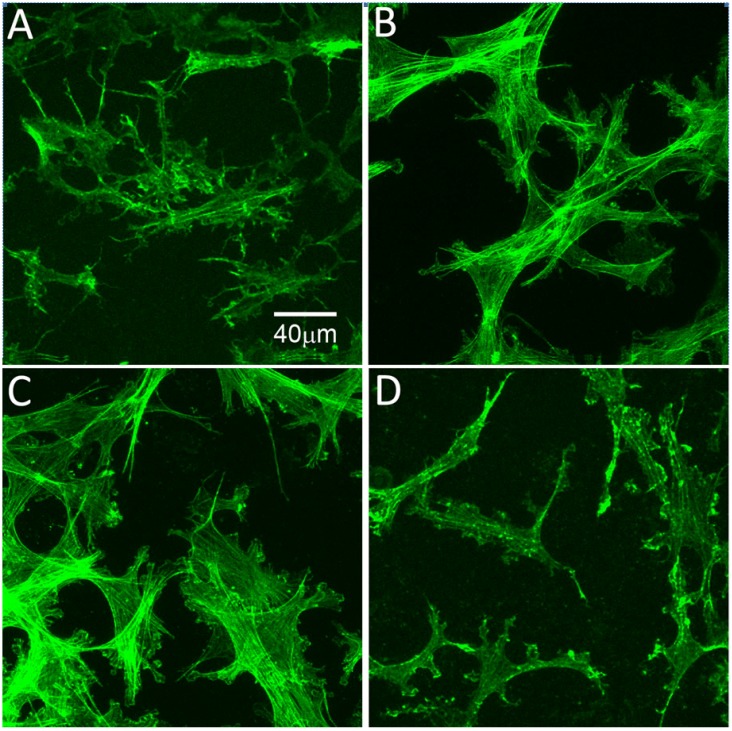
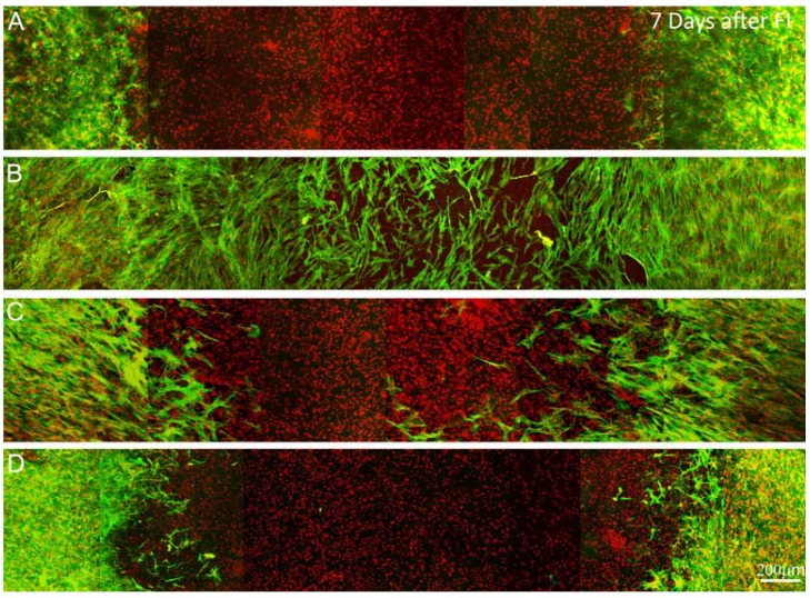
Following laser vision correction, corneal keratocytes must repopulate areas of cell loss by migrating through the intact corneal stroma, and this can impact corneal shape and transparency. In this study, we evaluate 3D culture models for simulating this process in vitro. Buttons (8 mm diameter) were first punched out of keratocyte populated compressed collagen matrices, exposed to a 3mm diameter freeze injury, and cultured in serum-free media (basal media) or media supplemented with 10% FBS, TGFβ1 or PDGF BB. Following freeze injury, a region of cell death was observed in the center of the constructs. Although cells readily migrated on top of the matrices to cover the wound area, a limited amount of cell migration was observed within the constructs. We next developed a novel "sandwich" model, which better mimics the native lamellar architecture of the cornea. Using this model, significant migration was observed under all conditions studied. In both models, cells in TGFβ and 10% FBS developed stress fibers; whereas cells in PDGF were more dendritic. PDGF stimulated the most inter-lamellar migration in the sandwich construct. Overall, these models provide insights into the complex interplay between growth factors, cell mechanical phenotypes and the structural properties of the ECM.
Keywords: 3D Culture; Cell Mechanics; Corneal Keratocytes; Extracellular Matrix; Growth Factors.
Figures
Similar articles
-
Growth factor regulation of corneal keratocyte differentiation and migration in compressed collagen matrices.Invest Ophthalmol Vis Sci. 2010 Feb;51(2):864-75. doi: 10.1167/iovs.09-4200. Epub 2009 Oct 8. Invest Ophthalmol Vis Sci. 2010. PMID: 19815729 Free PMC article.
-
Effect of HDAC Inhibitors on Corneal Keratocyte Mechanical Phenotypes in 3-D Collagen Matrices.Mol Vis. 2015 Apr 29;21:502-14. eCollection 2015. Mol Vis. 2015. PMID: 25999677 Free PMC article.
-
MMP regulation of corneal keratocyte motility and mechanics in 3-D collagen matrices.Exp Eye Res. 2014 Apr;121:147-60. doi: 10.1016/j.exer.2014.02.002. Epub 2014 Feb 14. Exp Eye Res. 2014. PMID: 24530619 Free PMC article.
-
The corneal fibrosis response to epithelial-stromal injury.Exp Eye Res. 2016 Jan;142:110-8. doi: 10.1016/j.exer.2014.09.012. Exp Eye Res. 2016. PMID: 26675407 Free PMC article. Review.
-
Keratocyte mechanobiology.Exp Eye Res. 2020 Nov;200:108228. doi: 10.1016/j.exer.2020.108228. Epub 2020 Sep 10. Exp Eye Res. 2020. PMID: 32919993 Free PMC article. Review.
Cited by
-
Corneal pain and experimental model development.Prog Retin Eye Res. 2019 Jul;71:88-113. doi: 10.1016/j.preteyeres.2018.11.005. Epub 2018 Nov 16. Prog Retin Eye Res. 2019. PMID: 30453079 Free PMC article. Review.
-
Mechanical boundary conditions bias fibroblast invasion in a collagen-fibrin wound model.Biophys J. 2014 Feb 18;106(4):932-43. doi: 10.1016/j.bpj.2013.12.002. Biophys J. 2014. PMID: 24559996 Free PMC article.
-
Transforming Growth Factor Beta 1 Modulates the Functional Expression of the Neurokinin-1 Receptor in Human Keratocytes.Curr Eye Res. 2016 Aug;41(8):1035-1043. doi: 10.3109/02713683.2015.1088954. Epub 2015 Dec 16. Curr Eye Res. 2016. PMID: 26673553 Free PMC article.
-
Platelet recruitment promotes keratocyte repopulation following corneal epithelial abrasion in the mouse.PLoS One. 2015 Mar 16;10(3):e0118950. doi: 10.1371/journal.pone.0118950. eCollection 2015. PLoS One. 2015. PMID: 25775402 Free PMC article.
-
Anti-Inflammatory and Anti-Fibrotic Effects of Human Amniotic Membrane Mesenchymal Stem Cells and Their Potential in Corneal Repair.Stem Cells Transl Med. 2018 Dec;7(12):906-917. doi: 10.1002/sctm.18-0042. Epub 2018 Sep 10. Stem Cells Transl Med. 2018. PMID: 30260581 Free PMC article.
References
-
- Pepose J.S., Ubels J.L. The cornea. In: Hart W.M., editor. Adler’s Physiology of the Eye. Mosby Year Book; St. Louis, MO, USA: 1992. pp. 29–70.
-
- Jester J.V., Barry P.A., Lind G.J., Petroll W.M., Garana R., Cavanagh H.D. Corneal keratocytes: In situ and in vitro organization of cytoskeletal contractile proteins. Invest. Ophthalmol. Vis. Sci. 1994;35:730–743. - PubMed
Grants and funding
LinkOut - more resources
Full Text Sources