Review
. 2013 Mar;35(3):182-94.
doi: 10.1007/s10059-013-0073-2. Epub 2013 Mar 11.

Orai1-NFAT signalling pathway triggered by T cell receptor stimulation

Affiliations
Review

Orai1-NFAT signalling pathway triggered by T cell receptor stimulation

Sonal Srikanth et al. Mol Cells. 2013 Mar.

Abstract

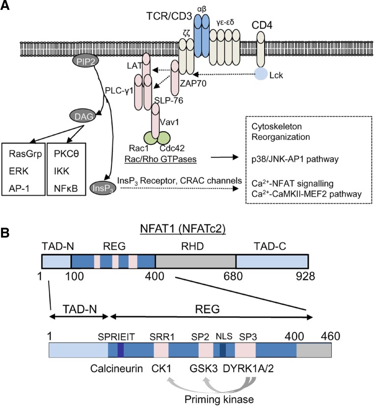
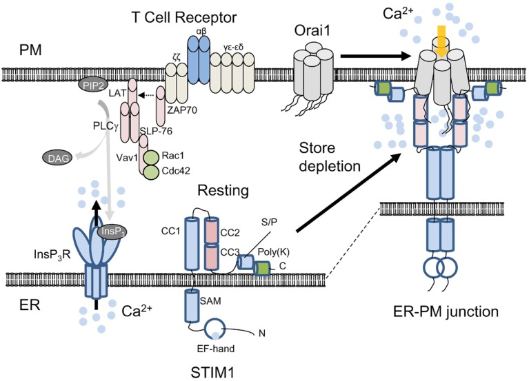
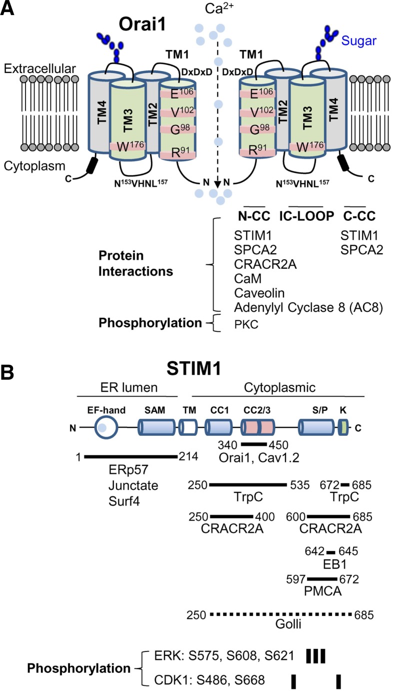
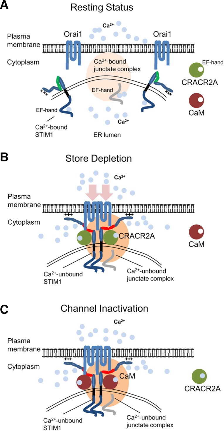
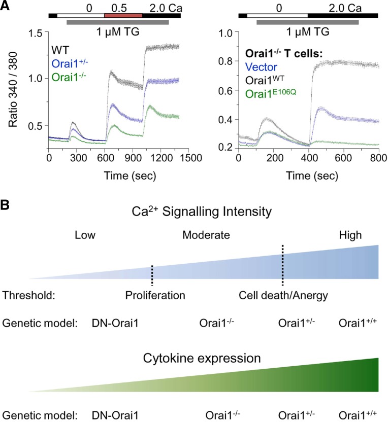
T cell receptor (TCR) stimulation plays a crucial role in development, homeostasis, proliferation, cell death, cytokine production, and differentiation of T cells. Thus, in depth understanding of TCR signalling is crucial for development of therapy targeting inflammatory diseases, improvement of vaccination efficiency, and cancer therapy utilizing T cell-based strategies. TCR activation turns on various signalling pathways, one of the important one being the Ca(2+)-calcineurin-nuclear factor of activated T cells (NFAT) signalling pathway. Stimulation of TCRs triggers depletion of intracellular Ca(2+) store and in turn, initiates store-operated Ca(2+) entry (SOCE), one of the major mechanisms to raise the intracellular Ca(2+) concentrations in T cells. Ca(2+)-release-activated-Ca(2+) (CRAC) channels are a prototype of store-operated Ca(2+) (SOC) channels in immune cells that are very well characterized. Recent identification of STIM1 as the endoplasmic reticulum (ER) Ca(2+) sensor and Orai1 as the pore subunit has dramatically advanced the understanding of CRAC channels and provides a molecular tool to investigate the physiological outcomes of Ca(2+) signalling during immune responses. In this review, we focus on our current understanding of CRAC channel activation, regulation, and downstream calcineurin-NFAT signaling pathway.


Figures

Fig. 1.
Fig. 1.
Signalling pathways of T cell receptor stimulation. (A) Antigen engagement of T cell receptor induces a series of phosphorylation events. Coreceptor (e.g. CD4) ligation in T cells activates protein tyrosine kinase Lck, which phosphorylates the ζ chain of TCR/CD3 complex to recruit ZAP-70 to the TCR/CD3 complex. ZAP70 phosphorylates two adaptor proteins LAT and SLP-76 that results in assembly of a signaling complex containing Vav1 and phospholipase C (PLC-γ1). This signaling complex recruits further downstream effector molecules including Rac1 and a Rho GTPase, cdc42 that have pleiotropic effects in cytoskeleton reorganization, p38/JNK, and Ca2+-NFAT signalling pathways. Cytoskeleton reorganization is important for formation of the immunological synapse between antigen presenting cells and T cells. Activated PLC-γ1 hydrolyzes PIP2 (phosphatidylinositol 4, 5-bisphosphate) into InsP3 (Inositol 1,4,5 trisphosphate) and DAG (diacyl glycerol). While DAG activates PKC (protein kinase C)-NF-κB and RasGRP1-AP-1 signalling pathways, InsP3 binds to the InsP3 receptor (InsP3 R) on the ER membrane to empty the ER Ca2+ store. ER Ca2+ depletion induces opening of CRAC channels, a prototype of store-operated Ca2+ channels. Eleva-ted [Ca2+]i triggers a broad range of downstream signalling pathways including the Ca2+-calmodulin/calcineurin-NFAT and the Ca2+-CaMKII-MEF2 signaling pathway. Ca2+-bound calmodulin forms a complex with a protein phosphatase calcineurin and dephosphorylates the heavily phosphorylated, cytoplasmic NFAT leading to its nuclear translocation. Nuclear NFAT forms a multi-meric protein complex with itself or with other transcription factors (e.g. AP-1) to induce gene transcription. (B) Schematic of the murine NFAT1 (NFATc2) protein. The transcription activation domains that interact with transcriptional cofactors are located at the N and C terminus (TAD-N and TAD-C). DNA binding domain shows the highest homology with the Rel homology domain of Rel-family transcription factors (RHD). The regulatory domain (REG) contains multiple phosphorylation sites to maintain cytoplasmic localization of NFAT under resting conditions and a docking site for Ca2+-calmodulin-calcineurin complex (SPRIET motif). After dephosphorylation by the protein phosphatase complex, the nuclear localization sequence (NLS) within the regulatory domain is exposed leading to nuclear import of NFAT. Serine-rich region (SRR) 1, Ser-Pro-X-X repeat motif (SP) 2, and SP3 within the regulatory domain are phosphorylated by casein kinase 1 (CK1), glycogen synthase kinase 3 (GSK3), and dual-specificity tyrosine-phosphorylation-regulated kinase (DYRK) family kinases, respectively. DYRKs play a role as a priming kinase for CK1 and GSK3-mediated phosphorylation.
Fig. 2.
Fig. 2.
Activation mechanism of Orai1 and STIM1. Schematic showing current understanding of CRAC channel activation. Under resting conditions, Orai1 and STIM1 are distributed at the PM and the ER membrane. The subunit stoichiometry of Orai1 under resting and stimulated conditions is currently unclear. For convenience, a tetrameric assembly of Orai1 is depicted here. Upon store depletion triggered by T cell receptor stimulation and InsP3 production via PLCγ1, STIM1 oligomerizes by sensing ER Ca2+ depletion with its ER-luminal EF-hand domain, clusters and translocates to the ER-PM junctions. By physical interaction with the cytoplasmic, N and C terminus of Orai1 through the CAD/SOAR domain (coiled coil domains 2 and 3), clustered STIM1 recruits and activates Orai1 in the ER-PM junctions. STIM1 contains an ER-luminal region comprising the EF-hand and SAM domains, a single transmembrane segment, and a cytoplasmic region. The cytoplasmic region has three coiled-coil domains (CC1, 2, and 3), serine/proline-rich domain (S/P) containing the residues involved in posttranslational modifications (see below), and a polybasic region (poly-K) at the C terminus that interacts with phosphoinositides after store depletion. The exact role of the poly-K tail of STIM1 in resting conditions is not known, but it may interact with phosphoinositides on the ER membrane to maintain its inactive, folded structure.
Fig. 3.
Fig. 3.
Interacting partners and posttranslational modification of Orai1 and STIM1. (A) Schematic of Orai1. Orai1 has four transmembrane segments (TM1-TM4). It has two extracellular domains and the second extracellular domain between TM3 and TM4 contains the asparagine (N223) residue involved in glycosylation (indicated in blue). The TM1 lines the pore and the residues in TM1 involved in Ca2+ selectivity and gating are depicted. The TM3 does not line the pore, but affects ion selectivity by possible interaction with TM1. Subunits of Orai1 form CRAC channels, but the molecular stoichiometry of CRAC channels is still in question. Schematic depicts a dimeric form for convenience of drawing. Orai1 contains three intracellular domains including the N terminus (N-CC), intracellular loop (IC-LOOP), and C-terminal coiled-coil domain (C-CC) that are important for protein interactions and channel activation/inactivation. Known molecular interactors of Orai1 in these intracellular domains are summarized. The N terminus of Orai1 is phosphorylated by protein kinase C (PKC). (B) Schematic of STIM1. STIM1 contains Ca2+-binding EF hands and a sterile α motif (SAM) domain in the ER-luminal region, a single transmembrane segment, and a long cytoplasmic region. The cytoplasmic region has three coiled-coil domains, serine/proline-rich domain, and a polybasic segment at the C terminus. Proteins associating with each of these domains are indicated. The fragment of STIM1 (340-450) involved in Orai1 interaction/gating is indicated. Golli proteins interact with the cytoplasmic region of STIM1, but the detailed interaction domain is not determined (dotted line). Residues phosphorylated by extracellular signal-regulated kinase (ERK) and cyclin-dependent kinase (CDK1) are indicated.
Fig. 4.
Fig. 4.
CRAC channel regulation by multiple Ca2+-sensing molecules in the ER and cytoplasm. (A) Schematic showing a possible mechanism of CRAC channel regulation. Under resting conditions, Orai1 and STIM1 are distributed at the PM and the ER membrane. Junctate is located at ER-PM junctions in a Ca2+-bound form via its ER-luminal EF hand domain (indicated in gray). Cytoplasmic Ca2+ sensors such as CRACR2A and calmodulin are not bound to Ca2+ in resting conditions. (B) Upon store depletion, STIM1 oligomerizes by sensing ER Ca2+ depletion with its ER-luminal EF-hand domain, and translocates to form clusters at the ER-PM junctions. By physical interactions with Orai1 through the CAD/SOAR domain (depicted in red), clustered STIM1 recruits and activates Orai1 in the ER-PM junctions. During the process, junctate loses bound Ca2+ and supports STIM1 recruitment into ER-PM junctions. CRACR2A is recruited into the Orai1-STIM1 complex to stabilize their interactions. (C) Following the increase of cytoplasmic [Ca2+], CRACR2A dissociates from the Orai1-STIM1 complex. Ca2+-bound calmodulin interacts with the N terminus of Orai1 and inactivates the channel via a mechanism called fast inactivation. The slow inactivation of CRAC channels depends on Ca2+ entry and interaction with SARAF. After channel inactivation, once the ER is refilled with Ca2+, Orai1 and STIM1 return to the resting status.
Fig. 5.
Fig. 5.
Roles of Ca2+ signalling in diverse aspects of T cell activation. (A) Gradual levels of store-operated Ca2+ entry generated by genetic modifications. Left-CRAC channel activity was measured in effector CD4+ T cells from wild type (WT), Orai1 heterozygous (Orai1+/−) and Orai1-deficient (Orai1−/−) mice after store depletion with thapsigargin (TG) in the presence of extracellular solution containing 0.5 and 2 mM Ca2+. Right-CRAC channel activity was measured in Orai1−/− CD4+ T cells transduced with retroviruses expressing empty vector (vector, blue trace), wild type (Orai1WT, black trace) or dominant negative mutant of Orai1 (Orai1E106Q). Data modified from article originally published in (Kim et al., 2011). (B) Ca2+ requirement for T cell death, cytokine production and proliferation differs. T cell proliferation does not need high Ca2+. Instead, excessive Ca2+ concentrations induce cell death and anergy in T cells. Therefore, T cell proliferation requires moderate intracellular Ca2+ concentrations as observed in Orai1-deficient (Orai1−/−) T cells and a further reduction in [Ca2+]i by overexpression of dominant negative Orai1 (DN-Orai1) in Orai1−/− T cells inhibits proliferation. Thus the threshold of [Ca2+]i necessary for proliferation is much lower than that for cell death and anergy. However, cytokine production gradually increases with increase in [Ca2+]i, with DN-Orai1 cells showing minimal cytokine production and Orai1+/+ cells showing maximal cytokine levels. Thus, the pattern of Ca2+ signalling can modulate the outcomes of T cell fates such as cytokine production, proliferation, anergy, and cell death in a digital or analogue manner.

References

    1. Aramburu J, Yaffe MB, Lopez-Rodriguez C, Cantley LC, Hogan PG, Rao A. Affinity-driven peptide selection of an NFAT inhibitor more selective than cyclosporin A. Science. 1999;285:2129–2133. - PubMed
    1. Arron JR, Winslow MM, Polleri A, Chang CP, Wu H, Gao X, Neilson JR, Chen L, Heit JJ, Kim SK, et al. NFAT dysregulation by increased dosage of DSCR1 and DYRK1A on chromosome 21. Nature. 2006;441:595–600. - PubMed
    1. Baine I, Abe BT, Macian F. Regulation of T-cell tolerance by calcium/NFAT signaling. Immunol Rev. 2009;231:225–240. - PubMed
    1. Balagopalan L, Coussens NP, Sherman E, Samelson LE, Sommers CL. The LAT story: a tale of cooperativity, coordination, and choreography. Cold Spring Harb Perspect Biol. 2010;2:a005512. - PMC - PubMed
    1. Barr VA, Bernot KM, Srikanth S, Gwack Y, Balagopalan L, Regan CK, Helman DJ, Sommers CL, Oh-Hora M, Rao A, et al. Dynamic movement of the calcium sensor STIM1 and the calcium channel Orai1 in activated T-cells: puncta and distal caps. Mol. Biol. Cell. 2008;19:2802–2817. - PMC - PubMed

Publication types