Mucoepidermoid carcinoma presenting as a retromolar mucocele
- PMID: 23483321
- PMCID: PMC3591044
- DOI: 10.4103/2231-0746.83161
Mucoepidermoid carcinoma presenting as a retromolar mucocele
Abstract
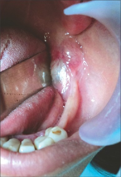
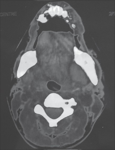
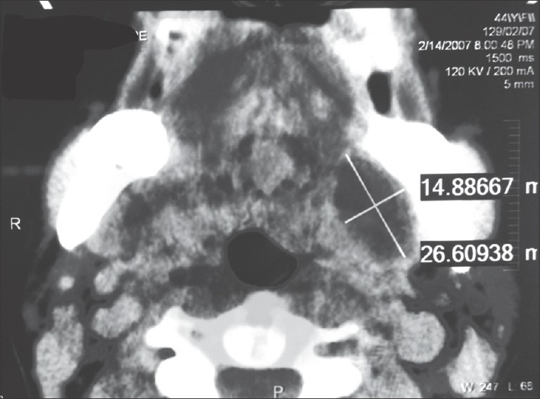
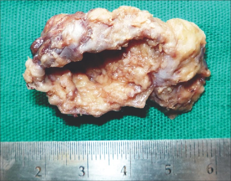
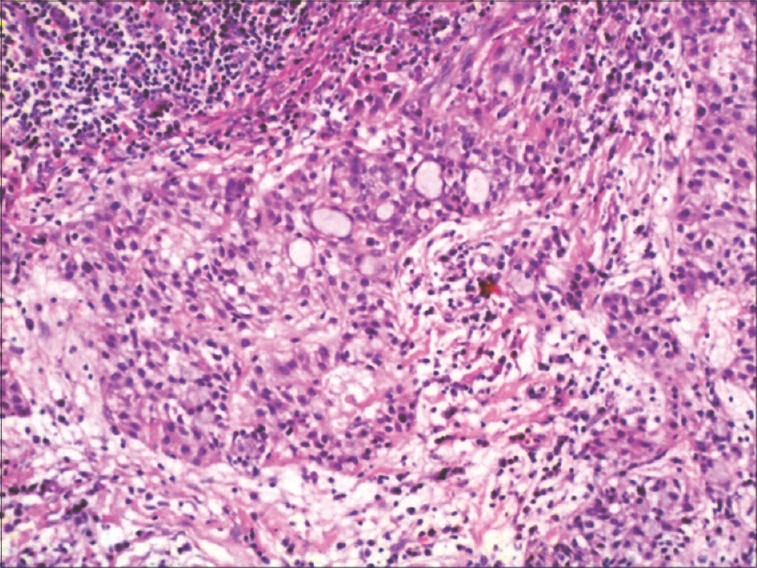
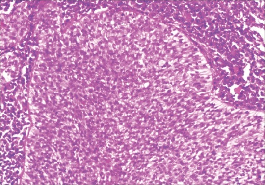
A 44 year old female presented with an intraoral soft tissue swelling in the retromolar region, which was painless, mobile and fluctuant in nature. Computed tomography as well as preoperative ultrasound revealed thick walled cystic lesion. The lesion was clinically diagnosed as mucocele. Ultrasound guided fine needle aspiration was done, which revealed turbid, straw colour fluid. This cystic swelling was completely excised and histopathologically identified to be low grade Mucoepidermoid carcinoma. This unusual presentation of Mucoepidermoid carcinoma as an intraoral cyst is one of the rare and unique reported case.
Keywords: Cyst; malignant minor salivary gland tumor; mucocele; mucoepidermoid carcinoma.
Conflict of interest statement
Figures
References
-
- Bernardes VF, Ramos-Jorge ML, Carmo MA, Cardoso SV, Mesquita RA, Aguiar MC. Intraoral mucoepidermoid carcinoma of salivary glands: Lack of association among clinicopathological features and immunoexpression of c-erbB-2 in 29 cases. Int J Morphol. 2008;26:1005–11.
-
- Baker SR, Malone B. Salivary Gland malignancy in children. Cancer. 1985;55:1730–6. - PubMed
-
- Margaret SB, Katya I, Derrick IW. Muco epidermoid carcinoma. A clinicopathologic study. Am J Surg Pathol. 2001;25:835–45. - PubMed
-
- Guzzo M, Andreola S, Sirizzotti G, Cantu G. Mucoepidermoid carcinoma of the salivary glands: Clinicopathologic review of 108 patients treated at the National Cancer Institute of Milan. Ann Surg Onocol. 2002;9:688–95. - PubMed
-
- Pires FR, de Almeida OP, de Araújo VC, Kowalski LP. Prognostic factors in head and neck mucoepidermoid carcinoma. Arch Otolaryngol Head Neck Surg. 2004;130:174–80. - PubMed