. 2013 Jul;31(7):1075-81.
doi: 10.1002/jor.22341. Epub 2013 Mar 11.

Absence of Cx43 selectively from osteocytes enhances responsiveness to mechanical force in mice

Affiliations

Absence of Cx43 selectively from osteocytes enhances responsiveness to mechanical force in mice

Nicoletta Bivi et al. J Orthop Res. 2013 Jul.

Abstract

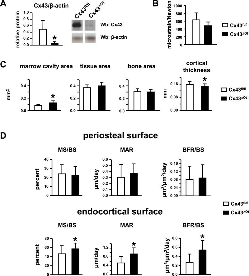
The osteocyte network is crucial for the response of bone to mechanical force. Within this network, connexin43 (Cx43) is thought to mediate the communication of osteocytes and osteoblasts among themselves and the exchange of small molecules with the extracellular milieu. Despite recent advances in understanding Cx43 role for the response of bone cells to mechanical stimulation, the contribution of Cx43 specifically in osteocytes to mechanotransduction in vivo is not well-known. We examined the anabolic response to ulnar axial loading of mice lacking Cx43 in osteocytes (Cx43(ΔOt)). Loading induced a greater increase in periosteal bone formation rate in Cx43(ΔOt) mice compared to control littermates, resulting from higher mineralizing surface and enhanced mineral apposition rate. Expression of β-catenin protein, a molecule implicated in mechanotransduction, was higher in bones from Cx43(ΔOt) mice, compared to littermate controls. In addition, MLO-Y4 osteocytic cells knocked-down for Cx43 exhibited higher β-catenin protein expression and enhanced response to mechanical stimulation. These findings suggest that osteocytes lacking Cx43 are "primed" to respond to mechanical stimulation and that absence of Cx43 in osteocytes unleashes bone formation, by a mechanism that might involve accumulation of β-catenin.


Figures

Figure 1
Figure 1. Cx43ΔOt mice exhibit reduced Cx43 levels and experience a lower strain compared to control mice in response to equal force
(A) Cx43 deficiency was confirmed by Western blot analysis on protein lysates prepared from the diaphysis of non-loaded ulnae of Cx43ΔOt and Cx43fl/fl mice. Bars represent mean ± SD, n=3. *: p<0.05 by t-test. (B) Ulnae from Cx43ΔOt mice exhibit 24% lower microstrain per unit load (ε/N) than ulnae from control mice, as measured using ex-vivo strain gauges applied to the ulna diaphysis. Bars represent mean ± SD. n=3, 6. (C) Geometrical properties of ulnar midshafts from Cx43fl/fl (control) and Cx43ΔOt mice assessed by histomorphometry. (D) Dynamic histomorphometric parameters of ulnae mid-diaphysis on the periosteal and endocortical surfaces of non-loaded control and Cx43ΔOt mice. Bars represent mean ± SD, n=17, 23. *: p<0.05 by unpaired t-test.
Figure 2
Figure 2. Deletion of Cx43 from osteocytes results in enhanced response to loading on the ulnae periosteal surface
(A) Mineralizing surface (MS/BS), mineral apposition rate (MAR) and bone formation rate (BFR/BS) in response to ulnar axial compression at low, medium and high strain. Bars are mean ± SD, *: p<0.05 by paired t-test, n 7-10; #: p<0.05 by two-way ANOVA, n=7-10; (B) Representative images of periosteal bone formation in ulnae of Cx43fl/fl and Cx43ΔOt mice, unloaded and loaded at high magnitude, taken at a 400X magnification. Arrows point at the calcein and alizarin labels.
Figure 3
Figure 3. Loading does not affect bone formation on the endocortical ulnae surface
Mineralizing surface (MS/BS), mineral apposition rate (MAR) and bone formation rate (BFR/BS) in response to ulna compression at low, medium and high strain. Bars represent mean ± SD, n=7-10.
Figure 4
Figure 4. Absence of Cx43 in osteocytes results in increased accumulation of β-catenin in vivo and in vitro
(A) β-catenin levels were evaluated by Western blotting of protein lysates from cortical bone and normalized to β-actin. Bars represent means ± SD, n=3,4. *: p<0.05 versus Cx43fl/fl mice by t-test. (B) Cx43, Axin2, and cyclin D1 mRNA levels normalized by GAPDH were evaluated by qPCR and β-catenin protein levels normalized by β-actin were evaluated by Western blotting in MLO-Y4 osteocytic cells expressing (scr shRNA) or not (Cx43 shRNA) Cx43. (B) Lef1-mediated trasncription was measured in cells treated with vehicle or 30 nM LiCl for 24h. Relative luciferase units (RLU) were normalized by Renilla activity. Bars represent means ± SD, n=4-6. *: p<0.05 versus cells treated with scrambled shRNA (scr) by t-test (B) or the corresponding vehicle-treated culture by one-way ANOVA (C).
Figure 5
Figure 5. Absence of Cx43 in osteocytes results in increased Wnt signaling in response to mechanical loading in vitro
Cells were mechanically stimulated by biaxial stretching at 3,400 microstrain for 5 hours. Gene expression was measured by qPCR on RNA isolated from MLO-Y4 cells expressing (scr shRNA) or not (Cx43 shRNA) Cx43 and normalized to GAPDH. Bars represent means ± SD, n=3. * indicates p<0.05 versus the corresponding non-loaded cells by t-test. #: p<0.05 versus non-loaded scr cells by t-test.

References

    1. Bonewald LF, Johnson ML. Osteocytes, mechanosensing and Wnt signaling. Bone. 2008;42:606–615. - PMC - PubMed
    1. Robinson JA, Chatterjee-Kishore M, Yaworsky PJ, et al. WNT/beta-catenin signaling is a normal physiological response to mechanical loading in bone. J Biol Chem. 2006;281:31720–31728. - PubMed
    1. Tu X, Rhee Y, Condon KW, et al. Sost downregulation and local Wnt signaling are required for the osteogenic response to mechanical loading. Bone. 2012;50:209–217. - PMC - PubMed
    1. Robling AG, Niziolek PJ, Baldridge LA, et al. Mechanical stimulation of bone in vivo reduces osteocyte expression of Sost/sclerostin. J Biol Chem. 2008;283:5866–5875. - PubMed
    1. Glass DA, Bialek P, Ahn JD, et al. Canonical Wnt signaling in differentiated osteoblasts controls osteoclast differentiation. Dev Cell. 2005;8:751–764. - PubMed

Publication types

MeSH terms

LinkOut - more resources