. 2013 Feb 18;23(7):854-861.
doi: 10.1002/adfm.201201636. Epub 2012 Sep 26.

Antibiotic-Releasing Silk Biomaterials for Infection Prevention and Treatment

Affiliations

Antibiotic-Releasing Silk Biomaterials for Infection Prevention and Treatment

Eleanor M Pritchard et al. Adv Funct Mater. .

Abstract

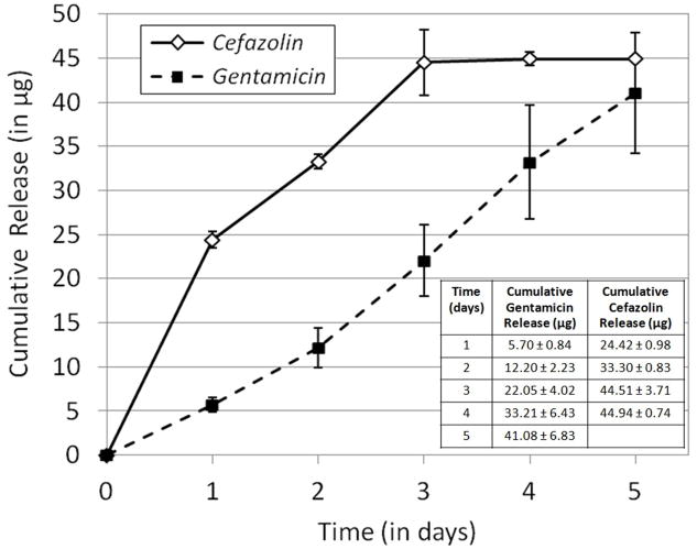
Effective treatment of infections in avascular and necrotic tissues can be challenging due to limited penetration into the target tissue and systemic toxicities. Controlled release polymer implants have the potential to achieve the high local concentrations needed while also minimizing systemic exposure. Silk biomaterials possess unique characteristics for antibiotic delivery including biocompatibility, tunable biodegradation, stabilizing effects, water-based processing and diverse material formats. We report on functional release of antibiotics spanning a range of chemical properties from different material formats of silk (films, microspheres, hydrogels, coatings). The release of penicillin and ampicillin from bulk-loaded silk films, drug-loaded silk microspheres suspended in silk hydrogels and bulk-loaded silk hydrogels was investigated and in vivo efficacy of ampicillin-releasing silk hydrogels was demonstrated in a murine infected wound model. Silk sponges with nanofilm coatings were loaded with gentamicin and cefazolin and release was sustained for 5 and 3 days, respectively. The capability of silk antibiotic carriers to sequester, stabilize and then release bioactive antibiotics represents a major advantage over implants and pumps based on liquid drug reservoirs where instability at room or body temperature is limiting. The present studies demonstrate that silk biomaterials represent a novel, customizable antibiotic platform for focal delivery of antibiotics using a range of material formats (injectable to implantable).

Keywords: antibiotics; biological applications of polymers; biomaterials; drug delivery systems; silk fibroin.


Conflict of interest statement

Competing Financial Interest Statement

The authors have no competing financial interests to declare.

Figures

Figure 1
Figure 1. Antibiotic release from bulk-loaded silk films
Optical density of S. aureus and E. coli liquid cultures at 600 nm (OD600) after 24 hours incubation at 37°C relative to the concentration of penicillin used in the preparation of antibiotic silk films. N=3, error bars represent standard deviations.
Figure 2
Figure 2. Antibiotic release from silk nanofilm coatings on silk sponges
Cumulative release of gentamicin and cefazolin from nanofilm-coated porous silk sponges on S. aureus lawns. N=3, error bars represent standard deviation.
Figure 3
Figure 3. In vitro testing of antibiotic-releasing silk hydrogels
Cumulative drug release from (A) penicillin and (B) ampicillin-loaded silk gels. Gels prepared either by mixing penicillin or ampicillin into the silk solution post-sonication/pre-gelation (bulk loaded) or by mixing antibiotic-loaded silk microspheres into the silk solution post-sonication/pre-gelation (microsphere loaded). N=3, error bars represent standard deviations. Where error bars are not shown, they fall into background.
Figure 4
Figure 4. In vivo testing of antibiotic-releasing silk hydrogels
Total S. aureus CFU counts for various wound treatments relative to total S. aureus CFU for untreated wound. Treatment groups shown: AMP+PBS (250 μg mL−1 mL-1 ampicillin in sterile phosphate buffered saline), AMP+SILK (250 μg mL-1 ampicillin bulk loaded into 4% (w/v) silk hydrogel) and SILK ALONE (unloaded 4% (w/v) silk hydrogel). N=5, error bars represent standard deviations. Data analyzed by two-tailed t-test, df =8; significance levels of individual tests are indicated: *P<0.05, **P<0.01.
Figure 5
Figure 5. Bacterial lawn growth inhibition induced by rifampicin-releasing silk biomaterials
(A) Silk fibers after immersion in a 20 mg mL-1 rifampicin in methanol solution overnight, rinsing in distilled water and drying. (B) Representative sample agar plate showing zone of inhibition in S. aureus lawns produced by rifampicin-releasing silk fibers. (C) Representative sample agar plate showing the zones of inhibition in S. aureus lawns produced by rifampicin-releasing silk film.
Figure 6
Figure 6. Rifampicin loading in silk biomaterials
(A) Rifampicin loading in silk sponges versus starting rifampicin soaking solution concentration for varied soaking solution solvents (B)Rifampicin loading in silk films versus starting rifampicin concentration in the methanol soak solution for varied film thickness (in mg of silk film)
Figure 7
Figure 7. Rifampicin release from silk biomaterials
(A) Cumulative rifampicin release from silk films and (B) silk sponges. N=3, error bars represent standard deviations, where error bars are not shown they fall into background.
Figure 8
Figure 8. Erythromycin release from silk biomaterials
(A) Cumulative erythromycin release from porous silk sponges loaded by soaking in 5 mg mL-1, 50 mg mL-1 and 500 mg mL-1 erythromycin in methanol solutions over 14 days. (B) Release from sponges prepared using the highest loading concentration (500 mg mL-1) over 31 days. N=3, error bars represent standard deviations.

References

    1. Sauermann R, Karch R, Langenberger H, Kettenbach J, Mayer-Helm B, Petsch M, Wagner C, Sautner T, Gattringer R, Karanikas G, Joukhadar C. Antimicrob agentsch. 2005;49:4448. - PMC - PubMed
    1. Gerding DN, Kozak AJ, Peterson LR, Hall WH. Antimicrob agentsch. 1980;17:1023. - PMC - PubMed
    1. Han G, Martinez LR, Mihu MR, Friedman AJ, Friedman JM, Nosanchuk JD. PLoS One. 2009;4:e7804. - PMC - PubMed
    1. Nandi SK, Mukherjee P, Roy S, Kundu B, De DK, Basu D. Mater Sci Eng C. 2009;29:2478.
    1. Englander L, Friedman A. J Clin Aesthet Dermatol. 2010;3:45. - PMC - PubMed

LinkOut - more resources