Staging colorectal cancer with the TNM 7(th): the presumption of innocence when applying the M category
- PMID: 23483791
- PMCID: PMC3587470
- DOI: 10.3748/wjg.v19.i8.1152
Staging colorectal cancer with the TNM 7(th): the presumption of innocence when applying the M category
Abstract
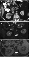
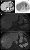
One of the main changes of the current TNM-7 is the elimination of the category MX, since it has been a source of ambiguity and misinterpretation, especially by pathologists. Therefore the ultimate staging would be better performed by the patient's clinician who can classify the disease M0 (no distant metastasis) or M1 (presence of distant metastasis), having access to the completeness of data resulting from clinical examination, imaging workup and pathology report. However this important change doesn't take into account the diagnostic value and the challenge of small indeterminate visceral lesions encountered, in particular, during radiological staging of patients with colorectal cancer. In this article the diagnosis of these lesions with multiple imaging modalities, their frequency, significance and relevance to staging and disease management are described in a multidisciplinary way. In particular the interplay between clinical, radiological and pathological staging, which are usually conducted independently, is discussed. The integrated approach shows that there are both advantages and disadvantages to abandoning the MX category. To avoid ambiguity arising both by applying and interpreting MX category for stage assigning, its abandoning seems reasonable. The recognition of the importance of small lesion characterization raises the need for applying a separate category; therefore a proposal for their categorization is put forward. By using the proposed categorization the lack of consideration for indeterminate visceral lesions with the current staging system will be overcome, also optimizing tailored follow-up.
Keywords: Colorectal cancer; Imaging; Indeterminate lesions; Metastases; Staging.
Figures


References
-
- Poston G, Adam R, Vauthey JN. Downstaging or downsizing: time for a new staging system in advanced colorectal cancer? J Clin Oncol. 2006;24:2702–2706. - PubMed
-
- Poston GJ, Figueras J, Giuliante F, Nuzzo G, Sobrero AF, Gigot JF, Nordlinger B, Adam R, Gruenberger T, Choti MA, et al. Urgent need for a new staging system in advanced colorectal cancer. J Clin Oncol. 2008;26:4828–4833. - PubMed
-
- Sobin LH, Gospodarowicz MK, Wittekind CH. TNM Classification of Malignant Tumors. New York: Wiley-Blackwell; 2009.
-
- Morris EJ, Forman D, Thomas JD, Quirke P, Taylor EF, Fairley L, Cottier B, Poston G. Surgical management and outcomes of colorectal cancer liver metastases. Br J Surg. 2010;97:1110–1118. - PubMed
Publication types
MeSH terms
LinkOut - more resources
Full Text Sources
Other Literature Sources
Medical

