. 2013 Mar 11:4:38.
doi: 10.3389/fphys.2013.00038. eCollection 2013.

Advanced glycation end products acutely impair ca(2+) signaling in bovine aortic endothelial cells

Affiliations

Advanced glycation end products acutely impair ca(2+) signaling in bovine aortic endothelial cells

Nadim Naser et al. Front Physiol. .

Abstract

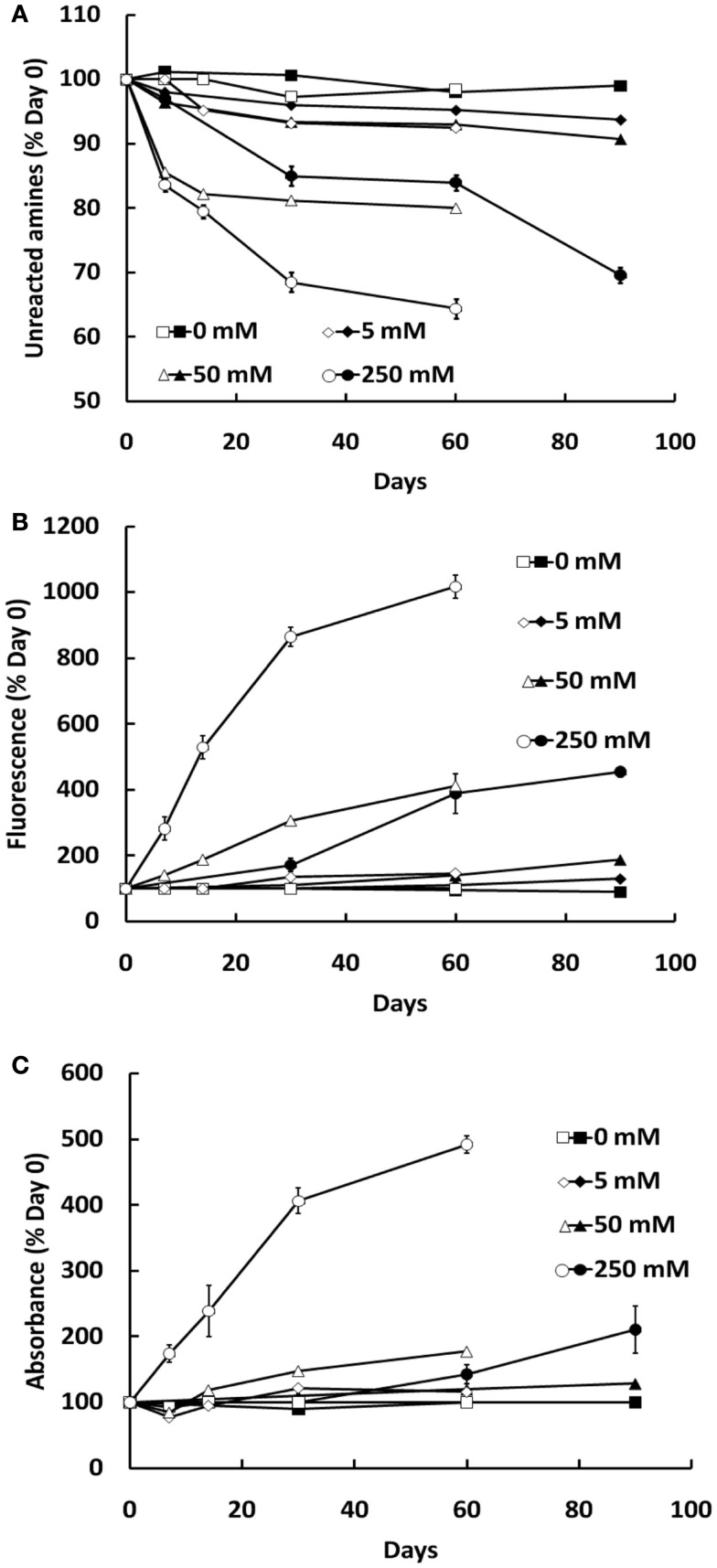
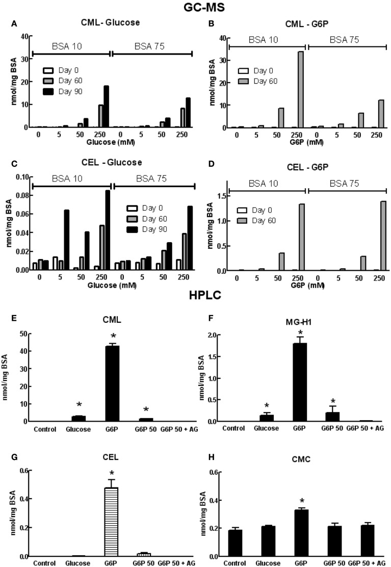
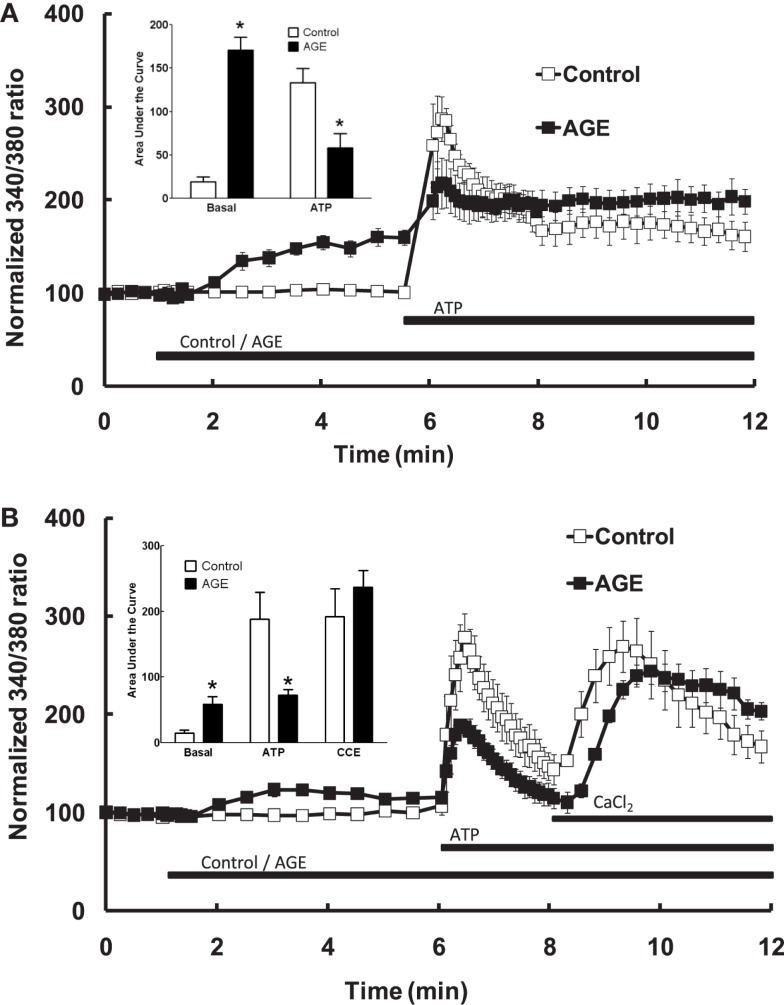
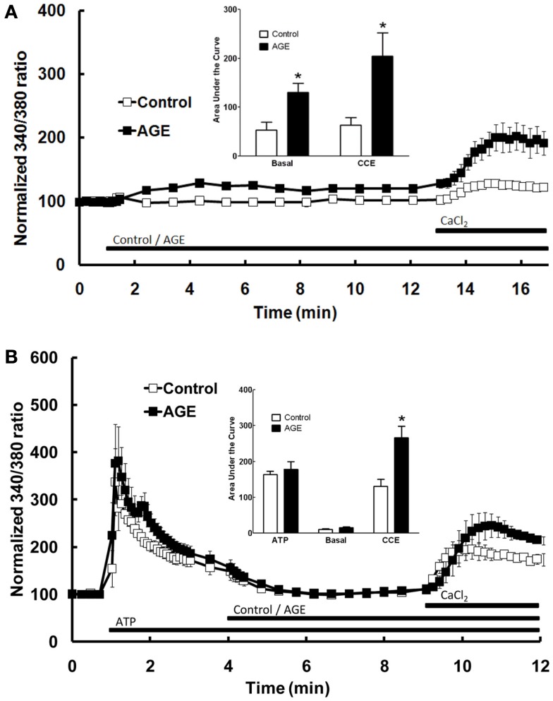
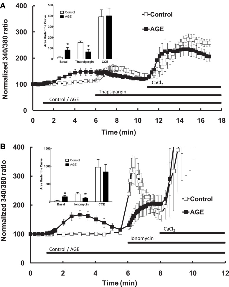
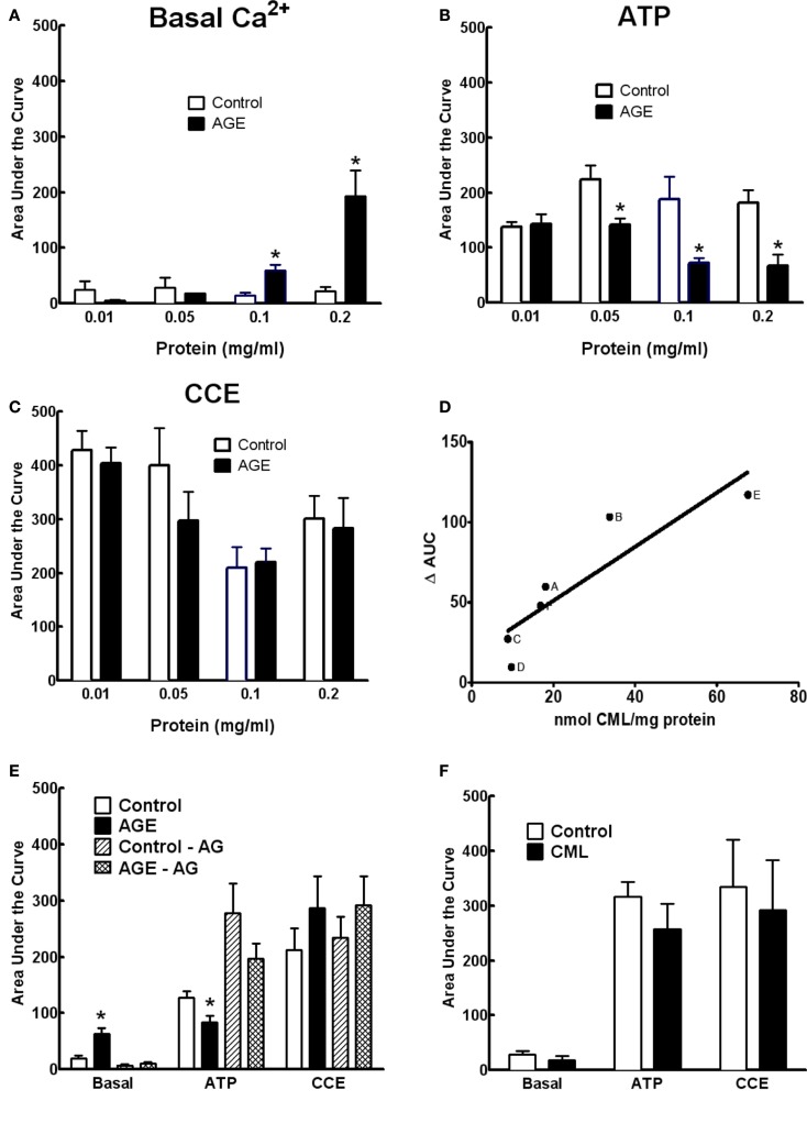
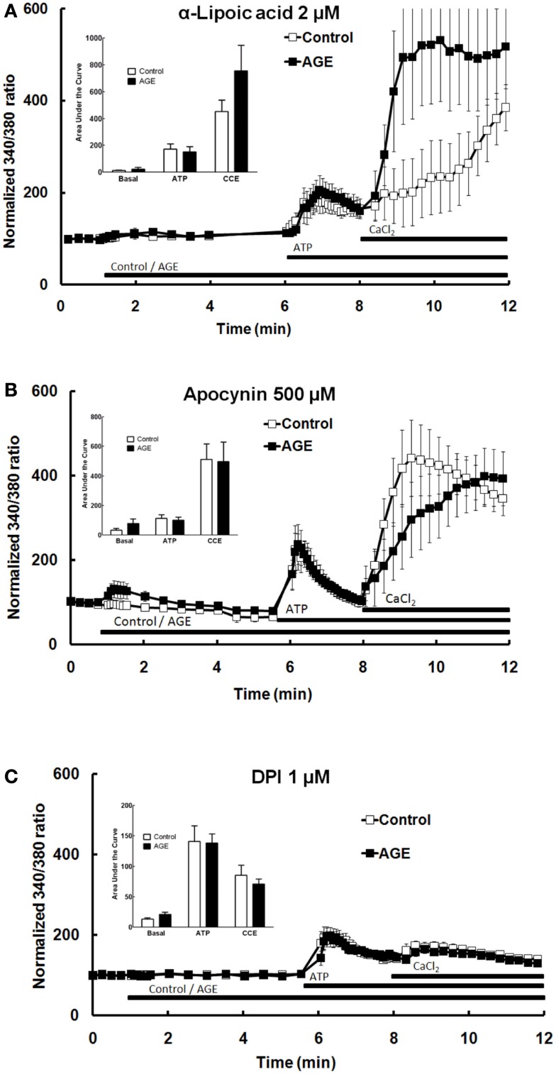
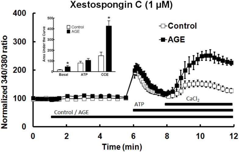
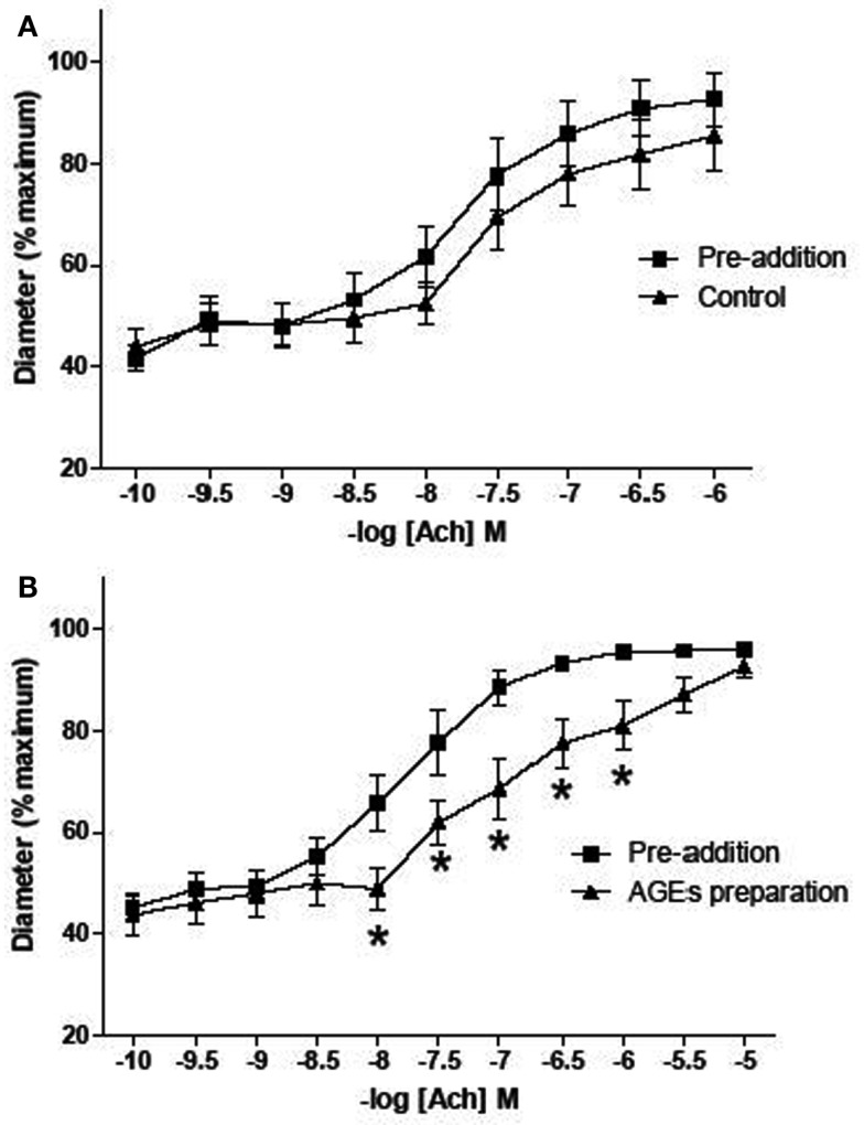
Post-translational modification of proteins in diabetes, including formation of advanced glycation end products (AGEs) are believed to contribute to vascular dysfunction and disease. Impaired function of the endothelium is an early indicator of vascular dysfunction in diabetes and as many endothelial cell processes are dependent upon intracellular [Ca(2+)] and Ca(2+) signaling, the aim of this study was to examine the acute effects of AGEs on Ca(2+) signaling in bovine aortic endothelial cells (BAEC). Ca(2+) signaling was studied using the fluorescent indicator dye Fura-2-AM. AGEs were generated by incubating bovine serum albumin with 0-250 mM glucose or glucose-6-phosphate for 0-120 days at 37°C. Under all conditions, the main AGE species generated was carboxymethyl lysine (CML) as assayed using both gas-liquid chromatograph-mass spectroscopy and high-performance liquid chromatography. In Ca(2+)-replete solution, exposure of BAEC to AGEs for 5 min caused an elevation in basal [Ca(2+)] and attenuated the increase in intracellular [Ca(2+)] caused by ATP (100 μM). In the absence of extracellular Ca(2+), exposure of BAEC to AGEs for 5 min caused an elevation in basal [Ca(2+)] and attenuated subsequent intracellular Ca(2+) release caused by ATP, thapsigargin (0.1 μM), and ionomycin (3 μM), but AGEs did not affect extracellular Ca(2+) entry induced by the re-addition of Ca(2+) to the bathing solution in the presence of any of these agents. The anti-oxidant α-lipoic acid (2 μM) and NAD(P)H oxidase inhibitors apocynin (500 μM) and diphenyleneiodonium (1 μM) abolished these effects of AGEs on BAECs, as did the IP3 receptor antagonist xestospongin C (1 μM). In summary, AGEs caused an acute depletion of Ca(2+) from the intracellular store in BAECs, such that the Ca(2+) signal stimulated by the subsequent application other agents acting upon this store is reduced. The mechanism may involve generation of reactive oxygen species from NAD(P)H oxidase and possible activation of the IP3 receptor.

Keywords: advanced glycation end products; calcium signaling; endothelium; reactive oxygen species.


Figures

Figure 1
Figure 1
Glycation of BSA (10 mg/ml) during co-incubation with G6P (open symbols) or glucose (filled symbols), as assessed by the decline in free amine groups (A), sample fluorescence (B), and sample absorbance (C). Symbols represent the mean ± SEM for three separate batches of BSA glycated under identical conditions and each sample was read in triplicate.
Figure 2
Figure 2
Concentrations of various AGE in samples of co-incubated BSA and glycating agent analyzed using GC-MS (A–D) or HPLC [(E–H); see list of abbreviations for individual AGE species definitions]. Columns in (A–D) represent a triplicate reading of a single sample; columns in (E–H) represent the mean ± SEM for three separate batches of BSA glycated under identical conditions and each sample was read in duplicate. *Indicates significant increase from control (P < 0.05, t-test).
Figure 3
Figure 3
Effect of AGEs on ATP-induced Ca2+ signaling in BAEC. (A) In the presence of extracellular Ca2+, AGEs alone induced an increase in [Ca2+] and inhibited the subsequent ATP-induced Ca2+ increase, as shown in the inset (Area Under Curve data). (B) In the absence of extracellular Ca2+, AGEs alone induced an increase in [Ca2+], inhibited ATP-induced intracellular Ca2+ release but did not affect CCE upon the re-addition of extracellular Ca2+ (CaCl2). Points and columns represent the mean ± SEM of five to six recordings. *Indicates significant effect of AGEs compared to control (P < 0.05, t-test).
Figure 4
Figure 4
Effect of AGEs on Ca2+ signaling in BAEC. (A) In the absence of extracellular Ca2+, AGEs alone induced an increase in [Ca2+] and induced significant CCE compared with control (unglycated BSA). (B) In the absence of extracellular Ca2+, AGEs added to the BAEC after ATP did not induce an increase in [Ca2+], but increased CCE (CaCl2). Points and columns represent the mean ± SEM of six recordings. *Indicates significant effect of AGEs compared to control (P < 0.05, t-test).
Figure 5
Figure 5
Effect of AGEs on thapsigargin (A) and ionomycin (B)-induced Ca2+ signaling in BAEC, in the absence of extracellular Ca2+. In both cases AGEs alone induced an increase in [Ca2+] and inhibited the subsequent agent-induced Ca2+ increase, but did not affect CCE upon the re-addition of extracellular Ca2+ (CaCl2). Points and columns represent the mean ± SEM of six recordings. *Indicates significant effect of AGEs compared to control (P < 0.05, t-test).
Figure 6
Figure 6
Concentration dependence of AGE effects on Ca2+ signaling in BAEC (A–C). (D) Correlation between CML content and inhibition of ATP-induced Ca2+ increase in BAEC of various AGE samples [(A), 250 mM glucose/90 days; (B), 250 mM G6P/60 days; (C), 50 mM G6P/60 days; (D), 250 mM glucose/60 days; (E), 0.2 mg/ml of 250 mM G6P/60 days; (F), 0.05 mg/ml of 250 mM G6P/60 days]. (E) AGE sample (50 mM G6P/60 days) co-incubated with aminoguanidine (AG, 25 mM) did not alter Ca2+ signaling, compared with AGE not containing AG. (F) CML (6 μM) did not alter Ca2+ signaling in BAEC. Points and columns represent the mean ± SEM of five to six recordings. *Indicates significant effect of AGE compared to control (P < 0.05, t-test).
Figure 7
Figure 7
α-Lipoic acid (A), apocynin (B), and DPI (C) each abolished the effects of AGEs on Ca2+ signaling in BAEC. Points and columns represent the mean ± SEM of 6–11 recordings.
Figure 8
Figure 8
Xestospongin C attenuated the effects of AGEs on basal [Ca2+] and abolished the effects on ATP-induced Ca2+ release, however AGEs, in the presence of xestospongin C, enhanced CCE. Columns represent the mean ± SEM of six recordings. *Indicates significant effect of AGEs compared to control (P < 0.05, t-test).
Figure A1
Figure A1
Effect of AGEs on ACh-induced vasodilatation of arterioles from rat cremaster muscles. Arterioles were cannulated and maintained at 70 mmHg. (B) Five minutes exposure to AGEs inhibited ACh-induced vasodilatation (n = 9; p-value 0.011, two-way ANOVA). (A) Arterioles infused with control AGEs solution did not affect ACh-induced vasodilatation (n = 9; p-value > 0.05, two-way ANOVA). AGEs were produced by exposing BSA (10 mg/ml) to 250 mM G6P for 60 days. For control AGEs solution, BSA (10 mg/ml) exposed to 0 mM G6P was used. Data are expressed relative to the maximum diameter obtained under passive conditions (in the absence of extracellular calcium) at 70 mmHg. Points represented mean ± SEM.

References

    1. Abdullaev I. F., Bisaillon J. M., Potier M., Gonzalez J. C., Motiani R. K., Trebak M. (2008). Stim1 and Orai1 mediate CRAC currents and store-operated calcium entry important for endothelial cell proliferation. Circ. Res. 103, 1289–129910.1161/01.RES.0000338496.95579.56 - DOI - PMC - PubMed
    1. Ahmed M. U., Brinkmann Frye E., Degenhardt T. P., Thorpe S. R., Baynes J. W. (1997). N-epsilon-(carboxyethyl)lysine, a product of the chemical modification of proteins by methylglyoxal, increases with age in human lens proteins. Biochem. J. 324(Pt 2), 565–570 - PMC - PubMed
    1. Alkhalaf A., Kleefstra N., Groenier K. H., Bilo H. J., Gans R. O., Heeringa P., et al. (2012). Effect of benfotiamine on advanced glycation endproducts and markers of endothelial dysfunction and inflammation in diabetic nephropathy. PLoS ONE 7:e40427.10.1371/journal.pone.0040427 - DOI - PMC - PubMed
    1. Antigny F., Jousset H., Konig S., Frieden M. (2011). Thapsigargin activates Ca(2)+ entry both by store-dependent, STIM1/Orai1-mediated, and store-independent, TRPC3/PLC/PKC-mediated pathways in human endothelial cells. Cell Calcium 49, 115–12710.1016/j.ceca.2010.12.001 - DOI - PubMed
    1. Antoniotti S., Fiorio Pla A., Barral S., Scalabrino O., Munaron L., Lovisolo D. (2006). Interaction between TRPC channel subunits in endothelial cells. J. Recept. Signal Transduct. Res. 26, 225–24010.1080/10799890600784050 - DOI - PubMed

LinkOut - more resources