

A histopathological exploration of the Madurella mycetomatis grain
- PMID: 23483927
- PMCID: PMC3590280
- DOI: 10.1371/journal.pone.0057774
A histopathological exploration of the Madurella mycetomatis grain
Abstract
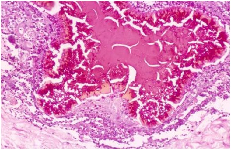
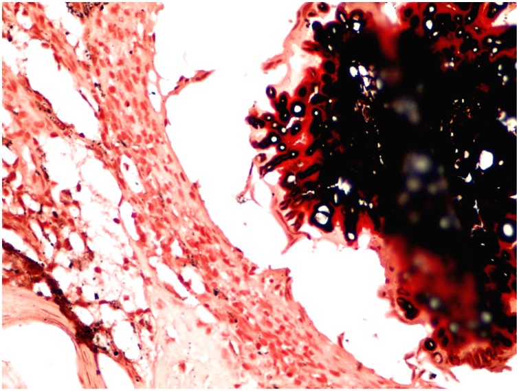
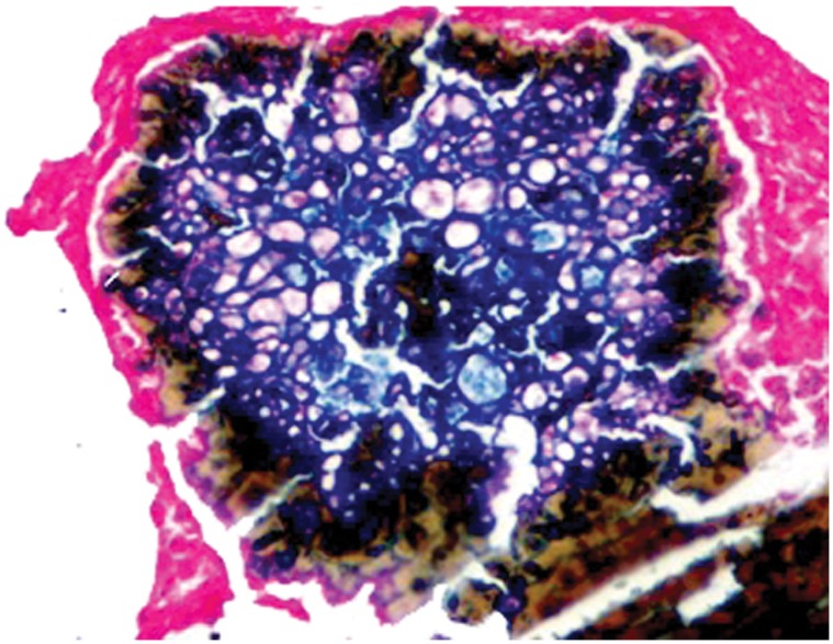
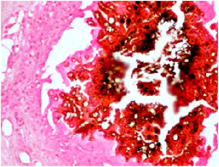
Although the Madurella mycetomatis grains seem to interfere with the host defense mechanisms and impede the antifungal drugs penetration, yet their histological features are not fully known and hence this study was set out to determine that. The study included 80 patients with confirmed M. mycetomatis eumycetoma. After informed written consent, surgical biopsies were obtained from the excised tissues during the patients' surgical treatment. All sections were stained with haematoxylin and eosin, Grocott's hexamine silver, Periodic Acid-Schiff's, Masson-Fontana, Perl's Prussian Blue, Von-kossa's, Formalin Inducing Fluorescence and Schmorl's stains. Modified bleaching technique was used. The concentrations of Zinc, Copper, Calcium, Iron, Lead, Cobalt and Nickel were determined by Atomic Absorption Spectrophotometer. The M. Mycetomatis grains appeared to consist of lipid, protein and melanin. The melanin was located on the hyphal wall as thick layers. The Zinc, Copper and Calcium concentrations in the grains were four, six, and sixteen folds higher than in normal tissue respectively, the other metals were found in the same concentrations as in normal tissue. In the grains, calcium was located in the melanin vicinity. From this study, it can be concluded that, the grains contain melanin, heavy metals, proteins, lipids and they contribute in the formation of the grain cement matrix. These elements seem to contribute in the organism pathogenicity and might impede the penetration of various anti-fungal agents.
Conflict of interest statement
Figures
References
-
- Fahal AH (2011) Mycetoma. Khartoum Med J. 4(1): 514–523.
-
- Ahmed AO, van Leeuwen W, Fahal AH, van de Sande W, Verbrugh H, et al. (2004) Mycetoma caused by Madurella mycetomatis. A neglected infectious burden. Lancet Infect Dis. 4: 566–574. - PubMed
-
- Fahal AH (2004) Mycetoma thorn on the flesh. Trans R Soc Trop Med Hyg. 98: 3–11. - PubMed
-
- EL Hassan AM, Fahal AH, EL Hag IA, Khalil EAG (1994) The pathology of mycetoma: Light microscopic and ultrastructural features. Sudan Med J. 32: 23–45.
-
- Elinov NP (1984) Carbohydrates and their role in the biology of pathogenic fungi. Zentralbl Bakteriol Mikrobiol Hyg A. 257(2): 246–56. - PubMed
MeSH terms
Substances
LinkOut - more resources
Full Text Sources
Other Literature Sources
