. 2013 May;24(10):1584-92.
doi: 10.1091/mbc.E12-08-0628. Epub 2013 Mar 13.

Arl8/ARL-8 functions in apoptotic cell removal by mediating phagolysosome formation in Caenorhabditis elegans

Affiliations

Arl8/ARL-8 functions in apoptotic cell removal by mediating phagolysosome formation in Caenorhabditis elegans

Ayaka Sasaki et al. Mol Biol Cell. 2013 May.

Abstract

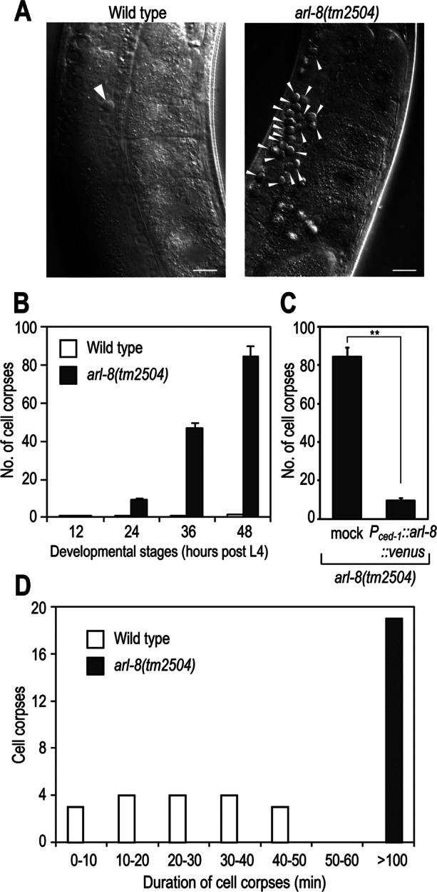
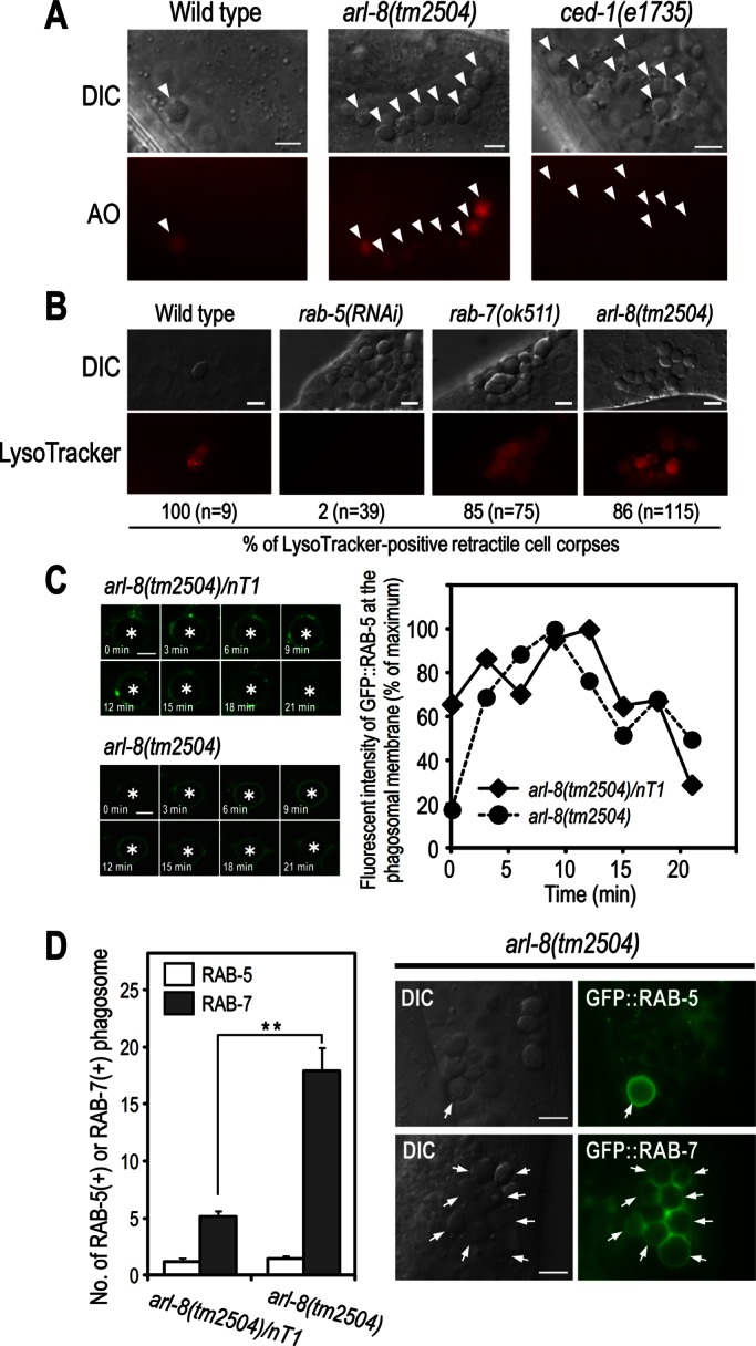
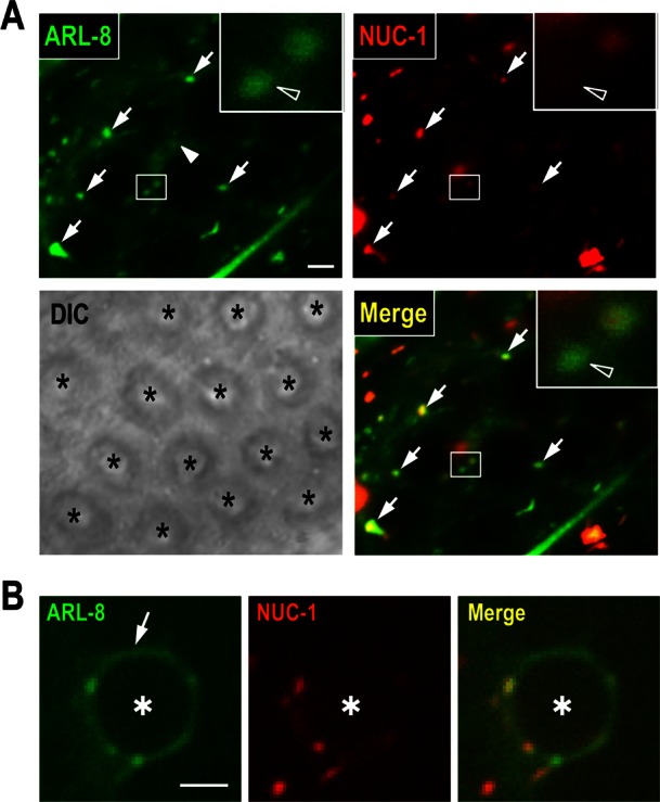
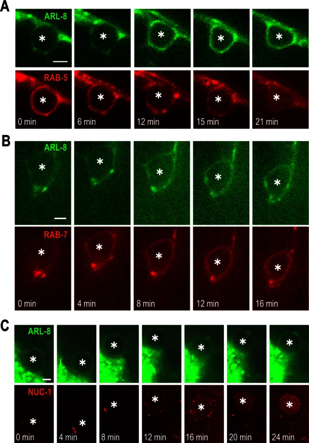
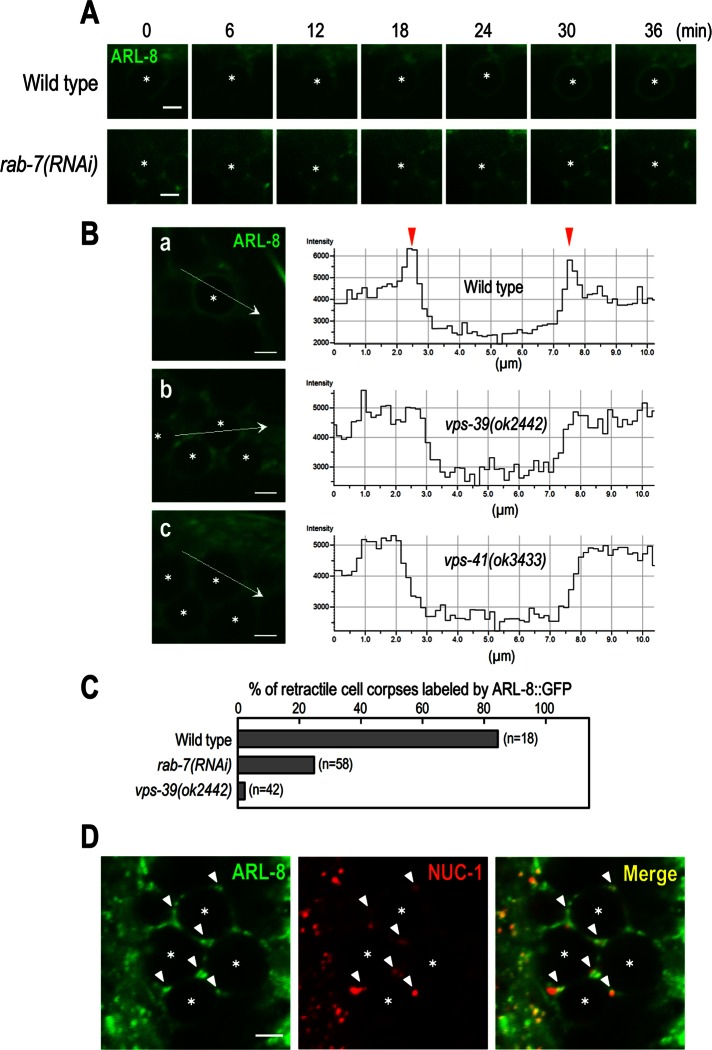
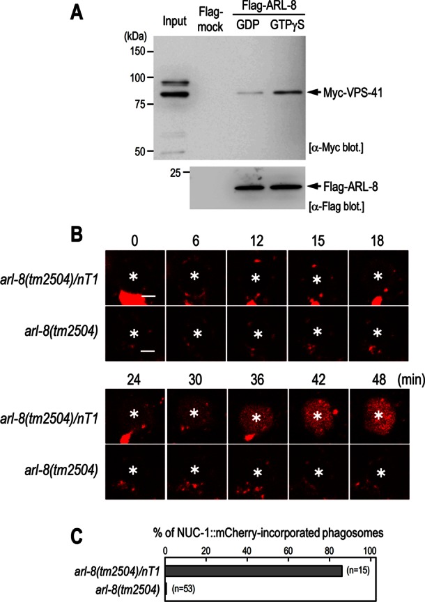
Efficient clearance of apoptotic cells by phagocytes is important for development, tissue homeostasis, and the prevention of autoimmune responses. Phagosomes containing apoptotic cells undergo acidification and mature from Rab5-positive early to Rab7-positive late stages. Phagosomes finally fuse with lysosomes to form phagolysosomes, which degrade apoptotic cells; however, the molecular mechanism underlying phagosome-lysosome fusion is not fully understood. Here we show that the Caenorhabditis elegans Arf-like small GTPase Arl8 (ARL-8) is involved in phagolysosome formation and is required for the efficient removal of apoptotic cells. Loss of function of arl-8 results in the accumulation of apoptotic germ cells. Both the engulfment of the apoptotic cells by surrounding somatic sheath cells and the phagosomal maturation from RAB-5- to RAB-7-positive stages occur in arl-8 mutants. However, the phagosomes fail to fuse with lysosomes in the arl-8 mutants, leading to the accumulation of RAB-7-positive phagosomes and the delayed degradation of apoptotic cells. ARL-8 localizes primarily to lysosomes and physically interacts with the homotypic fusion and protein sorting complex component VPS-41. Collectively our findings reveal that ARL-8 facilitates apoptotic cell removal in vivo by mediating phagosome-lysosome fusion during phagocytosis.


Figures

FIGURE 1:
FIGURE 1:
arl-8(tm2504) mutants are defective in the removal of germ cell corpses. (A) Nomarski images of gonad arms in wild-type or arl-8(tm2504) adult hermaphrodites 48 h after the L4 larval stage. Germ cell corpses are marked with arrowheads. Bars, 10 μm. (B) Germ cell corpses of one gonad arm from each animal were scored every 12 h after the L4 larval stage. Fifteen animals were scored for each strain, and data are expressed as mean ± SEM. (C) The increased germ cell corpse phenotype of arl-8(tm2504) animals was rescued by Pced-1::arl-8::venus. Germ cell corpses of one gonad arm from the indicated animals were scored after the L4 larval stage. More than 10 animals were scored for each strain, and the data are expressed as mean ± SEM. **p < 0.01 by Student's t test. (D) Histogram summarizing cell corpse duration the germline of the indicated animals. More than 15 cell corpses were monitored in each strain.
FIGURE 2:
FIGURE 2:
Phagosomes are arrested at an RAB-7–positive stage in arl-8(tm2504) mutants. (A) AO staining of germ cell corpses in the indicated animals. Germ cell corpses are marked with arrowheads. Most cell corpses were stained by AO in arl-8(tm2504) animals, whereas corpses were not stained in ced-1(e1735) animals in which cell corpses are not phagocytosed by sheath cells. Bars, 5 μm. (B) LysoTracker staining of germ cell corpses and quantitation of LysoTracker-positive cell corpses in the indicated animals. n, number of cell corpses scored. Bars, 5 μm. (C) Time-lapse images of GFP::RAB-5 expression at the phagosome (left) and quantitation of the fluorescence intensity (right) in arl-8(tm2504)/nT1 (as a control) and arl-8(tm2504) animals. Asterisks indicate germ cell corpses. Bars, 2.5 μm. (D) Quantitation of RAB-5–positive and RAB-7–positive phagosomes in the germline of the indicated animals (left). Fifteen animals were scored for each strain, and data are expressed as mean ± SEM. **p < 0.01 by Student's t test. Nomarski and fluorescence images of arl-8(tm2504) animals expressing GFP::RAB-5 or GFP::RAB-7 (right). Arrows indicate the germ cell corpses positive for the corresponding GFP-marker proteins. Bars, 5 μm.
FIGURE 3:
FIGURE 3:
Analysis of ARL-8::GFP localization in sheath cells. (A) Fluorescence and differential interference contrast images of the gonad arm of a wild-type animal coexpressing ARL-8::GFP and NUC-1::mCherry. Asterisks indicate germ cells. ARL-8::GFP was observed in punctate structures (arrows) and cytoplasm surrounding germ cells (arrowhead). Insets, enlarged views of the areas indicated by white rectangles. The open arrowheads in the insets indicate puncta of ARL-8::GFP with little NUC-1::mCherry. Bar, 2 μm. (B) Fluorescence images of a phagosome containing a germ cell corpse (asterisk) in a wild-type animal coexpressing ARL-8::GFP and NUC-1::mCherry. ARL-8::GFP was observed at the phagosomal membrane (arrow). Bar, 2.5 μm.
FIGURE 4:
FIGURE 4:
ARL-8 is recruited to phagosomes at late stages of phagosome maturation. Time-lapse images of fluorescence signal at a phagosome containing a germ cell corpse (asterisk) in a wild-type animal coexpressing ARL-8::GFP and mCherry::RAB-5 (A), ARL-8::GFP and mCherry::RAB-7 (B), or ARL-8::GFP and NUC-1::mCherry (C). Bars, 2.5 μm.
FIGURE 5:
FIGURE 5:
Analysis of ARL-8::GFP localization in rab-7 and the HOPS-complex mutants. (A) Time-lapse images of ARL-8::GFP signal in the gonad of wild-type and rab-7(RNAi) animals. Here 0 min represents the point at which mCherry::RAB-5 (not shown) was first detected on phagosomal membranes. Asterisks indicate germ cell corpses. Bars, 2.5 μm. (B) Fluorescence images of the gonad of wild-type (a), vps-39(ok2442) (b), or vps-41(ok3433) (c) animals expressing ARL-8::GFP and line-scan quantitation of the fluorescence signal at the position indicated by the white arrow in a–c. Asterisks indicate germ cell corpses. Bars, 2.5 μm. (C) The ratio of cell corpses labeled by ARL-8::GFP in the indicated animals. n, number of cell corpses scored. (D) Fluorescence images of the gonad arm of vps-39(ok2442) coexpressing ARL-8::GFP and NUC-1::mCherry. ARL-8::GFP-labeled punctate structures colocalized with NUC-1::mCherry (arrowheads). Asterisks indicate germ cell corpses. Bar, 2.5 μm.
FIGURE 6:
FIGURE 6:
ARL-8 physically interacts with VPS-41 and is required for phagosome–lysosome fusion. (A) Pull-down assay using Flag-ARL-8 and Myc-VPS-41. Purified Flag-ARL-8 proteins preloaded with either GDP or GTPγS were incubated with HEK293T cell lysates containing Myc-VPS-41 and immunoprecipitated using anti-Flag M2 agarose. The precipitated fractions were then subjected to Western blot analysis using the indicated antibodies. (B) Time-lapse images of NUC-1::mCherry signal in the gonad of arl-8(tm2504)/nT1 and arl-8(tm2504) animals. Asterisks indicate germ cell corpses. Here 0 min represents the point at which GFP::RAB-5 (not shown) was first detected on phagosomal membranes. Bars, 2.5 μm. (C) Quantitation of fusion events in the indicated animals. Data are expressed as the ratio of cell corpses that incorporated NUC-1::mCherry within 1 h after their appearance. n, number of cell corpses scored.

References

    1. Akbar MA, Tracy C, Kahr WH, Kramer H. The full-of-bacteria gene is required for phagosome maturation during immune defense in Drosophila. J Cell Biol. 2011;192:383–390. - PMC - PubMed
    1. Bagshaw RD, Callahan JW, Mahuran DJ. The Arf-family protein, Arl8b, is involved in the spatial distribution of lysosomes. Biochem Biophys Res Commun. 2006;344:1186–1191. - PubMed
    1. Brenner S. The genetics of Caenorhabditis elegans. Genetics. 1974;77:71–94. - PMC - PubMed
    1. Desjardins M, Huber LA, Parton RG, Griffiths G. Biogenesis of phagolysosomes proceeds through a sequential series of interactions with the endocytic apparatus. J Cell Biol. 1994;124:677–688. - PMC - PubMed
    1. Duclos S, Diez R, Garin J, Papadopoulou B, Descoteaux A, Stenmark H, Desjardins M. Rab5 regulates the kiss and run fusion between phagosomes and endosomes and the acquisition of phagosome leishmanicidal properties in RAW 264.7 macrophages. J Cell Sci. 2000;113:3531–3541. - PubMed

Publication types

Substances