. 2013 May:87:75-82.
doi: 10.1016/j.mvr.2013.02.002. Epub 2013 Feb 26.

Intussusceptive remodeling of vascular branch angles in chemically-induced murine colitis

Affiliations

Intussusceptive remodeling of vascular branch angles in chemically-induced murine colitis

Maximilian Ackermann et al. Microvasc Res. 2013 May.

Abstract

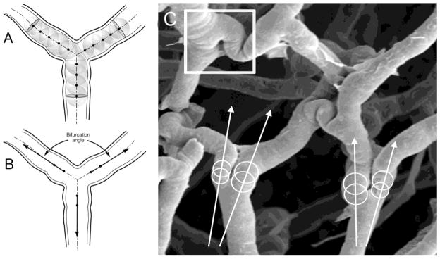
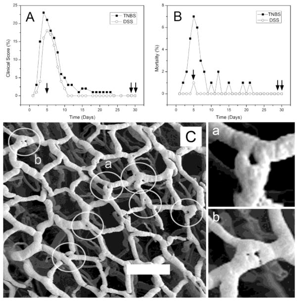
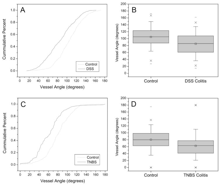
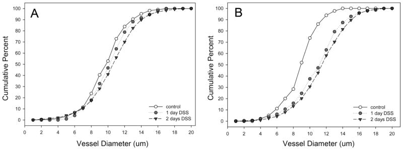
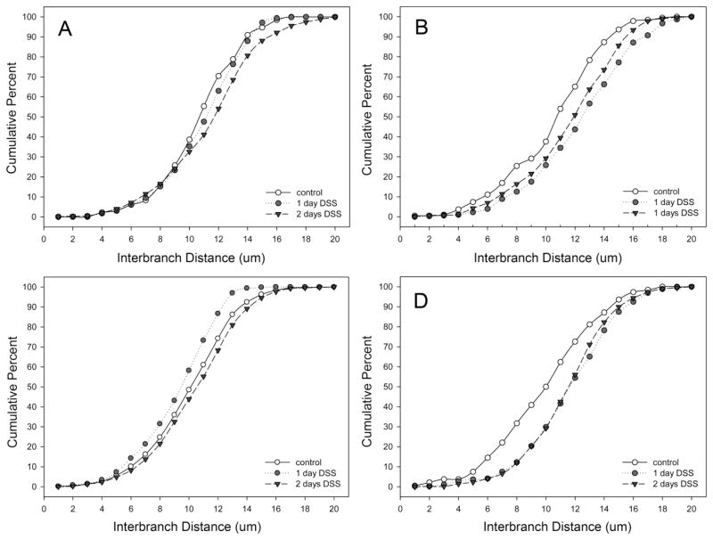
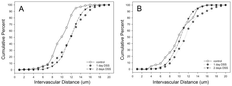
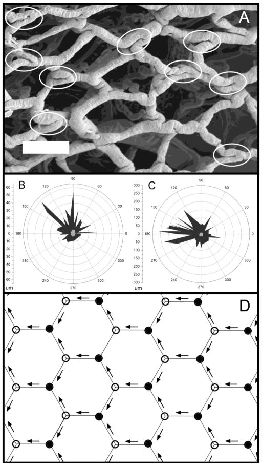
Intussusceptive angiogenesis is a developmental process linked to both blood vessel replication and remodeling in development. To investigate the prediction that the process of intussusceptive angiogenesis is associated with vessel angle remodeling in adult mice, we systematically evaluated corrosion casts of the mucosal plexus in mice with trinitrobenzesulfonic acid (TNBS)-induced and dextran sodium sulfate (DSS)-induced colitis. The mice demonstrated a significant decrease in vessel angles in both TNBS-induced and DSS-induced colitis within 4 weeks of the onset of colitis (p<.001). Corrosion casts 28-30 days after DSS treatment were studied for a variety of detailed morphometric changes. The vessel diameter and interbranch distance were significantly increased in the descending colon (p<.05). Also consistent with vessel growth, intervascular distance was decreased in the descending colon (p<.05). In contrast, no statistically significant morphometric changes were noted in the ascending colon. The morphometry of the corrosion casts also demonstrated 1) a similar orientation of the remodeled angles within the XY coordinate plane of the mucosal plexus, and 2) alternating periodicity of remodeled and unremodeled vessel angles. We conclude that inflammation-associated intussusceptive angiogenesis in adult mice is associated with vessel angle remodeling. Further, the morphometry of the vessel angles suggests the influence of blood flow on the location and orientation of remodeled vessels.


Figures

Figure 1
Figure 1
The method of branch angle morphometry was designed to be sensitive to geometric variation at the apex of the bifurcation. A) Maximally-sized spheres were inscribed in each vessel at the bifurcation. Sequential spheres within each vessel were inscribed so that the surface of the sphere intersected the centerpoint of the preceding sphere. B) The centerline tract of the first two spheres was used to define the vessel coordinates. C) Example of two branch angles in colon mucosal plexus vessels. Note the intussusceptive pillar (square).
Figure 2
Figure 2
The stimulation of intussusceptive angiogenesis by chemically-induced colitis. A) Frequency distribution of mice with a colitis score (see Methods) greater than 1 for TNBS- and DSS-induced colitis. Single arrow shows the timing of corrosion casts demonstrating intussusceptive pillars; double arrows identify the timing of corrosion casts examined for branch angle remodeling. B) Frequency distribution of deaths after the onset of TNBS- or DSS-induced colitis (TNBS N=100; DSS N=100). C) Corrosion casts of the mucosal plexus (descending colon) 5 days after the onset of TNBS-induced colitis. Circles identify “holes” that demonstrate the site of intussusceptive pillars. Two of the vessel bifurcations (a,b) are shown in higher magnification. Bar = 150 μm.
Figure 3
Figure 3
The branch angles were measured in both DSS-induced (A,B) and TNBS- induced (C,D) colitis. The branch angles (N=750) of the DSS-induced colitis were examined by stereo-pair SEM morphometry; branch angles (N=350) of the TNBS-induced colitis mice were examined using 2D planar images. In the TNBS mice measurements, two investigators, blinded to the conditions, measured the bifurcation angle using the geometric technique (see Methods) to control for potential projection-dependent distortions. The median value between investigators was calculated and the histogram plotted for both colitis (square) and control (circle) mice (p<.001, t-test for samples of unequal variance).
Figure 4
Figure 4
The diameters of mucosal plexus vessels in the ascending (A) and descending (B) colons in DSS colitis. Murine colitis was stimulated with either 1 or 2 days of DSS exposure. At 28–30 days, corrosion casting demonstrated a significant increase in vessel diameter in the descending colon; there was no significant change in the ascending colon.
Figure 5
Figure 5
The interbranch distance of mucosal plexus vessels in the cecum (A), ascending (B), transverse (C) and descending (D) colons in DSS colitis. Murine colitis was stimulated with either 1 or 2 days of DSS exposure. At 28–30 days, corrosion casting demonstrated a significant increase in interbranch distance in the descending colon; there was no significant change in the cecum, ascending or transverse colon. Of note, the control values in the transverse colon panel (C) represent aggregate control values from all regions.
Figure 6
Figure 6
The intervascular distance between mucosal plexus vessels in the ascending (A) and descending (B) colons in DSS colitis. Murine colitis was stimulated with either 1 or 2 days of DSS exposure. At 28–30 days, corrosion casting demonstrated a significant decrease in vessel distance in the descending colon; there was an increased distance in the ascending colon.
Figure 7
Figure 7
The orientation of remodeled branch angles. A) A SEM photomicrograph of a corrosion cast obtained 30 days after the onset of TNBS-induced colitis (bar=100um). Acute branch angles, likely reflecting structural remodeling, are highlighted (elipses). Note the similar length and orientation of the parallel segments within the highlighted regions; specifically, the parallel segements of vessels at the vessel bifurcation. Two other examples (N=2 mice) of nonrandom remodeling are shown in the polar graphs (B,C). The length and orientation of the parallel segments are shown for colitis (black) and control (gray) mice. D) Schematic illustration of the periodic variation in converging (closed circles) and diverging (open circles) bifurcations created by a unidirectional flow field (arrows) in the colon mucosal plexus.

References

    1. Baumbach GL, Ghoneim S. Vascular remodeling in hypertension. Scanning Microsc. 1993;7:137–42. - PubMed
    1. Burri PH, et al. Intussusceptive angiogenesis: its emergence, its characteristics, and its significance. Dev Dyn. 2004;231:474–88. - PubMed
    1. Djonov V, et al. Vascular remodelling during the normal and malignant life cycle of the mammary gland. Microsc Res Tech. 2001;52:182–9. - PubMed
    1. Djonov V, et al. Intussusceptive angiogenesis: its role in embryonic vascular network formation. Circ Res. 2000;86:286–92. - PubMed
    1. Djonov VG, et al. Optimality in the developing vascular system: branching remodeling by means of intussusception as an efficient adaptation mechanism. Dev Dyn. 2002;224:391–402. - PubMed

Publication types

Substances

LinkOut - more resources