

Cystic fibrosis transmembrane conductance regulator recruitment to phagosomes in neutrophils
- PMID: 23486169
- PMCID: PMC3717385
- DOI: 10.1159/000346568
Cystic fibrosis transmembrane conductance regulator recruitment to phagosomes in neutrophils
Abstract
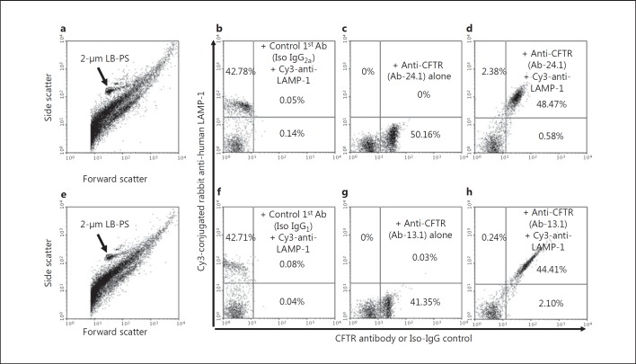
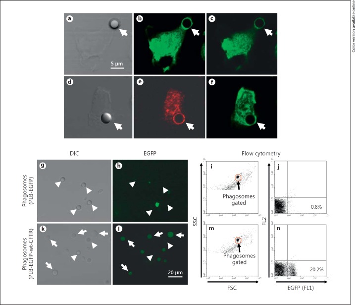
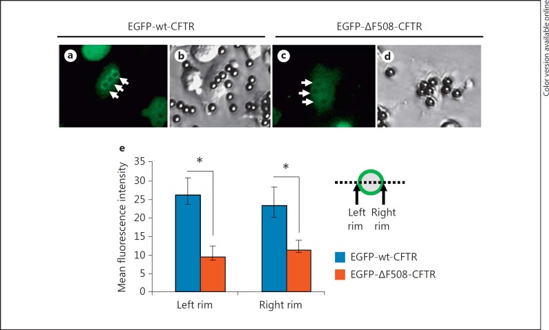
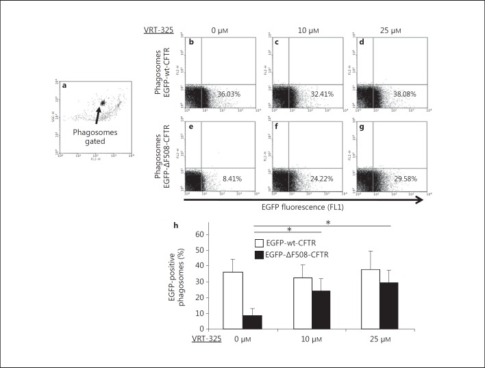
Optimal microbicidal activity of human polymorphonuclear leukocytes (PMN) relies on the generation of toxic agents such as hypochlorous acid (HOCl) in phagosomes. HOCl formation requires H2O2 produced by the NADPH oxidase, myeloperoxidase derived from azurophilic granules, and chloride ion. Chloride transport from cytoplasm into phagosomes requires chloride channels which include cystic fibrosis transmembrane conductance regulator (CFTR), a cAMP-activated chloride channel. However, the phagosomal targeting of CFTR in PMN has not been defined. Using human peripheral blood PMN, we determined that 95-99% of lysosomal-associated membrane protein 1 (LAMP-1)-positive mature phagosomes were CFTR positive, as judged by immunostaining and flow cytometric analysis. To establish a model cell system to evaluate CFTR phagosomal recruitment, we stably expressed enhanced green fluorescent protein (EGFP) alone, EGFP-wt-CFTR and EGFP-DF508-CFTR fusion proteins in promyelocytic PLB-985 cells, respectively. After differentiation into neutrophil-like cells, CFTR presentation to phagosomes was examined. EGFP-wt-CFTR was observed to associate with phagosomes and colocalize with LAMP-1. Flow cytometric analysis of the isolated phagosomes indicated that such a phagosomal targeting was determined by the CFTR portion of the fusion protein. In contrast, significantly less EGFP-DF508-CFTR was found in phagosomes, indicating a defective targeting of the molecule to the organelle. Importantly, the CFTR corrector compound VRT-325 facilitated the recruitment of DF508-CFTR to phagosomes. These data demonstrate the possibility of pharmacologic correction of impaired recruitment of mutant CFTR, thereby providing a potential means to augment chloride supply to the phagosomes of PMN in patients with cystic fibrosis to enhance their microbicidal function.
Copyright © 2013 S. Karger AG, Basel.
Figures

Comment in
-
Neutrophils in the innate immunity conundrum of cystic fibrosis: a CFTR-related matter?J Innate Immun. 2013;5(3):195-6. doi: 10.1159/000350215. Epub 2013 Mar 23. J Innate Immun. 2013. PMID: 23548924 Free PMC article. No abstract available.
Similar articles
-
Neutrophils in the innate immunity conundrum of cystic fibrosis: a CFTR-related matter?J Innate Immun. 2013;5(3):195-6. doi: 10.1159/000350215. Epub 2013 Mar 23. J Innate Immun. 2013. PMID: 23548924 Free PMC article. No abstract available.
-
Chloride transport in functionally active phagosomes isolated from Human neutrophils.Free Radic Biol Med. 2012 Dec 15;53(12):2308-17. doi: 10.1016/j.freeradbiomed.2012.10.542. Epub 2012 Oct 23. Free Radic Biol Med. 2012. PMID: 23089227 Free PMC article.
-
CFTR Expression in human neutrophils and the phagolysosomal chlorination defect in cystic fibrosis.Biochemistry. 2006 Aug 29;45(34):10260-9. doi: 10.1021/bi060490t. Biochemistry. 2006. PMID: 16922501 Free PMC article.
-
Salt, chloride, bleach, and innate host defense.J Leukoc Biol. 2015 Aug;98(2):163-72. doi: 10.1189/jlb.4RU0315-109R. Epub 2015 Jun 5. J Leukoc Biol. 2015. PMID: 26048979 Free PMC article. Review.
-
Chloride flux in phagocytes.Immunol Rev. 2016 Sep;273(1):219-31. doi: 10.1111/imr.12438. Immunol Rev. 2016. PMID: 27558337 Review.
Cited by
-
Role of Cystic Fibrosis Bronchial Epithelium in Neutrophil Chemotaxis.Front Immunol. 2020 Aug 4;11:1438. doi: 10.3389/fimmu.2020.01438. eCollection 2020. Front Immunol. 2020. PMID: 32849500 Free PMC article. Review.
-
Virulence Mechanisms of Mycobacterium abscessus: Current Knowledge and Implications for Vaccine Design.Front Microbiol. 2022 Mar 3;13:842017. doi: 10.3389/fmicb.2022.842017. eCollection 2022. Front Microbiol. 2022. PMID: 35308378 Free PMC article. Review.
-
Revisiting the Role of Leukocytes in Cystic Fibrosis.Cells. 2021 Dec 1;10(12):3380. doi: 10.3390/cells10123380. Cells. 2021. PMID: 34943888 Free PMC article. Review.
-
Dysfunctions of Neutrophils in the Peripheral Blood of Children with Cystic Fibrosis.Biomedicines. 2023 Jun 15;11(6):1725. doi: 10.3390/biomedicines11061725. Biomedicines. 2023. PMID: 37371820 Free PMC article.
-
CFTR Modulators Dampen Aspergillus-Induced Reactive Oxygen Species Production by Cystic Fibrosis Phagocytes.Front Cell Infect Microbiol. 2020 Jul 24;10:372. doi: 10.3389/fcimb.2020.00372. eCollection 2020. Front Cell Infect Microbiol. 2020. PMID: 32793514 Free PMC article.
References
-
- Welsh MJ, Ramsey BW, Accurso F, Cutting G, Cystic fibrosis . Metabolic and Molecular Basis of Inherited Disease. In: Scriver CR, editor. New York: McGraw-Hill; 2001. pp. pp 5121–5188.
-
- Davis PB, Drumm M, Konstan MW. Cystic fibrosis. Am J Respir Crit Care Med. 1996;154:1229–1256. - PubMed
-
- Downey DG, Bell SC, Elborn JS. Neutrophils in cystic fibrosis. Thorax. 2009;64:81–88. - PubMed
-
- Hayes E, Pohl K, McElvaney NG, Reeves EP. The cystic fibrosis neutrophil: a specialized yet potentially defective cell. Arch Immunol Ther Exp (Warsz) 2011;59:97–112. - PubMed
Publication types
MeSH terms
Substances
Grants and funding
LinkOut - more resources
Full Text Sources
Other Literature Sources
Medical
Research Materials
Miscellaneous
