

Morpho-elasticity of intestinal villi
- PMID: 23486174
- PMCID: PMC3627092
- DOI: 10.1098/rsif.2013.0109
Morpho-elasticity of intestinal villi
Abstract
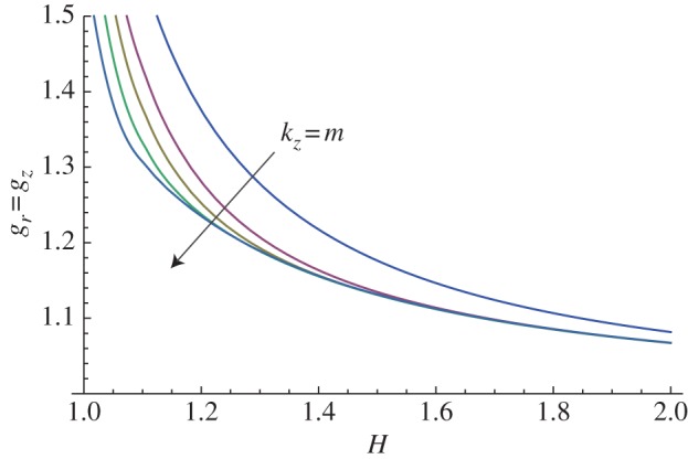
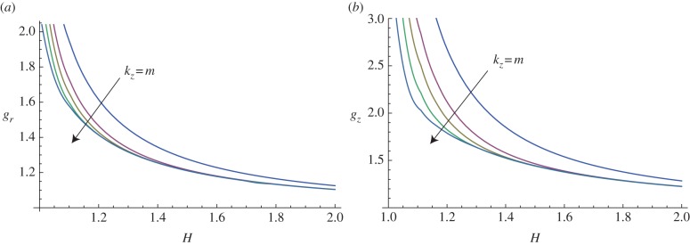
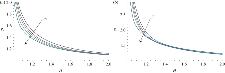
Villi are ubiquitous structures in the intestine of all vertebrates, originating from the embryonic development of the epithelial mucosa. Their morphogenesis has similar stages in living organisms but different forming mechanisms. In this work, we model the emergence of the bi-dimensional undulated patterns in the intestinal mucosa from which villi start to elongate. The embryonic mucosa is modelled as a growing thick-walled cylinder, and its mechanical behaviour is described using an hyperelastic constitutive model, which also accounts for the anisotropic characteristics of the reinforcing fibres at the microstructural level. The occurrence of surface undulations is investigated using a linear stability analysis based on the theory of incremental deformations superimposed on a finite deformation. The Stroh formulation of the incremental boundary value problem is derived, and a numerical solution procedure is implemented for calculating the growth thresholds of instability. The numerical results are finally discussed with respect to different growth and materials properties. In conclusion, we demonstrate that the emergence of intestinal villi in embryos is triggered by a differential growth between the mucosa and the mesenchymal tissues. The proposed model quantifies how both the geometrical and the mechanical properties of the mucosa drive the formation of previllous structures in embryos.
Figures

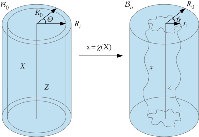
 into the position x in the actual configuration
  . (Online version in colour.)
 in which geometrical incompatibilities are allowed, and the elastic component Fe restores the compatibility of the tissue deformation. (Online version in colour.)
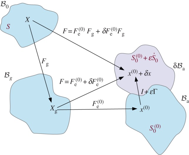
 represents the projection of the incremental nominal stress in the perturbed configuration
  . (Online version in colour.)
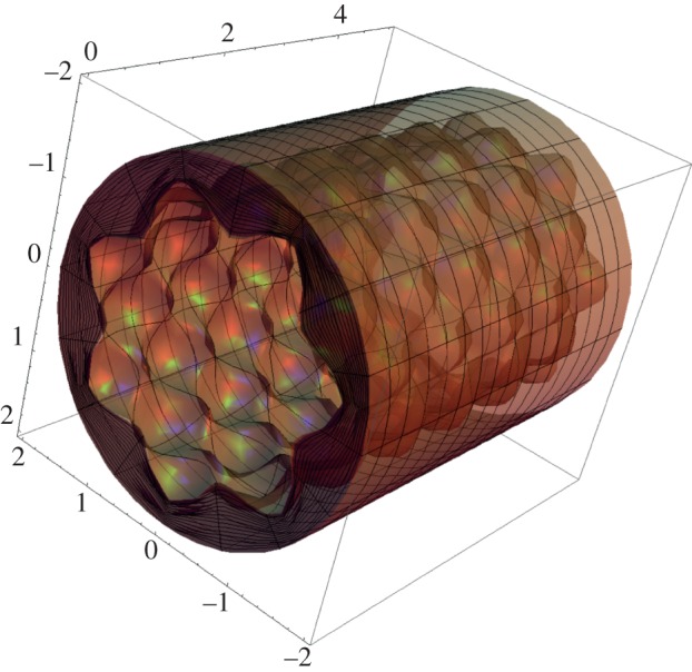
References
-
- Rao JN, Wang JY. 2010. Regulation of gastrointestinal mucosal growth. San Rafael, CA: Morgan & Claypool Life Sciences - PubMed
-
- Grey RD. 1972. Morphogenesis of intestinal villi I: scanning electron microscopy of the duodenal epithelium of the developing chick embryo. J. Morphol. 137, 193–214 10.1002/jmor.1051370206 (doi:10.1002/jmor.1051370206) - DOI - PubMed
-
- Johnson FP. 1910. The development of the mucous membrane of the esophagus, stomach and small intestine in the human embryo. Am. J. Anat. 10, 521–561 10.1002/aja.1000100116 (doi:10.1002/aja.1000100116) - DOI
-
- Bohórquez DV, Bohórquez NE, Ferket PR. 2011. Ultrastructural development of the small intestinal mucosa in the embryo and turkey poult: a light and electron microscopy study. Poultry Sci. 90, 842–855 10.3382/ps.2010-00939 (doi:10.3382/ps.2010-00939) - DOI - PubMed
-
- Burgess DR. 1975. Morphogenesis of intestinal villi II: mechanism of formation of previllous ridges. J. Embryol. Exp. Morphol. 34, 723–740 - PubMed
Publication types
MeSH terms
LinkOut - more resources
Full Text Sources
Other Literature Sources