. 2013 Jun;109(11):2803-14.
doi: 10.1152/jn.00500.2012. Epub 2013 Mar 13.

Mutant SOD1-expressing astrocytes release toxic factors that trigger motoneuron death by inducing hyperexcitability

Affiliations

Mutant SOD1-expressing astrocytes release toxic factors that trigger motoneuron death by inducing hyperexcitability

Elsa Fritz et al. J Neurophysiol. 2013 Jun.

Abstract

Amyotrophic lateral sclerosis (ALS) is a devastating paralytic disorder caused by dysfunction and degeneration of motoneurons starting in adulthood. Recent studies using cell or animal models document that astrocytes expressing disease-causing mutations of human superoxide dismutase 1 (hSOD1) contribute to the pathogenesis of ALS by releasing a neurotoxic factor(s). Neither the mechanism by which this neurotoxic factor induces motoneuron death nor its cellular site of action has been elucidated. Here we show that acute exposure of primary wild-type spinal cord cultures to conditioned medium derived from astrocytes expressing mutant SOD1 (ACM-hSOD1(G93A)) increases persistent sodium inward currents (PC(Na)), repetitive firing, and intracellular calcium transients, leading to specific motoneuron death days later. In contrast to TTX, which paradoxically increased twofold the amplitude of calcium transients and killed motoneurons, reduction of hyperexcitability by other specific (mexiletine) and nonspecific (spermidine and riluzole) blockers of voltage-sensitive sodium (Na(v)) channels restored basal calcium transients and prevented motoneuron death induced by ACM-hSOD1(G93A). These findings suggest that riluzole, the only FDA-approved drug with known benefits for ALS patients, acts by inhibiting hyperexcitability. Together, our data document that a critical element mediating the non-cell-autonomous toxicity of ACM-hSOD1(G93A) on motoneurons is increased excitability, an observation with direct implications for therapy of ALS.

Keywords: amyotrophic lateral sclerosis; hyperexcitability; motoneuron degeneration; sodium channel.


Figures
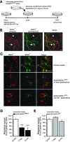
Fig. 1.
Fig. 1.
Long-term and short-term treatment of primary spinal cultures with ACM-hSOD1G93A equally trigger cell death of motoneurons. A: flow diagram of experiment. Medium was conditioned for 7 days by astrocytes derived from transgenic mice overexpressing human (h)SOD1G93A (ACM-hSOD1G93A). Primary wild-type (WT) rat spinal cord cultures (3 DIV) were exposed to ACM-hSOD1G93A for 4 days or for 30 min; all were fixed at 7 DIV to assay cell survival with immunocytochemistry. B: fixed 7 DIV primary spinal cultures were double-labeled with anti-microtubule-associated protein 2 (MAP2) antibody (red) to visualize interneurons (arrowhead) and motoneurons (arrow) and with the SMI-32 antibody (green) to identify motoneurons (arrow). Scale bar, 25 μm. C: spinal cultures were treated with control medium for 4 days (top) or ACM-hSOD1G93A for either 4 days (middle) or 30 min (bottom), fixed at 7 DIV, and labeled with MAP2 and SMI-32. Note that a single, short-term 30-min exposure of spinal cord neurons to ACM-hSOD1G93A is as effective in triggering motoneuron cell death as chronic application. Interneurons are spared in both conditions. Scale bar, 200 μm. D: graph of the ratio of SMI-32+/MAP2+ neurons showing % of surviving motoneurons at 7 DIV after treatment with control medium or ACM-hSOD1G93A applied for 4 days or 30 min. E: % of surviving motoneurons at 7 DIV after 4-day or 30-min application of 3 DIV cultures of medium that was conditioned by astrocytes derived from transgenic mice overexpressing hSOD1WT (ACM-hSOD1WT). Values represent means ± SE from at least 3 independent experiments performed in duplicate, analyzed by 1-way ANOVA followed by a Tukey post hoc test. ***P < 0.001 relative to control medium at 7 DIV.
Fig. 2.
Fig. 2.
ACM-hSOD1G93A increases persistent sodium inward current (PCNa). A: representative persistent inward current traces generated by a slow triangular voltage-clamp command (14 mV/s) from a holding potential of −60 to +10 mV (4 s) and back (4 s) (bottom) in the absence (colored traces) or presence (black traces) of the voltage-sensitive sodium (Nav) channel blocker TTX (1 μM); recordings from a 6 DIV control spinal neuron (top) and a sister neuron incubated with ACM-hSOD1G93A for ≥30 min (middle). As can be observed from the merge (bottom), PCNa (arrowhead; TTX-sensitive inward current activated at voltages starting at around −40 mV) is significantly larger in neurons treated with ACM-hSOD1G93A (red traces) relative to the control cell (green traces). B: typical patched interneuron with cell membrane area [membrane capacitance (Cm)] of ∼10 pF (left) and typical patched motoneuron with Cm of ∼20 pF (right). C: PCNa of individual spinal cord neurons (5–7 DIV) were generated under control conditions (dashed line) or after application of ACM-hSOD1G93A for 30–90 min (solid line) by a slow voltage-clamp command (as in A) and plotted against its Cm. The best-fitted slopes (S) highlight that PCNa is dependent on the size of the neurons. D: averaged mean peak PCNa amplitude normalized to Cm (pA/pF) for 5–7 DIV control neurons and those incubated with ACM-hSOD1G93A. Values represent means ± SE from at least 9 neurons, analyzed by t-test. **P < 0.01 relative to control.
Fig. 3.
Fig. 3.
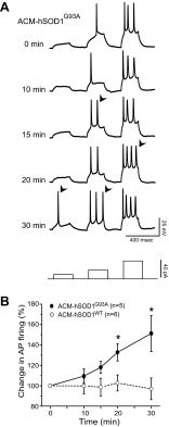
ACM-hSOD1G93A rapidly increases neuronal excitability. A: whole cell current-clamp recordings of control primary spinal neuron in the presence of ACM-hSOD1G93A. Action potentials (APs) are evoked by injection of rectangular depolarizing current pulses (bottom; 10, 20, and 40 pA for 300 ms). Representative membrane potential traces show that bath application of ACM-hSOD1G93A to 5–7 DIV primary spinal neurons rapidly increases the firing frequency (arrowheads). B: average change in AP firing frequency produced by 30-min application of ACM-hSOD1G93A or ACM-hSOD1WT. Values represent means ± SE from at least 5 neurons/time point, analyzed by t-test. *P < 0.05 compared with ACM-hSOD1WT at same time point.
Fig. 4.
Fig. 4.
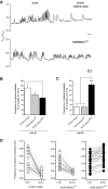
ACM-hSOD1G93A rapidly increases the frequency of calcium transients in cultured spinal neurons. A: representative ratiometric intracellular calcium transient ([Ca2+]i) traces of 2 independent spinal neurons (5–7 DIV) measured before (minute 0, left) and 30 min after application of either control medium or ACM-hSOD1G93A (minute 30, right). Parallel lines correspond to 30-min incubation periods. B and C: mean fraction of calcium transient amplitudes (B) and frequencies (C) at 30 min after treatment with the different media, relative to control activity measured at minute 0. Values represent means ± SE from 23–45 neurons/condition, analyzed by 1-way ANOVA followed by a Tukey post hoc test. *P < 0.05 and ***P < 0.001 relative to control medium and ACM-mSOD1WT at minute 30. D: line series graphs of calcium transients of individual neurons at 0 and 30 min under control conditions (left) or after treatment with ACM-mSOD1WT (center) or ACM-hSOD1G93A (right).
Fig. 5.
Fig. 5.
Calcium transients in cultured spinal cord neurons correlate with AP firing. A: simultaneous recording of spontaneous AP firing in cell-attached mode (top) and calcium transients (bottom) in a spinal cord neuron. B: expanded timescale for the neuron in A. Note the coincidence of calcium transients with bursts of AP firing. C: bar graph showing the mean frequency of AP firing from 198 transients recorded in 4 cultured spinal cord neurons. Note that during intertransient (inter-t.) periods, AP firing is virtually absent. Values represent means ± SE. ***P < 0.001, t-test. D: action currents in an expanded timescale from the last burst in B (dotted box).
Fig. 6.
Fig. 6.
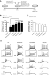
Nav channel blockers prevent ACM-hSOD1G93A-induced motoneuron death at doses that slightly reduce excitability. A: flow diagram of experiment. Medium (ACM-hSOD1G93A or control medium) was applied chronically at 3 DIV (as in Fig. 1) alone or together with the Nav blockers TTX (1 nM), mexiletine (25 nM), spermidine (10 μM), or riluzole (100 nM), and cell survival was assayed at 7 DIV. B: % of surviving motoneurons at 7 DIV treated with the different Nav blockers, relative to sister neurons treated with control medium (indicated with *) or to ACM-hSOD1G93A alone (indicated with #). Note that the drugs mexiletine, spermidine, and riluzole prevented motoneuron death induced by ACM-hSOD1G93A. TTX trends toward prevention but is unable to significantly reduce ACM-hSOD1G93A-mediated motoneuron death. C: effects of the same doses of Nav channel blockers, coapplied with control medium, on motoneuron survival. Note that the drugs did not improve survival of control neurons. Values represent means ± SE from at least 3 independent experiments, analyzed by 1-way ANOVA followed by a Tukey post hoc test. *P < 0.05, **P < 0.01, ***P < 0.001 relative to survival with control medium at 7 DIV; ##P < 0.01, ###P < 0.001 compared with survival with ACM-hSOD1G93A at 7 DIV. D: representative membrane potential traces (rectangular current pulse of 100 pA for 500 ms) of control 5–7 DIV primary spinal neurons treated with increasing concentrations of TTX, mexiletine, spermidine, or riluzole; the boxes highlight the doses used for the above motoneuron survival studies. Number stated above the last AP in each trace indicates the number of spikes in the train. Note that at the doses at which mexiletine, spermidine, and riluzole were able to prevent the ACM-hSOD1G93A-induced cell death, these compounds only eliminated a few spikes within the train of APs. At higher doses, spike suppression was strong and drugs killed motoneurons (data not shown). Also, while even high concentrations of mexiletine and spermidine do not suppress the initial firing, increasing doses of riluzole and TTX abruptly reduce the firing pattern and rate and ultimately abolish the first AP (arrows).
Fig. 7.
Fig. 7.
Acute application of Nav channel blockers has differential effects on calcium dynamics modified by ACM-hSOD1G93A. A: flow diagram of experiment. ACM-hSOD1G93A was applied on 5–7 DIV spinal cultures for 30 min (as in Fig. 3) to measure calcium transients (ACM − Nav blockers). Thereafter, Nav channel blockers TTX (1 nM), mexiletine (25 nM), spermidine (10 μM), and riluzole (100 nM) were applied to measure calcium transients of the same neurons 7 min later (ACM + Nav blockers); Nav channel blockers were used at the same concentrations as in Fig. 4. B and C: mean fraction of calcium transient frequencies (B) and amplitudes (C) after ACM-hSOD1G93A and drug treatments (ACM + Nav blockers) relative to ACM-hSOD1G93A alone (ACM − Nav blockers). Note that all Nav channel blockers markedly decreased mean frequency (to 20–30%). However, whereas mexiletine, spermidine, and riluzole significantly decreased the mean amplitude of the calcium transients (to 60%), TTX increases this parameter by ∼225%. Values represent means ± SE from 17–19 neurons/condition, analyzed by 1-way ANOVA followed by a Tukey post hoc test. *P < 0.05, ***P < 0.001 relative to 30-min ACM-hSOD1G93A.

Similar articles

Cited by

References

    1. Aebischer J, Cassina P, Otsmane B, Moumen A, Seilhean D, Meininger V, Barbeito L, Pettmann B, Raoul C. IFNγ triggers a light-dependent selective death of motoneurons contributing to the non-cell autonomous effects of mutant SOD1. Cell Death Differ 18: 754–768, 2011 - PMC - PubMed
    1. Aman TK, Grieco-Calub TM, Chen C, Rusconi R, Slat EA, Isom LL, Raman IM. Regulation of persistent Na current by interactions between beta subunits of voltage-gated Na channels. J Neurosci 29: 2027–2042, 2009 - PMC - PubMed
    1. Beckman JS, Estévez AG, Crow JP, Barbeito L. Superoxide dismutase and the death of motoneurons in ALS. Trends Neurosci 24: S15–S20, 2001 - PubMed
    1. Bellingham MC. A review of the neural mechanisms of action and clinical efficiency of riluzole in treating amyotrophic lateral sclerosis: what have we learned in the last decade? CNS Neurosci Ther 17: 4–31, 2011 - PMC - PubMed
    1. Bento-Abreu A, Van Damme P, Van Den Bosch L, Robberecht W. The neurobiology of amyotrophic lateral sclerosis. Eur J Neurosci 31: 2247–2265, 2010 - PubMed

Publication types

MeSH terms

LinkOut - more resources