. 2013 Jun;109(11):2815-26.
doi: 10.1152/jn.00711.2012. Epub 2013 Mar 13.

The μ-opioid receptor agonist DAMGO presynaptically suppresses solitary tract-evoked input to neurons in the rostral solitary nucleus

Affiliations

The μ-opioid receptor agonist DAMGO presynaptically suppresses solitary tract-evoked input to neurons in the rostral solitary nucleus

Alison J Boxwell et al. J Neurophysiol. 2013 Jun.

Abstract

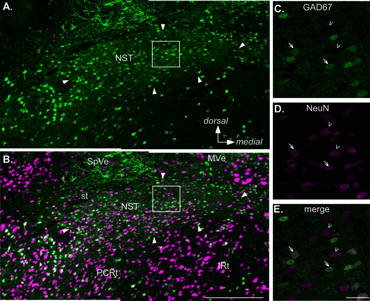
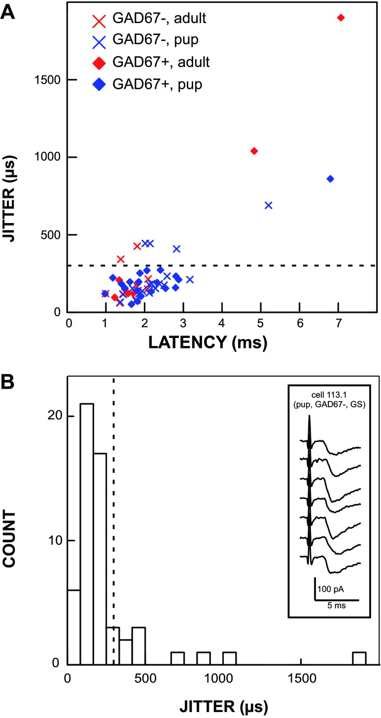
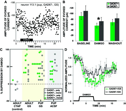
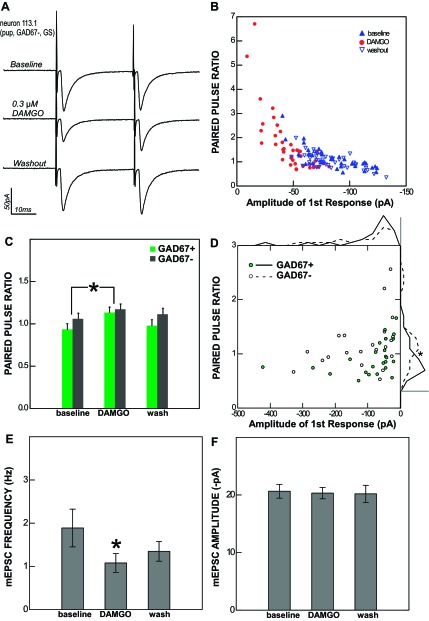
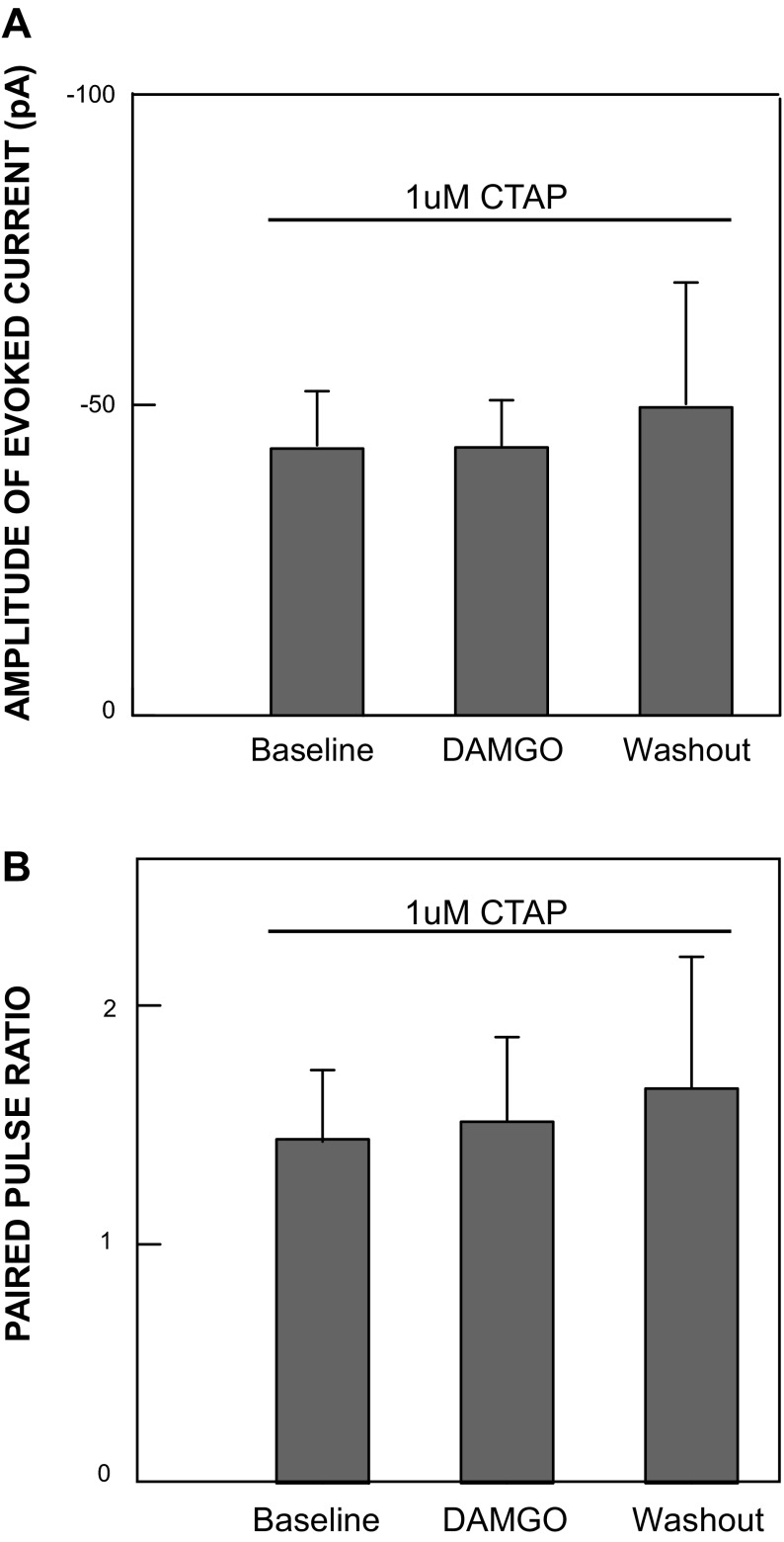
Taste processing in the rostral nucleus of the solitary tract (rNST) is subject to modulatory influences including opioid peptides. Behavioral pharmacological studies suggest an influence of μ-opioid receptors in rNST, but the underlying mechanism is unknown. To determine the cellular site of action, we tested the effects of the μ-opioid receptor agonist DAMGO in vitro. Whole cell patch-clamp recordings were made in brain stem slices from GAD67-GFP knockin mice expressing enhanced green fluorescent protein (EGFP) under the control of the endogenous promoter for GAD67, a synthetic enzyme for GABA. Neuron counts showed that ∼36% of rNST neurons express GABA. We recorded monosynaptic solitary tract (ST)-evoked currents (jitter ≤ 300 μs) in both GAD67-EGFP-positive (GAD67+) and GAD67-EGFP-negative (GAD67-) neurons with equal frequency (25/31; 22/28), but the inputs to the GAD67+ neurons had significantly smaller paired-pulse ratios compared with GAD67- neurons. DAMGO (0.3 μM) significantly suppressed ST-evoked currents in both cell types (mean suppression = 46 ± 3.3% SE), significantly increased the paired-pulse ratio of these currents, and reduced the frequency of spontaneous miniature excitatory postsynaptic currents but did not diminish their amplitude, indicating a presynaptic site of action. Under inhibitory amino acid receptor blockade, DAMGO was significantly more suppressive in GAD67+ neurons (59% reduction) compared with GAD67- neurons (35% reduction), while the reverse was true in normal artificial cerebrospinal fluid (GAD67+: 35% reduction; GAD67-: 57% reduction). These findings suggest that DAMGO suppresses activity in rNST neurons predominantly via a presynaptic mechanism, and that this effect may interact significantly with tonic or evoked inhibitory activity.

Keywords: GABA; GAD67-GFP knockin mouse; disinhibition; taste.


Figures

Fig. 1.
Fig. 1.
Confocal images of a section from a level of the rostral nucleus of the solitary tract (rNST) in an adult GAD67+ mouse approximately midway between the rostral pole of the nucleus and where the NST abuts the IVth ventricle. Sections were also immunostained for the neuronal marker NeuN. A and B: low-power maximum-intensity projections showing the distribution of GAD67+, enhanced green fluorescent protein (EGFP)-stained neurons and fibers (green, A) and EGFP expression merged with NeuN staining (magenta, B). Arrowheads indicate the borders of the nucleus. Note, however, that the most medial pole contains sparse somal label for either marker and corresponds with a region occupied by preganglionic parasympathetic neurons that constitute the rostral pole of the dorsal motor nucleus of the vagus. Aside from this region, GAD67+ neurons are distributed throughout the nucleus; likewise, the nucleus is characterized by profuse EGFP staining of the neuropil. C–E. higher-magnification images at a single z-plane. Arrows indicate examples of double-stained neurons; arrowheads indicate neurons stained only with NeuN. Scale bars, 250 μm (A and B), 25 μm (C–E). IRt, intermediate subdivision of the medullary reticular formation; MVe, medial vestibular nucleus; PCRt, parvocellular reticular formation; SpVe, spinal vestibular nucleus; st, solitary tract.
Fig. 2.
Fig. 2.
Many cells, including GAD67+ neurons, received monosynaptic afferent input from the solitary tract (ST), and this innervation persisted into adulthood. A plot of latency vs. jitter (A) shows that most responses elicited by ST stimulation had a jitter value < 300 μs. A histogram of jitter values (B) shows the cutoff (dashed line) that we used to define mono- vs. polysynaptic innervation at 300 μs. Inset in B shows an example of raw data from a single neuron with an average latency of 1.69 ms and a jitter of 150 μs. GS, gabazine-strychnine.
Fig. 3.
Fig. 3.
Photomicrographs of a ventrally located site where a GAD67+ neuron was recorded. A: low-magnification DIC image showing the stimulating electrode on the solitary tract (ST) and the recording electrode in the ventral 1/3 of the nucleus; the position of the tip is indicated by *. The approximate outlines of the rNST are indicated by the dashed line. Pieces of debris (x) cut across the medial part of rNST and medial vestibular nucleus (MeV). PCRt, parvocellular reticular formation. B: high-magnification DIC image showing the pipette on the recorded neuron. C: high-magnification fluorescent image showing that the cell is GAD67+. Scale bars, 250 μm (A), 25 μm (B and C).
Fig. 4.
Fig. 4.
DAMGO suppressed the response of rNST neurons to ST-evoked input. A: effect of DAMGO on the amplitude of a ST-evoked current in a single GAD67− neuron under gabazine-strychnine (GS) blockade in a pup. ST-evoked responses were measured at 3.33-s intervals. B: mean ± SE amplitudes of rNST responses in GAD67+ and GAD67− neurons across the population before (baseline), during (DAMGO), and after (washout) DAMGO application (n = 53; the 1 response facilitated by DAMGO was omitted). ANOVA indicated an overall effect of DAMGO (*P < 0.0005) but no effect of GAD67-EGFP status or DAMGO × GAD67-EGFP interaction; Bonferroni-adjusted post hoc comparisons revealed significant differences between all 3 conditions: baseline, DAMGO, and washout (all P < 0.005). C: dot density plot showing the degree to which individual responses were suppressed by DAMGO (response increment omitted). Responses are grouped by animal age and bath solution: artificial cerebral spinal fluid (ACSF), gabazine and strychnine (GS), and gabazine and strychnine with the μ-opioid receptor (MOR) antagonist CTAP (CTAP). Symbols indicate monosynaptic vs. polysynaptic responses and GAD67-EGFP status. D: effect of DAMGO shown for normalized responses over time for GAD67+ (n = 15) and GAD67− (n = 15) neurons recorded when gabazine and strychnine were included in the bath. Individual measurements are for the moving average of 5 adjacent time points. When inhibitory amino acid receptors were blocked, DAMGO had a significantly larger effect on ST-evoked responses in GAD67+ than in GAD67− neurons, although responses in both cell types were affected.
Fig. 5.
Fig. 5.
The effect of DAMGO on rNST neurons is presynaptic. A: the response of an rNST neuron to ST stimulation (averages of 40 sweeps in voltage clamp) before, during, and after the application of 0.3 μM DAMGO. B: single data points showing the relationship between the amplitude of the 1st response to ST stimulation and the paired-pulse ratio (PPR) for the same cell in each condition (baseline, DAMGO, and washout). C: across the population, the average PPR increases during DAMGO application for both GAD67+ and GAD67− neurons. *P < 0.001. D: scatterplot of average baseline amplitude and PPR values for individual monosynaptic ST-evoked responses (n = 44) showing an inverse relationship between the 1st response amplitude and PPR for both GAD67+ and GAD67− neurons. The PPRs of monosynaptic responses to ST stimulation were significantly smaller in GAD67+ neurons than in GAD67− neurons. This effect may be seen most clearly in the frequency polygons (binned in 15 equal segments) displayed outside the graph area; scale bars correspond to a frequency of 10 neurons. E: the frequency of miniature excitatory postsynaptic current (mEPSCs; tested in 7 neurons, 2 GAD67+ and 5 GAD67−) was significantly reduced by DAMGO. *P < 0.014. F: the average amplitude of mEPSCs was unaffected by DAMGO. Taken together, these results indicate a presynaptic site of action.
Fig. 6.
Fig. 6.
All DAMGO effects are abolished in the presence of the μ-opioid antagonist CTAP: mean amplitudes of evoked responses (A) and PPRs (B) from 5 neurons recorded under MOR receptor blockade accomplished by adding 1 μM CTAP to a bathing solution containing normal ACSF with gabazine (5 μM) and strychnine (5 μM). Under these conditions, no significant change in the amplitude of the evoked response or PPR was observed when 0.3 μM DAMGO was added, indicating that DAMGO effects in the absence of CTAP blockade were specifically mediated by MORs.

Similar articles

Cited by

References

    1. Appleyard SM, Bailey TW, Doyle MW, Jin YH, Smart JL, Low MJ, Andresen MC. Proopiomelanocortin neurons in nucleus tractus solitarius are activated by visceral afferents: regulation by cholecystokinin and opioids. J Neurosci 25: 3578–3585, 2005 - PMC - PubMed
    1. Bailey TW, Appleyard SM, Jin YH, Andresen MC. Organization and properties of GABAergic neurons in solitary tract nucleus (NTS). J Neurophysiol 99: 1712–1722, 2008 - PubMed
    1. Bradley BE. The Role of the Nucleus of the Solitary Tract in Gustatory Processing. Boca Raton, FL: CRC, 2007 - PubMed
    1. Brown RE, McKenna JT, Winston S, Basheer R, Yanagawa Y, Thakkar MM, McCarley RW. Characterization of GABAergic neurons in rapid-eye-movement sleep controlling regions of the brainstem reticular formation in GAD67-green fluorescent protein knock-in mice. Eur J Neurosci 27: 352–363, 2008 - PMC - PubMed
    1. Chen Z, Travers JB. Inactivation of amino acid receptors in medullary reticular formation modulates and suppresses ingestion and rejection responses in the awake rat. Am J Physiol Regul Integr Comp Physiol 285: R68–R83, 2003 - PubMed

Publication types

MeSH terms

Substances