Review
. 2013 Apr;13(4):258-71.
doi: 10.1038/nrc3484. Epub 2013 Mar 14.

The effects of PEDF on cancer biology: mechanisms of action and therapeutic potential

Affiliations
Review

The effects of PEDF on cancer biology: mechanisms of action and therapeutic potential

S Patricia Becerra et al. Nat Rev Cancer. 2013 Apr.

Abstract

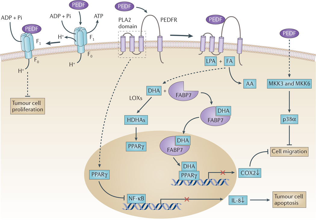
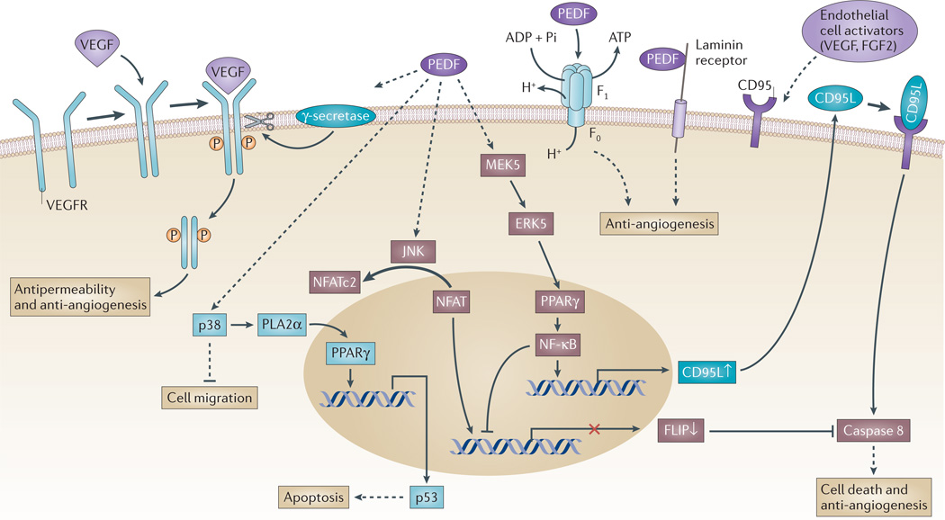
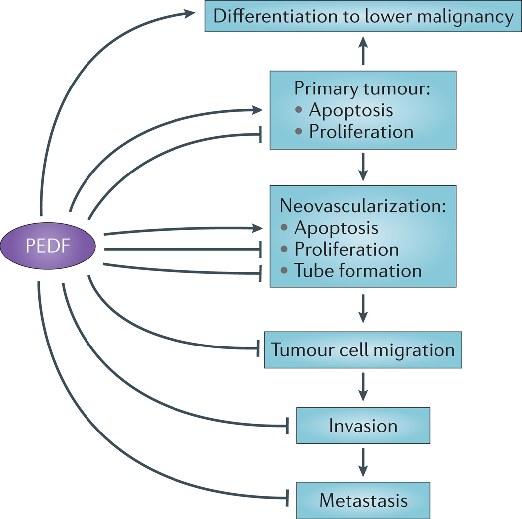
The potent actions of pigment epithelium-derived factor (PEDF) on tumour-associated cells, and its extracellular localization and secretion, stimulated research on this multifunctional serpin. Such studies have identified several PEDF receptors and downstream signalling pathways. Known cellular PEDF responses have expanded from the initial discovery that PEDF induces retinoblastoma cell differentiation to its anti-angiogenic, antitumorigenic and antimetastatic properties. Although the diversity of PEDF activities seems to be complex, they are consistent with the varied mechanisms that regulate this multimodal factor. If PEDF is to be used for cancer management, a deeper appreciation of its many functions and mechanisms of action is needed.


Figures

Figure 1
Figure 1. PEDF in tumour cells
Pigment epithelium-derived factor (PEDF) is a ligand for several receptors, and its interaction with these receptors is thought to trigger the signalling pathways illustrated here. PEDF binds to PED F receptor (PEDFR) and stimulates its phospholipase activity,. When PEDFR is at the membrane, its phospholipase A2 (PLA2) active site is located close to the phospholipid bilayer where it can use phospholipids as substrates. Depending on the relative abundance of the fatty acids omega-3 docosahexaenoic acid (DHA) and omega-6 arachidonic acid (AA) in phospholipid membranes, free DHA or AA can be liberated by PEDFR. DHA is a precursor of the anti-angiogenic and neuroprotector neuroprotectin D1 (NPD1). Other DHA metabolites, such as hydroxy-DHAs (HDHAs), which are produced by lipoxygenases (LOXs), can act on peroxisome proliferator-activated receptor-γ (PPARγ),,. Upregulation of PPARγ leads subsequently to the suppression of nuclear factor-κB (NF-κB)-mediated transcriptional activation, reduced production of interleukin 8 (IL-8) and limited proliferation of prostate cancer cells. Cytosolic fatty-acid-binding protein 7 (FABP7) can bind DHA with higher affinity than AA, translocate DHA to the nucleus and transfer it to PPARγ, thus resulting in the downregulation of promigratory genes, such as cyclooxygenase 2 (COX2). PEDF is a ligand of cell-surface F ATP synthase, and its 34-mer peptide region inhibits ATP production and reduces endothelial and tumour cell viability and angiogenesis,. PEDF, through interaction with a yet unknown receptor, can sequentially activate MKK3, MKK6 and p38α MAPK to inhibit cell migration. FA, fatty acid; LPA, lysophosphatidic acid.
Figure 2
Figure 2. Signalling events of PEDF in endothelial cells
Vascular endothelial growth factor (VEGF) binds to a homodimerized VEGF receptor (VEGFR), which becomes phosphorylated and activated. Pigment epithelium-derived factor (PEDF) can increase γ-secretase-mediated cleavage of VEGFR1 and VEGFR2 at the transmembrane region to generate an intracellular domain fragment,. At the same time, PEDF can inhibit VEGF-induced phosphorylation and activation of VEGFR1 (REF. 86). PEDF inhibits VEGF-driven angiogenesis and permeability through the regulated intracellular proteolysis of VEGFR. PEDF can activate the p38 MAPK pathway to inhibit endothelial cell migration. It can also activate peroxisome proliferator-activated receptor-γ (PPARγ) through cytosolic phospholipase A2α (PLA2α) to induce the expression of TP53, which encodes the pro-apoptotic protein p53 (REF. 155). At the same time, endothelial activators such as VEGF and fibroblast growth factor 2 (FGF2) stimulate and expose CD95 (also known as FAS) on the endothelial plasma membrane. PEDF can sequentially activate MEK5 (which is a MAPK kinase), ERK5, PPARγ and nuclear factor-κB (NF-κB), which induces the expression of the pro-apoptotic gene CD95 ligand (CD95L), the protein product of which translocates to the plasma membrane. The resulting CD95L-CD95 complex induces the binding and activation of caspase 8 that under certain conditions triggers the cell death cascade,. At the same time, NF-κB activation has a negative impact on cellular FLICE-like inhibitory protein (FLIP) expression, which decreases the capacity of FLIP to inhibit caspase 8. Conversely, PEDF triggers JUN N-terminal kinase (JNK)-mediated phosphorylation of nuclear factor of activated T-cells, cytoplasmic 2 (NFATc2) and sequesters it in the cytoplasm, thus blocking FLIP expression. In this manner, PEDF leads to apoptosis in activated endothelial cells. PEDF is a ligand of two known proteins on endothelial cells that result in anti-angiogenic responses. PEDF binds to laminin receptor and cell-surface F1 ATP synthase to inhibit ATP production and inhibit angiogenesis,,.
Figure 3
Figure 3. The effects of PEDF on tumour progression
A linear representation of different pigment epithelium-derived factor (PEDF) targets that form a concerted set of activities to inhibit cancer progression. PEDF can differentiate tumours to a less-malignant phenotype. Tumour cells secrete angiogenic factors to activate endothelial cells. PEDF can block angiogenesis-mediated activities and neovascularization. PEDF can also block tumour migration, invasion and metastasis.

References

    1. Steele FR, Chader GJ, Johnson LV, Tombran-Tink J. Pigment epithelium-derived factor: neurotrophic activity and identification as a member of the serine protease inhibitor gene family. Proc. Natl Acad. Sci. USA. 1993;90:1526–1530.The first article to report the complete sequence of the human PEDF mRNA and protein, and its identification as a member of the serpin gene family with neurotrophic activity on retinoblastoma tumour cells.

    1. Tombran-Tink J, Chader GG, Johnson LV. PEDF: a pigment epithelium-derived factor with potent neuronal differentiative activity. Exp. Res. 1991;53:411–414. - PubMed
    1. Doggett DL, Rotenberg MO, Pignolo RJ, Phillips PD, Cristofalo VJ. Differential gene expression between young and senescent, quiescent WI-38 cells. Mech. Ageing Dev. 1992;65:239–255. - PubMed
    1. Pignolo RJ, Cristofalo VJ, Rotenberg MO. Senescent WI-38 cells fail to express EPC-1, a gene induced in young cells upon entry into the G0 state. J. Biol. Chem. 1993;268:8949–8957.This report describes how SERPINF1 is a gene that is induced to ≥100-fold levels in young lung fibroblast cells relative to senescent cells, which fail to express it, and is a first association of PEDF with ageing.

    1. Francis MK, et al. Loss of EPC-1/PEDF expression during skin aging in vivo. J. Invest. Dermatol. 2004;122:1096–1105. - PubMed

Publication types

MeSH terms