Review
. 2013 Jun 1;9(6):830-49.
doi: 10.4161/auto.24132. Epub 2013 Mar 13.

Autophagy in stem cells

Affiliations
Review

Autophagy in stem cells

Jun-Lin Guan et al. Autophagy. .

Abstract

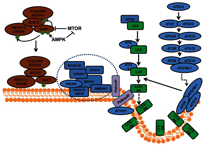
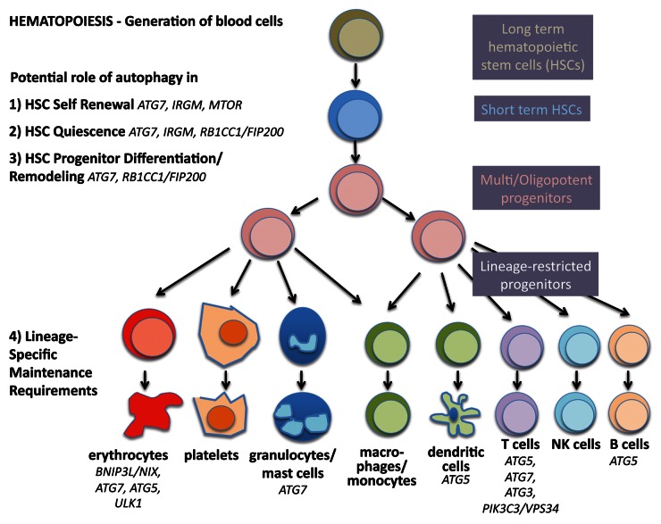
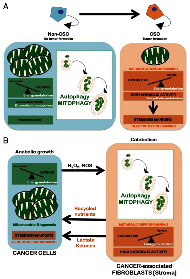
Autophagy is a highly conserved cellular process by which cytoplasmic components are sequestered in autophagosomes and delivered to lysosomes for degradation. As a major intracellular degradation and recycling pathway, autophagy is crucial for maintaining cellular homeostasis as well as remodeling during normal development, and dysfunctions in autophagy have been associated with a variety of pathologies including cancer, inflammatory bowel disease and neurodegenerative disease. Stem cells are unique in their ability to self-renew and differentiate into various cells in the body, which are important in development, tissue renewal and a range of disease processes. Therefore, it is predicted that autophagy would be crucial for the quality control mechanisms and maintenance of cellular homeostasis in various stem cells given their relatively long life in the organisms. In contrast to the extensive body of knowledge available for somatic cells, the role of autophagy in the maintenance and function of stem cells is only beginning to be revealed as a result of recent studies. Here we provide a comprehensive review of the current understanding of the mechanisms and regulation of autophagy in embryonic stem cells, several tissue stem cells (particularly hematopoietic stem cells), as well as a number of cancer stem cells. We discuss how recent studies of different knockout mice models have defined the roles of various autophagy genes and related pathways in the regulation of the maintenance, expansion and differentiation of various stem cells. We also highlight the many unanswered questions that will help to drive further research at the intersection of autophagy and stem cell biology in the near future.

Keywords: autophagy; cancer stem cells; embryonic stem cells; tissue stem cells.


Figures

None
Figure 1. The process and regulation of autophagosome formation in mammalian cells. Mammalian autophagosome formation is induced by the activation of the ULK1 complex (comprising ULK1, ATG13, RB1CC1/FIP200 and C12orf44/ATG101), which is promoted by suppression of MTOR complex 1 (MTORC1) under starvation conditions or AMPK (both directly as well as by repressing MTORC1) in low-energy states. Once the activated ULK1 complex translocates to part of the endoplasmic reticulum (or possibly other intracellular membranes), it coordinates with the class III phosphatidylinositol-3-phosphate (PtdIns3P) kinase complex (including PIK3C3, BECN1, PIK3R4/p150, AMBRA1, UVRAG, ATG14 and SH3GLB1) for the initiation of autophagosomes upon formation of PtdIns3P and recruitment of double ZFYVE 1. Two ubiquitin-like systems including the ATG12–ATG5-ATG16L1 complex (containing ATG5, ATG7, ATG10, ATG12 and ATG16L1) and the LC3–phosphatidylethanolamine (PE) conjugation system (including ATG3, ATG4, ATG7 and MAP1LC3) will then promote the elongation of phagophore membranes for the formation of autophagosomes.
None
Figure 2. Autophagy in stemness of hematopoietic stem cells. During hematopoiesis, differentiated blood cells are generated from hematopoietic stem cells (HSCs). Long-term HSCs develop into short-term HSCs, multi- and then oligopotent progenitors and lineage-restricted progenitors, which give rise to the differentiated blood cells. These include erythrocytes, platelets, granulocytes, macrophages, dendritic cells and the lymphocytes T, B and NK cells. Autophagy is thought to play a role in self-renewal and quiescence in HSCs, the two hallmarks of stemness. Moreover, during differentiation autophagy is hypothesized to help multipotency and remodeling. Last, evidence is increasing that autophagy is required to maintain the healthy differentiated lineages.
None
Figure 3. Autophagy/mitophagy and the CSC cellular state: Two models. Mitochondria can be eliminated through nonspecific autophagic processes or selectively removed by mitophagy. The former is generally related to metabolic responses that are imposed by a lack of nutrients. Mitophagy, however, permits a tight adjustment in the number of cellular mitochondria by regulating the selective degradation of damaged, dysfunctional and superfluous mitochondria and adjust to changing physiological demands. Mitophagy, therefore, can play a crucial role in adapting the number and quality of mitochondria to new microenvironmental conditions. (A) Cell-autonomous model. Upregulation of mitophagy leading to significant reductions in both the number and the size of mitochondria (i.e., the “mitochondrial phenotype” that is commonly associated with stem cells) concurrently enhances a bioenergetic shift from somatic oxidative mitochondria toward an alternative glycolytic phenotype (“direct Warburg effect”), an energetic infrastructure associated with the transcriptional networks responsible for stemness and pluripotency. (B) Non-cell-autonomous model (“autophagic tumor stroma model of cancer metabolism”). Cancer cells’ mitochondrial oxidative phosphorylation (OXPHOS) can induce oxidative stress in adjacent fibroblasts (cancer-associated fibroblasts [CAFs] via H2O2 and reactive oxygen species (ROS), resulting in the onset of a myofibroblastic autophagic phenotype in CAFs. The CAFs autophagic phenotype leads to a loss of mitochondria via mitophagy, forcing CAFs to undergo aerobic glycolysis (“reverse Warburg effect”). The products of aerobic glycolysis (such as L-lactate and ketones) are then reused by cancer cells for OXPHOS, resulting in increased mitochondrial mass in cancer cells. The utilization of the high-energy metabolites L-lactate and ketones in cancer cells can promote the transcriptional activation of CSC-like phenotypes.

References

    1. He C, Klionsky DJ. Regulation mechanisms and signaling pathways of autophagy. Annu Rev Genet. 2009;43:67–93. doi: 10.1146/annurev-genet-102808-114910. - DOI - PMC - PubMed
    1. Mizushima N, Levine B. Autophagy in mammalian development and differentiation. Nat Cell Biol. 2010;12:823–30. doi: 10.1038/ncb0910-823. - DOI - PMC - PubMed
    1. Behrends C, Sowa ME, Gygi SP, Harper JW. Network organization of the human autophagy system. Nature. 2010;466:68–76. doi: 10.1038/nature09204. - DOI - PMC - PubMed
    1. Mizushima N, Komatsu M. Autophagy: renovation of cells and tissues. Cell. 2011;147:728–41. doi: 10.1016/j.cell.2011.10.026. - DOI - PubMed
    1. Rubinsztein DC, Mariño G, Kroemer G. Autophagy and aging. Cell. 2011;146:682–95. doi: 10.1016/j.cell.2011.07.030. - DOI - PubMed

Publication types

LinkOut - more resources