. 2013 Apr 26;288(17):12295-304.
doi: 10.1074/jbc.M113.450973. Epub 2013 Mar 12.

Crystal structure of an insect antifreeze protein and its implications for ice binding

Affiliations

Crystal structure of an insect antifreeze protein and its implications for ice binding

Aaron Hakim et al. J Biol Chem. .

Abstract

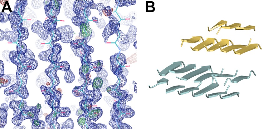
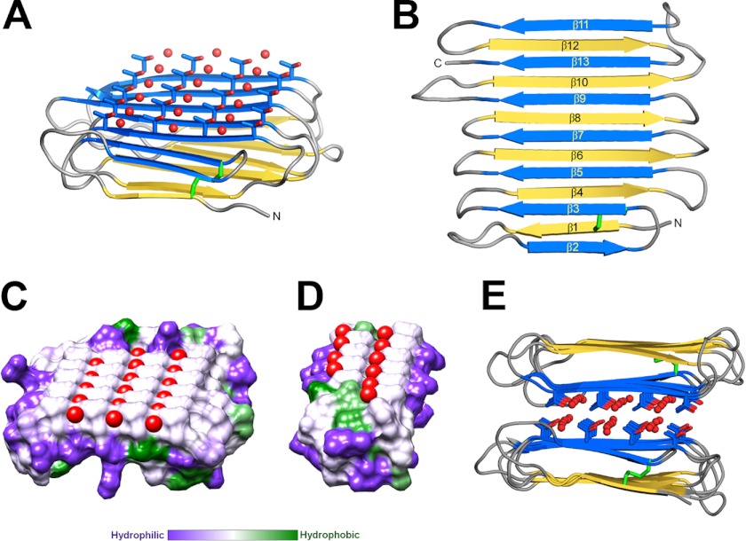
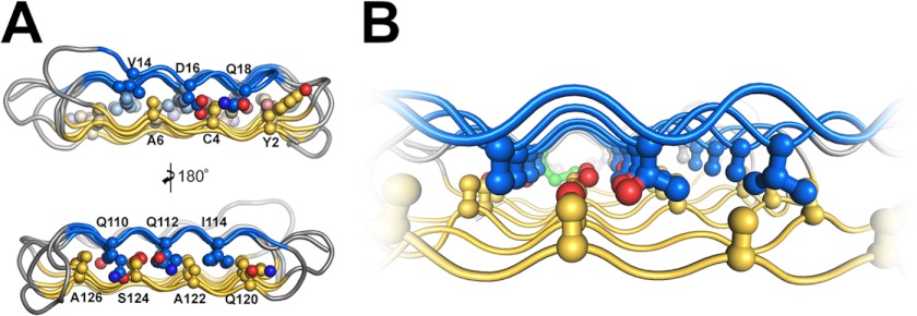
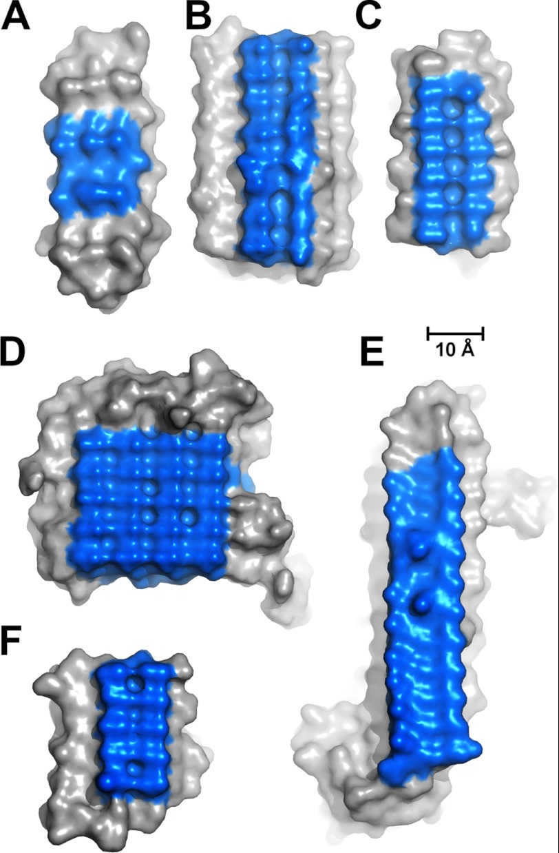
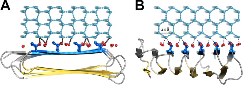
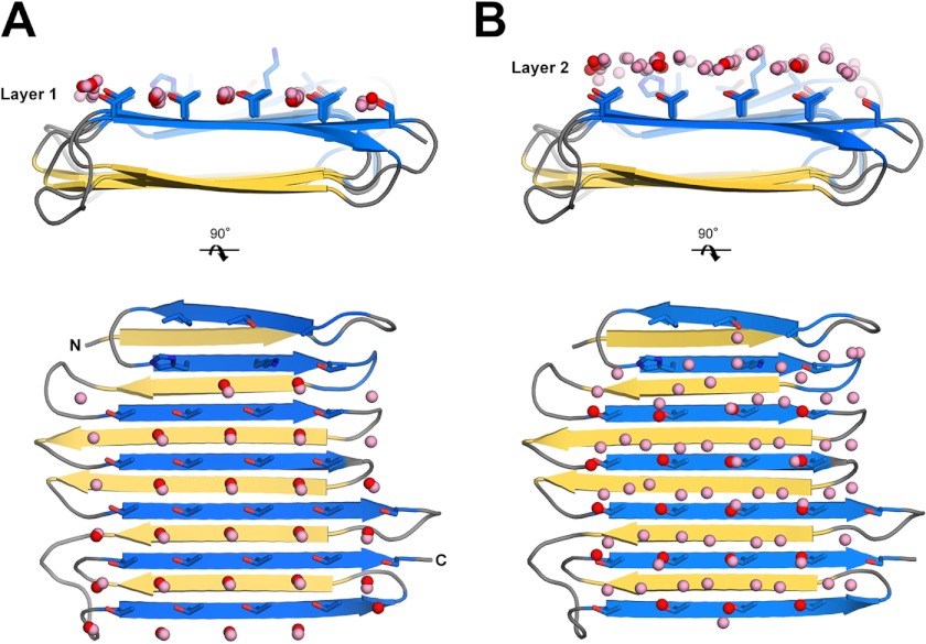
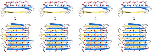
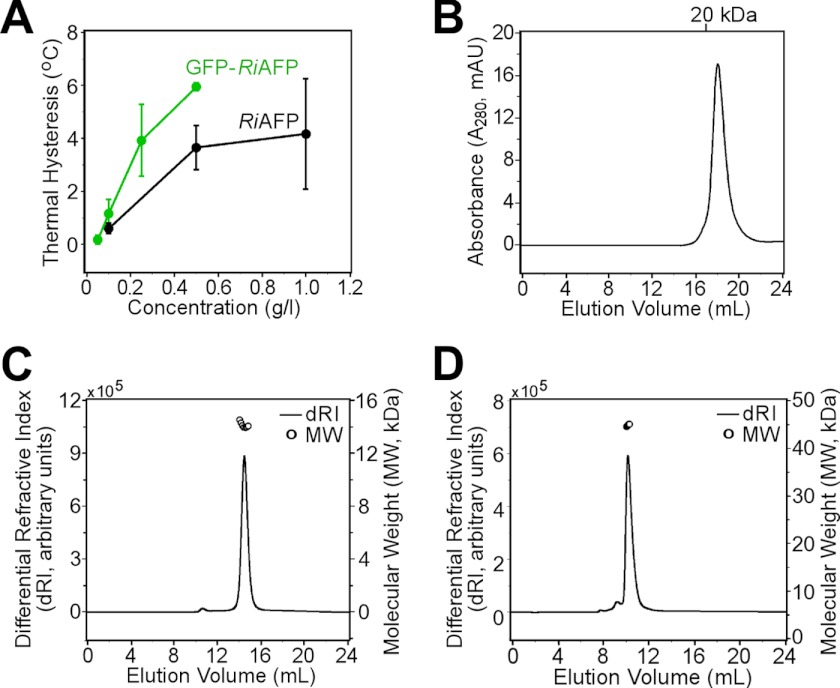
Antifreeze proteins (AFPs) help some organisms resist freezing by binding to ice crystals and inhibiting their growth. The molecular basis for how these proteins recognize and bind ice is not well understood. The longhorn beetle Rhagium inquisitor can supercool to below -25 °C, in part by synthesizing the most potent antifreeze protein studied thus far (RiAFP). We report the crystal structure of the 13-kDa RiAFP, determined at 1.21 Å resolution using direct methods. The structure, which contains 1,914 nonhydrogen protein atoms in the asymmetric unit, is the largest determined ab initio without heavy atoms. It reveals a compressed β-solenoid fold in which the top and bottom sheets are held together by a silk-like interdigitation of short side chains. RiAFP is perhaps the most regular structure yet observed. It is a second independently evolved AFP type in beetles. The two beetle AFPs have in common an extremely flat ice-binding surface comprising regular outward-projecting parallel arrays of threonine residues. The more active, wider RiAFP has four (rather than two) of these arrays between which the crystal structure shows the presence of ice-like waters. Molecular dynamics simulations independently reproduce the locations of these ordered crystallographic waters and predict additional waters that together provide an extensive view of the AFP interaction with ice. By matching several planes of hexagonal ice, these waters may help freeze the AFP to the ice surface, thus providing the molecular basis of ice binding.


Figures

FIGURE 1.
FIGURE 1.
Structure determination of RiAFP. A, preliminary electron density map generated by Arp/wArp from initial phases. The 2Fo − 2Fc map is in blue and contoured at 1.3σ, whereas the FoFc map is in green and red (indicating positive and negative peaks, respectively) and contoured at +3.0σ. B, model used in P31 molecular replacement. Despite building most of the final model, no connectivity was observed between individual β-strands in the higher (P3121) space group.
FIGURE 2.
FIGURE 2.
Crystal structure of RiAFP. A, overall fold of RiAFP. β-Strands are shown in blue and yellow. The threonine side chains and coordinated water molecules (red) on the IBS are shown in ball-and-stick representation. The disulfide bond is indicated in green. B, secondary structure diagram with sequentially numbered ice-binding β-strands in blue, hydrophilic β-strands in yellow. N and C represent the protein termini. C and D, flat ice-binding surface of RiAFP (C) and TmAFP (D), represented by a hydrophobic surface diagram produced in Chimera, which relies on the Kyte-Doolittle scale to rank amino acid hydrophobicity. Water molecules coordinated by the threonine hydroxyls on the IBS are shown in red. E, RiAFP dimers in the crystallographic asymmetric unit, with the ice-binding surfaces packed face-to-face.
FIGURE 3.
FIGURE 3.
The thin core of RiAFP spans less than 6 Å and is tightly packed with interdigitating Ala, Ser, and Thr residues. A, end-on and 180° view of the core residues in RiAFP contributing to the capping motifs. B, close up of the interdigitating alanine, serine, and threonine residues within the tightly packed, thin, stable core.
FIGURE 4.
FIGURE 4.
Comparisons of ice-binding surfaces, colored blue, among sfAFP (A), LpAFP (B), TmAFP (C), RiAFP (D), MpAFP (E) and CfAFP (F).
FIGURE 5.
FIGURE 5.
Docking of RiAFP to the primary prism plane of a hexagonal ice lattice (light cyan) based on Sc calculations. Dashed lines (black) represent potential hydrogen bonds (within 2.4–3.2 Å). A, end-on view of one of four possible binding modes (Sc = 0.78) shown for illustrative purposes. The c-axis of the ice lattice is perpendicular to the plane of the page. B, 90° side view corresponding to all four binding modes of RiAFP to the primary prism plane. The c-axis of the ice lattice runs from left to right.
FIGURE 6.
FIGURE 6.
Ordered surface waters on the IBS of RiAFP. Crystallographic waters are in red, simulation waters are in pink. A, correspondence of water simulation and crystallographic waters in the threonine layer (layer 1) representing bound water molecules considered intrinsic to RiAFP. B, correspondence of water simulation and crystallographic waters in layer 2.
FIGURE 7.
FIGURE 7.
Dashed lines indicate a quorum of anchored clathrate waters organized by each of the four ranks of threonine residues. All four water patterns, any of which could potentially independently bind the AFP to ice, are illustrated. Crystallographic waters are in red; simulation waters are in pink. Dashed lines (black) represent potential hydrogen bonds (within 2.4–3.5 Å). It is unlikely that more than two ranks of anchored clathrate waters can simultaneously fit well to either the primary prism or basal plane of ice.
FIGURE 8.
FIGURE 8.
Ice-binding characteristics of RiAFP. A, an ice crystal formed in a solution containing RiAFP has a rounded, oval shape with the c-axis perpendicular to (coming out of) the plane of the page. B, at temperatures below the thermal hysteresis gap, the ice crystal “bursts” laterally along the a-axes in a 6-point dendritic pattern. C and D, top-down and side views of a single ice crystal hemisphere grown in the presence of GFP-RiAFP are shown. The direction of the c-axis is indicated by a white arrow. Green color represents the binding to ice and overgrowth of the GFP-tagged RiAFP, which in this experiment occurs over the entire hemisphere with no indication of preferential ice plane binding. E, an ice hemisphere grown in GFP-LpIBP with its c-axis perpendicular to the page shows binding to the basal plane (center) and six equivalent primary prism planes. F, an ice hemisphere grown in GFP-LpIBP reveals binding to three of the six equivalent primary prism planes, with the other three planes being hidden by the hemisphere. The images in E and F are reprinted with permission from Middleton et al. (38).
FIGURE 9.
FIGURE 9.
Biophysical analysis of RiAFP in solution. A, the thermal hysteresis activities of recombinant RiAFP (black) and GFP-RiAFP (green) compared as a function of concentration. B, monodisperse peak of RiAFP elution from a Superdex 200 (10/300) column at 4 °C. C and D, multiangle light scattering profile of recombinant RiAFP (C) and GFP-RiAFP (D) at 25 °C.

References

    1. Jia Z., Davies P. L. (2002) Antifreeze proteins: an unusual receptor-ligand interaction. Trends Biochem. Sci. 27, 101–106 - PubMed
    1. Raymond J. A., DeVries A. L. (1977) Adsorption inhibition as a mechanism of freezing resistance in polar fishes. Proc. Natl. Acad. Sci. U.S.A. 74, 2589–2593 - PMC - PubMed
    1. Knight C. A. (2000) Structural biology: adding to the antifreeze agenda. Nature 406, 249, 251 - PubMed
    1. Nada H., Furukawa Y. (2011) Growth inhibition at the ice prismatic plane induced by a spruce budworm antifreeze protein: a molecular dynamics simulation study. Phys. Chem. Chem. Phys. 13, 19936–19942 - PubMed
    1. Hayward J. A., Haymet A. D. J. (2001) Ice/water interface: molecular dynamics simulations of the basal, prism, {20–21}, and {2–1-10} interfaces of ice Ih. J. Chem. Phys. 114, 3713–3726

Publication types

Associated data