. 2013 Apr 19;288(16):11135-43.
doi: 10.1074/jbc.M113.457440. Epub 2013 Mar 13.

RIF1 counteracts BRCA1-mediated end resection during DNA repair

Affiliations

RIF1 counteracts BRCA1-mediated end resection during DNA repair

Lin Feng et al. J Biol Chem. .

Abstract

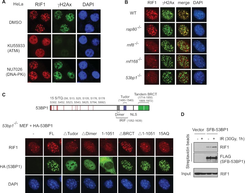
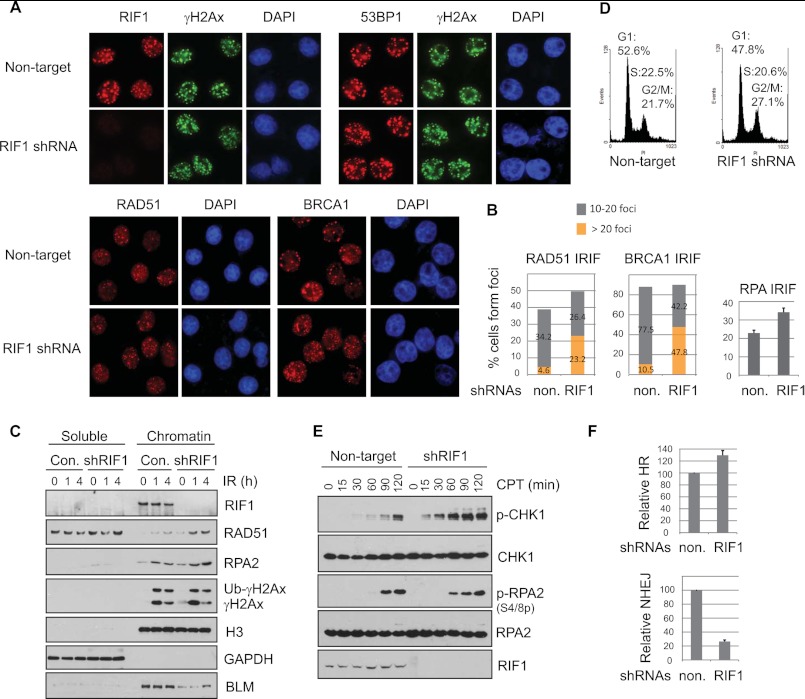
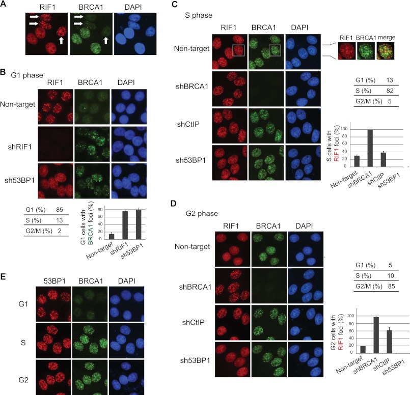
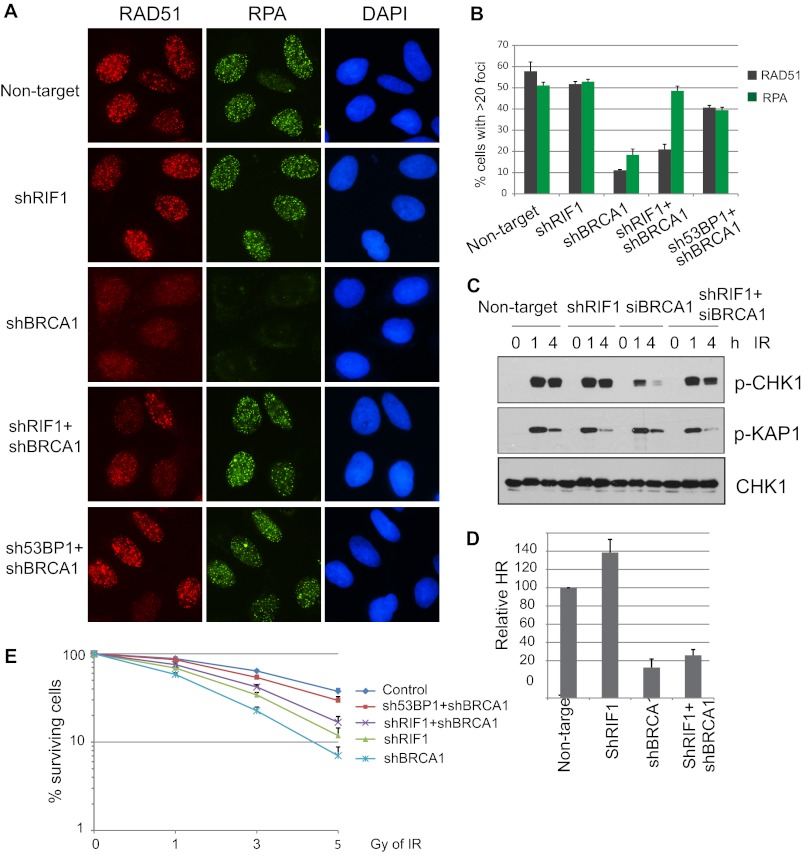
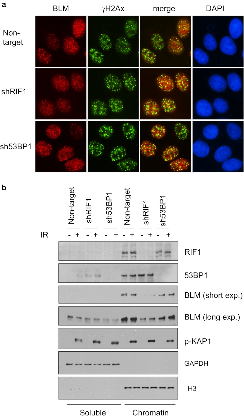
BRCA1 promotes homologous recombination repair and antagonizes 53BP1-dependent nonhomologous end joining (NHEJ) pathway. However, the molecular basis of the competition between BRCA1 and 53BP1 pathways remains elusive. Here we report that RIF1 protein translocates to damage sites via ATM-dependent 53BP1 phosphorylation. Strikingly, loss of RIF1 rescues initial DNA end resection and checkpoint activation in BRCA1-depleted cells. Interestingly RIF1 accumulation at damage sites is antagonized by BRCA1 in S and G2 phases. Conversely, the translocation of BRCA1 to damage sites is inhibited by RIF1 in G1 phase. However, loss of RIF1 differs from that of 53BP1 deficiency, as it cannot fully rescue RAD51 foci formation, homologous recombination defect, and radio-hypersensitivity in BRCA1-deficient cells. This is likely because RIF1, but not 53BP1, also regulates the foci formation and chromatin loading of BLM (the Bloom syndrome helicase). Thus, RIF1 not only acts downstream of 53BP1 and counteracts BRCA1-mediated end resection but also has a secondary role in promoting BLM function in DNA repair.


Figures

FIGURE 1.
FIGURE 1.
Localization of mammalian RIF1 in response to DNA double-strand breaks. A, ATM, but not DNA-PK, mediates IRIF of RIF1. HeLa cells were pretreated with DMSO, ATM inhibitor KU55933 (10 μm), or DNA-PK inhibitor NU7026 (2.5 μm) and then irradiated (10 Gy). 2 h after irradiation, cells were fixed and immunostained with anti-RIF1 and anti-γH2AX antibodies. B, RIF1 re-localization after DNA damage in wild-type (WT), rnf8−/− (25), rnf168−/− (49), 53bp1−/− (50), and rap80−/− (51) MEF cells is shown. Immunostaining experiments were performed using anti-RIF1 and anti-γH2AX antibodies. C, 53BP1 DSB localization and N-terminal ATM phosphorylation are both required for targeting RIF1 to DNA damage sites. Top, shown is schematic diagram of domain organization of human 53BP1. Bottom, shown are 53bp1−/− MEF cells transfected with HA-tagged wild-type or various mutants of human 53BP1. 48 h later cells were irradiated (10 Gy) and immunostained with anti-RIF1 and anti-HA antibodies. 15AQ is a mutant that abolishes all 15 ATM-phosphorylation sites in 53BP1. FL, full-length. IR induced RIF1–53BP1 interaction. 293T cells were transiently transfected with empty vector or plasmids encoding S-FLAG-SBP (SFB)-tagged 53BP1. 48 h later cells were irradiated (30 Gy) or left untreated, and cell lysates were subjected to pulldown using streptavidin-conjugated beads and blotted with anti-RIF1 antibody.
FIGURE 2.
FIGURE 2.
Impact of RIF1 on accumulation of DNA repair proteins at sites of DNA damage. A, HeLa cells depleted of endogenous RIF1 were irradiated (5 Gy) and recovered for 4 h before fixation and permeabilization. Immunostaining experiments were performed as described under “Experimental Procedures.” Three different, non-overlapping lentiviral shRNAs for RIF1 were used, and similar results were obtained. B, left, a histogram shows the percentage of cells shown in A containing low (<20) or high (>20) levels of RAD51 or BRCA1 foci. Right, shown is quantification of cells that show RPA foci. At least 300 cells were counted in each experiment. C, HeLa cells as described in A were irradiated or left untreated and harvested at the indicated time points. Cells were lysed, and soluble and chromatin fractions were prepared as described under “Experimental Procedures.” Samples were immunoblotted with antibodies as indicated. D, a cell cycle profile of RIF1-proficient and -deficient HeLa cells is shown. E, HeLa cells were infected with lentivirus carrying non-target or RIF1-specific shRNA. Cells were either left untreated or treated with 0.5 μm camptothecin (CPT) for the indicated times and then harvested and immunoblotted with the indicated antibodies. F, top, increased HR repair efficiency was observed in RIF1-depleted cells. DR-U2OS cells were infected with non-target or RIF1-specific lentiviral shRNAs and then transfected with I-SceI expression plasmid (pCBASce); the latter induced double-strand breaks. Successful repair by HR resulted in the appearance of GFP+ cells. The relative HR frequencies in RIF1-depleted cells are shown in comparison to those in control cells. Results are the means (±S.D.) of three independent experiments. Bottom, loss of RIF1 decreased NHEJ repair. HeLa cells infected with non-targeting shRNA or RIF1-specific lentiviral shRNA were transfected with linearized plasmid pcDNA3.1/hygro. Cells were incubated in selective media containing 100 μg ml−1 hygromycin for 14 days, and the numbers of colonies were determined. Results are the means (±S.D.) of three independent experiments.
FIGURE 3.
FIGURE 3.
RIF1 and BRCA1 accumulate at DNA damage sites in a mutually exclusive manner. A, shown is IR-induced RIF1 and BRCA1 foci formation in asynchronous cells. Arrows indicate the cells with RIF1 but no BRCA1 foci. B, depletion of RIF1 or 53BP1 promoted BRCA1 localization to DNA damage sites in G1 cells. HeLa cells infected with lentiviral particles carrying the indicated shRNAs were enriched in G1 phase by treating cells with nocodazole and then released for 8 h. 2 h before the desired time point, cells were irradiated with 5 Gy of IR. Quantification of BRCA1 foci formation is shown in the bottom panel, and the data are represented as the mean ± S.E. (n = 3). C, BRCA1 deficiency enabled RIF1 foci formation in S phase cells. HeLa cells were manipulated as described in B except that they were enriched in S phase by double thymidine block and released for 4 h. Quantification of RIF1 foci formation is shown in the right panel, and data are represented as the mean ± S.E. (n = 3). Please note that the majority of S phase RIF1 foci did not co-localize with BRCA1, and they form independent of DNA damage. D, HeLa cells were manipulated as described in C except they were enriched in G2 phase by a double thymidine block and released for 8 h. Quantification of RIF foci formation was shown in the bottom panel, and results are presented as the means (±S.D.) of three independent experiments. Cell cycle distributions were analyzed by flow cytometry and summarized in B–D. E, 53BP1 IRIF in G1, S, and G2 phases are shown. Cell synchronization was carried out as described in B–D.
FIGURE 4.
FIGURE 4.
Depletion of RIF1 alleviates DNA repair defect in BRCA1-knockdown cells. A, U2OS cells were infected with lentiviral particles carrying the indicated shRNAs. 48 h post-infection, cells were irradiated with 10 Gy IR and recovered for 4 h. Immunostaining was performed using antibodies as indicated. B, quantification of RAD51 and RPA foci formation in cells was as described in A. More than 150 cells were counted to determine the percentages of foci forming cells in each sample. C, checkpoint activation in single- or double-knockdown cells is shown. Control or RIF1 stable knockdown cells were transfected with control or BRCA1-specific siRNAs. 48 h later cells were exposed to 0 or 10 Gy IR and harvested at the indicated time points. Total lysates were subjected to Western blot analysis with the indicated antibodies. D, DR-GFP reporter (U2OS-DR) cells were infected with indicated lentivival shRNAs for 48 h and then electroporated with I-SceI expression plasmid (pCBASce). The percentage of GFP-positive cells was determined by flow cytometry 48 h after electroporation. The data were normalized to those obtained from cells infected with non-targeting shRNA. Data represent the means (±S.D.) of three independent experiments. E, shown is clonogenic survival of cells as described in A after exposed to the indicated doses of IR. Results are the means (±S.D.) of three independent experiments.
FIGURE 5.
FIGURE 5.
RIF1 regulates BLM DSB recruitment and chromatin loading. a, defective BLM IRIF formation in RIF1-depleted cells is shown. HeLa cells were infected with shRNAs as indicated. 4 h after irradiation, cells were fixed and immunostained with anti-BLM and anti-γH2Ax antibodies. Three different, nonoverlapping lentiviral shRNAs for RIF1 were used, and the same results were obtained. b, depletion of RIF1 reduces the amount of chromatin-associated BLM. RIF1 and 53BP1 knockdown cells were mocked-treated or treated with ionizing radiation. 1 h after IR, the cells were harvested, and soluble and chromatin fractions of cell lysates were analyzed by immunoblotting using the indicated antibodies.

References

    1. Yu X., Chen J. (2004) DNA damage-induced cell cycle checkpoint control requires CtIP, a phosphorylation-dependent binding partner of BRCA1 C-terminal domains. Mol. Cell Biol. 24, 9478–9486 - PMC - PubMed
    1. Yun M. H., Hiom K. (2009) CtIP-BRCA1 modulates the choice of DNA double-strand-break repair pathway throughout the cell cycle. Nature 459, 460–463 - PMC - PubMed
    1. Gravel S., Chapman J. R., Magill C., Jackson S. P. (2008) DNA helicases Sgs1 and BLM promote DNA double-strand break resection. Genes Dev. 22, 2767–2772 - PMC - PubMed
    1. Nimonkar A. V., Ozsoy A. Z., Genschel J., Modrich P., Kowalczykowski S. C. (2008) Human exonuclease 1 and BLM helicase interact to resect DNA and initiate DNA repair. Proc. Natl. Acad. Sci. U.S.A. 105, 16906–16911 - PMC - PubMed
    1. Nimonkar A. V., Genschel J., Kinoshita E., Polaczek P., Campbell J. L., Wyman C., Modrich P., Kowalczykowski S. C. (2011) BLM-DNA2-RPA-MRN and EXO1-BLM-RPA-MRN constitute two DNA end resection machineries for human DNA break repair. Genes Dev. 25, 350–362 - PMC - PubMed

Publication types

MeSH terms