. 2013 Apr 9;80(15):1415-23.
doi: 10.1212/WNL.0b013e31828c2e9e. Epub 2013 Mar 13.

Molecular and pathologic insights from latent HIV-1 infection in the human brain

Affiliations

Molecular and pathologic insights from latent HIV-1 infection in the human brain

Paula Desplats et al. Neurology. .

Abstract

Objective: We aimed to investigate whether HIV latency in the CNS might have adverse molecular, pathologic, and clinical consequences.

Methods: This was a case-control comparison of HIV-1 seropositive (HIV+) patients with clinical and neuropathologic examination. Based on the levels of HIV-1 DNA, RNA, and p24 in the brain, cases were classified as controls, latent HIV CNS infection, and HIV encephalitis (HIVE). Analysis of epigenetic markers including BCL11B, neurodegeneration, and neuroinflammation was performed utilizing immunoblot, confocal microscopy, immunochemistry/image analysis, and qPCR. Detailed antemortem neurocognitive data were available for 23 out of the 32 cases.

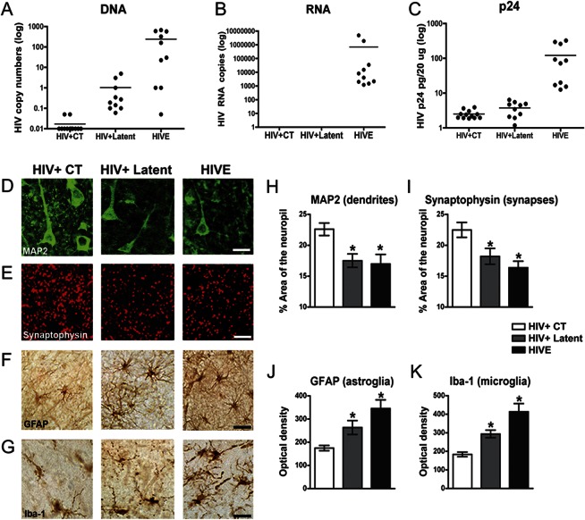
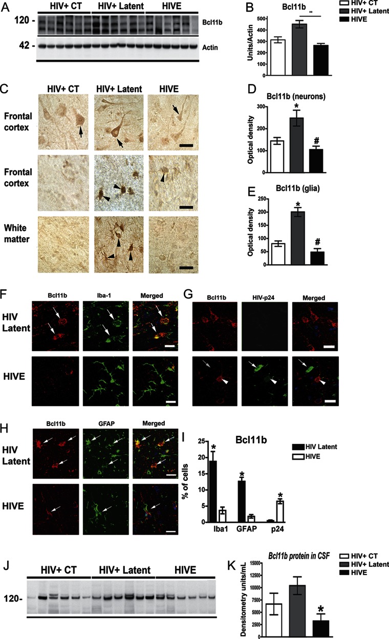
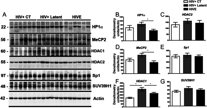
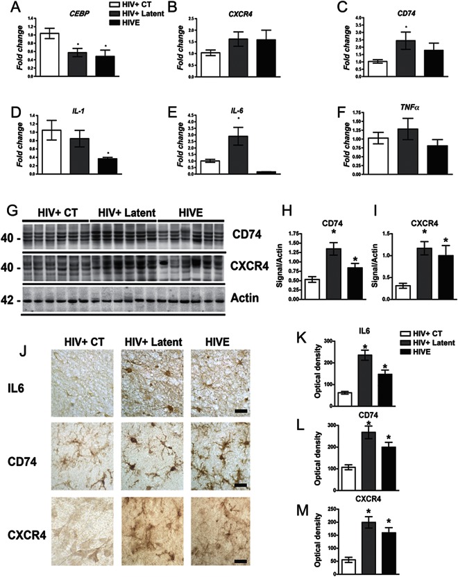
Results: HIV+ controls (n = 12) had no detectable HIV-1 DNA, RNA, or p24 in the CNS; latent HIV+ cases (n = 10) showed high levels of HIV-1 DNA but no HIV RNA or p24; and HIVE cases (n = 10) had high levels of HIV-1 DNA, RNA, and p24. Compared to HIV+ controls, the HIV+ latent cases displayed moderate cognitive impairment with neurodegenerative and neuroinflammatory alterations, although to a lesser extent than HIVE cases. Remarkably, HIV+ latent cases showed higher levels of BCL11B and other chromatin modifiers involved in silencing. Increased BCL11B was associated with deregulation of proinflammatory genes like interleukin-6, tumor necrosis factor-α, and CD74.

Conclusion: Persistence of latent HIV-1 infection in the CNS was associated with increased levels of chromatin modifiers, including BCL11B. Alteration of these epigenetic factors might result in abnormal transcriptomes, leading to inflammation, neurodegeneration, and neurocognitive impairment. BCL11B and other epigenetic factors involved in silencing might represent potential targets for HIV-1 involvement of the CNS.


Figures

Figure 1
Figure 1. Analysis of HIV-1 expression and neurodegenerative pathology in HIV+ controls, HIV+ latent, and HIVE brains
HIV-1 DNA and RNA were analyzed by PCR and viral p24 protein by ELISA in frontal cortex samples from HIV+ controls (CT), HIV+ latent, and HIV encephalitis (HIVE) groups. (A) Detection of HIV-1 DNA corroborated the presence of the provirus in the brains of all infected groups. (B, C) HIV-1 active replication, as indicated by viral RNA and p24 detection, is only evident for the HIVE group. Student t test unpaired, 2-tailed, p < 0.001. (D) Immunolabeling of neuronal cells and dendritic structures with anti-MAP2 antibody. (E) Presynaptic terminals were immunostained with anti-synaptophysin antibody. (F) Astroglial cells were immunolabeled for glial fibrillary acidic protein (GFAP), and microglial cells were identified by anti-Iba1 antibody (G). Bar represents 10 μm. (H, I) Image analysis for the percentage of the area of the neuropil covered by the dendritic and synaptic markers. (J, K) Image analysis for levels of GFAP and Iba1 immunoreactivity expressed as corrected optical density per group. One-way analysis of variance with post hoc Dunnet was used to compare control vs latent and HIVE groups, *p < 0.05.
Figure 2
Figure 2. BCL11B is highly expressed in the brain of latent HIV+ cases and colocalizes with glial markers
(A) Representative immunoblot shows a duplex around 120 kDa corresponding to BCL11B. (B) Image analysis of BCL11B immunoreactive bands normalized to actin. (C) Patterns of BCL11B immunoreactivity in the frontal cortex and white matter. Arrows indicate BCL11B immunoreactivity in neurons and arrowheads in glial cells. Bar represents 10 μm. (D–E) Image analysis of BCL11B immunoreactivity levels in neuronal or glial cells expressed as corrected optical density per group. One-way analysis of variance (ANOVA) with post hoc Dunnet when compared to control, *p < 0.05; **p < 0.01; and with post hoc Tukey-Krammer when compared to latent HIV #p < 0.05. (F–H) Double labeling studies were performed in the frontal cortex with antibodies against BCL11B (red channel) and glial fibrillary acidic protein (GFAP), Iba1, or p24 (green channel) and analyzed by laser scanning confocal microscopy. (F) BCL11B colocalized with the microglial marker Iba1 (arrows) in HIV+ latent but not in HIV encephalitis (HIVE) cases. (G) Cells expressing high levels of viral p24 protein in HIVE cases exhibit the lower content of BCL11B, while latent cases show no viral expression and higher BCL11B immunoreactive signal. Arrows indicate p24-positive cells that are BCL11B-negative. (H) Colocalization of BCL11B to GFAP astroglial cells in HIV+ latent cases in comparison to HIVE cases. (I) Image analysis shows the percentage of BCL11B positive from the total cells recorded positive for Iba1, GFAP, and p24 in HIV latent or HIVE cases. Unpaired, 2-tailed, Student t test, *p < 0.05. (J) Representative blot shows BCL11B immunoreactive bands in CSF samples. (K) Densitometric analysis of BCL11B in CSF. One-way ANOVA with post hoc Dunnet to compare control (CT) vs latent and HIVE groups, *p < 0.05.
Figure 3
Figure 3. The levels of chromatin-remodeling proteins are altered in HIV+ latent cases
(A) Representative immunoblot analysis shows HP1α, MeCP2, HDAC1, HDAC2, SP1, and SUV39H1 immunoreactivity by Western blotting in frontal cortex samples. (B–G) Densitometric quantification of chromatin-remodeling protein immunoreactivity, expressed as signal to actin ratios. One-way analysis of variance with post hoc Dunnet, *p < 0.05. CT = control; HIVE = HIV encephalitis.
Figure 4
Figure 4. Altered expression of BCL11B-regulated proinflammatory targets in HIV+ latent cases
(A–C) Quantification of BCL11B-downstream targets CEBP, CXCR4, and CD74 mRNAs by qPCR in frontal cortex samples. (D–F) Quantification of the relative abundance of inflammatory markers mRNA by qPCR revealed alterations in the HIV+ latent and the HIV encephalitis (HIVE) groups, suggestive of proinflammatory processes. One-way analysis of variance (ANOVA) with post hoc Dunnet to compare control (CT) vs latent and HIVE groups, *p < 0.05. (G) Representative immunoblot analysis of CXCR4 and CD74 proteins in frontal cortex samples. (H, I) Densitometric analysis of CXCR4 and CD74 immunoreactivity expressed as a ratio to actin. One-way ANOVA with post hoc Dunnet to compare CT vs latent and HIVE groups, *p < 0.05. (J) Immunodetection of interleukin (IL)–6, CD74, and CXCR4 in neuronal and glial cells in frontal cortex sections. Bar represents 10 μm. (K–M) Image analysis of neuronal or glial cells shows corrected optical density of each studied protein per group.

Comment in

  • The cost of silencing HIV in the brain.
    Wiley CA, Cysique LA. Wiley CA, et al. Neurology. 2013 Apr 9;80(15):1363-4. doi: 10.1212/WNL.0b013e31828c3077. Epub 2013 Mar 13. Neurology. 2013. PMID: 23486870 No abstract available.

References

    1. Fauci AS. The human immunodeficiency virus: infectivity and mechanisms of pathogenesis. Science 1988;239:617–622 - PubMed
    1. Ellis R, Langford D, Masliah E. HIV and antiretroviral therapy in the brain: neuronal injury and repair. Nat Rev Neurosci 2007;8:33–44 - PubMed
    1. Price RW, Spudich S. Antiretroviral therapy and central nervous system HIV type 1 infection. J Infect Dis 2008;197(suppl 3):S294–S306 - PMC - PubMed
    1. Everall I, Vaida F, Khanlou N, et al. Cliniconeuropathologic correlates of human immunodeficiency virus in the era of antiretroviral therapy. J Neurovirol 2009;15:360–370 - PMC - PubMed
    1. Langford TD, Letendre SL, Larrea GJ, Masliah E. Changing patterns in the neuropathogenesis of HIV during the HAART era. Brain Pathol 2003;13:195–210 - PMC - PubMed

Publication types

MeSH terms