. 2013 Mar 13;33(11):4804-14.
doi: 10.1523/JNEUROSCI.4674-12.2013.

The organization of prefrontal-subthalamic inputs in primates provides an anatomical substrate for both functional specificity and integration: implications for Basal Ganglia models and deep brain stimulation

Affiliations

The organization of prefrontal-subthalamic inputs in primates provides an anatomical substrate for both functional specificity and integration: implications for Basal Ganglia models and deep brain stimulation

William I A Haynes et al. J Neurosci. .

Abstract

The identification of a hyperdirect cortico-subthalamic nucleus connection highlighted the important role of the subthalamic nucleus (STN) in regulating behavior. However, this pathway was shown primarily from motor areas. Hyperdirect pathways associated with cognitive and motivational cortical regions are particularly relevant given recent data from deep brain stimulation, both for neurologic and psychiatric disorders. Our experiments were designed to demonstrate the existence and organization of prefrontal-STN projections, help delineate the "limbic" STN, and determine whether convergence between cortico-STN fibers from functionally diverse cortical areas exists in the STN. We injected anterograde tracers in the ventromedial prefrontal, orbitofrontal, anterior cingulate, and dorsal prefrontal cortices of Macaca nemestrina and Macaca fascicularis to analyze the organization of terminals and passing fibers in the STN. Results show a topographically organized prefrontal hyperdirect pathway in primates. Limbic areas project to the medial tip of the nucleus, straddling its border and extending into the lateral hypothalamus. Associative areas project to the medial half, motor areas to the lateral half. Limbic projections terminated primarily rostrally and motor projections more caudally. The extension of limbic projections into the lateral hypothalamus, suggests that this region be included in the STN. A high degree of convergence exists between projections from functionally diverse cortical areas, creating potentially important interfaces between terminal fields. Taken together, the results provide an anatomical substrate to extend the role of the hyperdirect pathway in models of basal ganglia function, and new keys for understanding deep brain stimulation effects on cognitive and motivational aspects of behavior.


Figures

Figure 1.
Figure 1.
Ventral pallidal connections to the STN. A, Coronal section illustrating VP terminal fields at the edge of the medial STN border, extending medially into the hypothalamus following an injection in the subcommissural ventral pallidum (inset). B, A coronal section illustrating labeled cells (and terminals) at the edge of the medial STN border, extending medially into the hypothalamus after an injection that includes the ventral pallidum. AC, Anterior commissure; Cd, caudate nucleus; MB, mammillary body; Pu, putamen; SN, substantia nigra; STN, subthalamic nucleus; Thal, thalamus.
Figure 2.
Figure 2.
Schematic of the STN divisions. The STN was divided into thirds along the rostrocaudal axis, the rostral and caudal poles were considered as additional, distinct entities as they had specific properties. The medial tip also had specific properties and was isolated from the medial half.
Figure 3.
Figure 3.
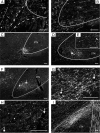
Photomicrographs of STN labeling after cortical injections of anterograde tracers (dark field microscopy of coronal sections). A, Projections from OFC are mostly located medial to the STN′s conventional boundaries (solid white line), but are contained within the STN′s limits according to Dejerine (dashed white lines). B, Projections from vmPFC straddle the conventional medial boundary of the STN (white line). C, The dense projection from dACC is concentrated in the medial tip of the STN and is in a position to overlap with the vmPFC projection (B). It also extends beyond the medial boundary, creating a potential interface with OFC projections (A). D, The projection from area 9 is located in the medial half of the STN. Although the dense projection does not occupy the medial tip, diffuse projections do, providing an interface with dACC (C). This is visible in E, a micrograph from the same case at a higher magnification (the white arrows in D and E indicate the same blood vessel). F, Projections from the rostral area 6 are more caudal and somewhat more dorsal than DPFC projections. Nonetheless, DPFC and area 6 projections overlap extensively. This is demonstrated in G the same section as F at a higher magnification, and in H the section adjacent to F and G, showing the result of a DPFC injection in the same monkey. Matching blood vessels in F–H are indicated by the white arrows. Lesser intensity of staining in H is likely the result of variations in transport (see Results). I, Projections from M1 are dorsal and lateral. Scale bars, 200 μm.
Figure 4.
Figure 4.
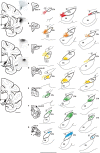
Charts of frontal projections to the STN. Three coronal sections, evenly spaced along the rostrocaudal axis, are illustrated in the left panel of the figure to indicate the approximate anterior (AP = 11.10), central (AP = 10.2), and posterior thirds (AP = 9.0) levels depicted for each case (A–G). Scale bar, 5 mm. The schematic for the injection sites illustrate the center of the injection. Photographs of the prefrontal injection sites complement the prefrontal cases schematics to illustrate the extent of the halo around the injection sites. A, Projections from the vmPFC/OFC (red) are mainly outside of the conventional medial borders of the STN, and concentrated in the anterior third. B, Projections from the dACC (orange) are concentrated in the medial tip of the STN and extend over its medial border. C, D, Projections from DPFC (areas 9 and 46, respectively) (yellow) lie in the medial half of the STN, dorsal and lateral to projections from dACC (B). E, Projections from the rostral area 6 (green) appear caudally to other PFC projections, lateral but overlapping with area 46 dense projections (D). F, Projections from caudal area 6 (green) are located in the ventrolateral STN. G, Projections from M1 (blue) occupy the dorsolateral STN and seem to overlap primarily with caudal area 6 dense projections. Scale bar: (left, bottom) A–G, 1 mm. SN, Substantia nigra; STN, subthalamic nucleus; ZI, zona incerta.
Figure 5.
Figure 5.
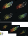
Overlap of dense projections. A–C, Coronal view at anterior (A), central (B), and posterior (C) thirds of the 3D model. Approximate AP levels are similar to those in Fig. 3. Colored meshes represent the outer surface of the combined dense projections from each cortical area. Overlaps occur mainly between projections from functionally close cortical regions. D, Axial, superior view of the same dense projections. Scale bar, 1 mm.
Figure 6.
Figure 6.
Overlap of diffuse projections. Posterior, coronal 3D views of diffuse and dense cortical projections. Colored volumes/surfaces, Dense projection fields; colored lines, diffuse projections. A, All diffuse projections. The topography is the same as for dense projections, although there is more overlap. B, All dense projections (surfaces). Note, compared to the diffuse projections in A, the dense terminal fields show less overlap. C, Diffuse and dense projections from prefrontal areas, vmPFC, ACC, and DPFC. Diffuse projections increase the interface between the different prefrontal inputs. D, Diffuse and dense projections from premotor and motor regions. E, Diffuse projections derived from DPFC and dACC injections extend into area 6 and M1 territory, thus increasing their interface. F, Reciprocally, area 6 diffuse projections extend into the prefrontal projection territory. They are also able to interface with diffuse projections from vmPFC/OFC. Scale bar, 1 mm.
Figure 7.
Figure 7.
Passing fibers. A, Passing fibers have a topographic organization similar to diffuse projections (Fig. 6A). B, Fibers from vmPFC, OFC, and dACC travel in the medial tip of the STN and in the adjacent lateral hypothalamus. C, Fibers from DPFC travel in the medial half of the STN. D, Fibers from rostral and caudal area 6 travel, respectively, in the medial and lateral halves of the STN. Fibers from M1 travel in the dorsal portion of the lateral half. E–G, Views of the anterior, central, and posterior thirds of the STN illustrate the rostrocaudal topography of passing fibers. Prefrontal fibers enter the STN rostrally (E) to those from area 6 and M1 (F). However, fibers from area 6 and M1 travel further caudally (G). Scale bar, 1 mm.

Comment in

References

    1. Berthoud HR, Münzberg H. The lateral hypothalamus as integrator of metabolic and environmental needs: from electrical self-stimulation to opto-genetics. Physiol Behav. 2011;104:29–39. - PMC - PubMed
    1. Bevan MD, Clarke NP, Bolam JP. Synaptic integration of functionally diverse pallidal information in the entopeduncular nucleus and subthalamic nucleus in the rat. J Neurosci. 1997;17:308–324. - PMC - PubMed
    1. Bogacz R, Larsen T. Integration of reinforcement learning and optimal decision-making theories of the basal ganglia. Neural computation. 2011;23:817–851. - PubMed
    1. Cavanagh JF, Wiecki TV, Cohen MX, Figueroa CM, Samanta J, Sherman SJ, Frank MJ. Subthalamic nucleus stimulation reverses mediofrontal influence over decision threshold. Nat Neurosci. 2011;14:1462–1467. - PMC - PubMed
    1. Chaturvedi A, Butson CR, Lempka SF, Cooper SE, McIntyre CC. Patient-specific models of deep brain stimulation: influence of field model complexity on neural activation predictions. Brain stimulation. 2010;3:65–67. - PMC - PubMed

Publication types

LinkOut - more resources