

Ultra-high 7T MRI of structural age-related changes of the subthalamic nucleus
- PMID: 23486960
- PMCID: PMC6619019
- DOI: 10.1523/JNEUROSCI.3241-12.2013
Ultra-high 7T MRI of structural age-related changes of the subthalamic nucleus
Abstract
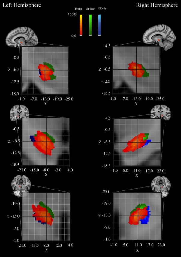
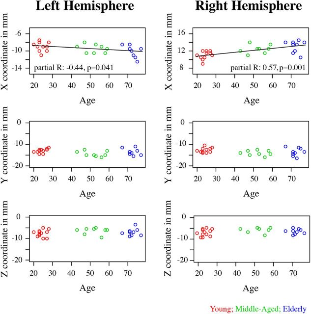
The subthalamic nucleus (STh) is a small subcortical structure which is involved in regulating motor as well as cognitive functions. Due to its small size and close proximity to other small subcortical structures, it has been a challenge to localize and visualize it using magnetic resonance imaging (MRI). Currently there are several standard atlases available that are used to localize the STh in functional MRI studies and clinical procedures such as deep brain stimulation (DBS). DBS is an increasingly common neurosurgical procedure that has been successfully used to alleviate motor symptoms present in Parkinson's disease. However, current atlases are based on low sample sizes and restricted age ranges (Schaltenbrand and Wahren, 1977), and hence the use of these atlases effectively ignores the substantial structural brain changes that are associated with aging. In the present study, ultra-high field 7 tesla (T) magnetic resonance imaging (MRI) in humans was used to visualize and segment the STh in young, middle-aged, and elderly participants. The resulting probabilistic atlas maps for all age groups show that the STh shifts in the lateral direction with increasing age. In sum, the results of the present study suggest that age has to be taken into account in atlases for the optimal localization of the STh in healthy and diseased brains.
Figures
References
-
- Alho E, Grinberg L, Heinsen H. Review of printed and electronic stereotactic atlases of the human brain. In: Peres JFP, editor. Neuroimaging for clinicians—combining research and practice. New York: InTech; 2011.
-
- Aquino D, Bizzi A, Grisoli M, Garavaglia B, Bruzzone MG, Nardocci N, Savoiardo M, Chiapparini L. Age-related iron deposition in the basal ganglia: quantitative analysis in healthy subjects. Radiology. 2009;252:165–172. - PubMed
-
- Aron AR. The neural basis of inhibition in cognitive control. Neuroscientist. 2007;13:214–228. - PubMed
MeSH terms
LinkOut - more resources
Full Text Sources
Other Literature Sources
Medical