

Detection of Sentinel Lymph Nodes in Gynecologic Tumours by Planar Scintigraphy and SPECT/CT
- PMID: 23486989
- PMCID: PMC3590971
- DOI: 10.4274/Mirt.236
Detection of Sentinel Lymph Nodes in Gynecologic Tumours by Planar Scintigraphy and SPECT/CT
Abstract
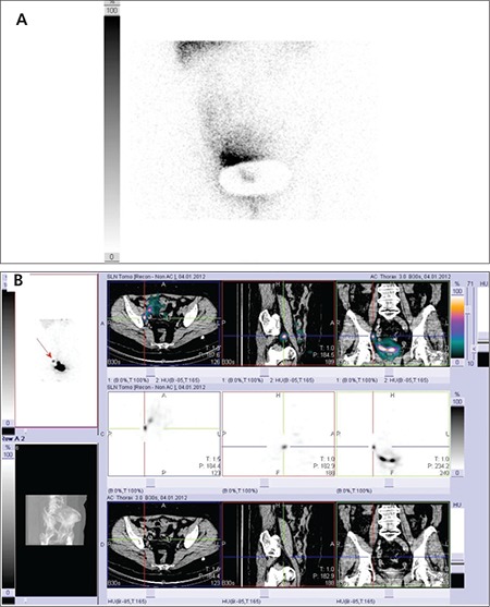
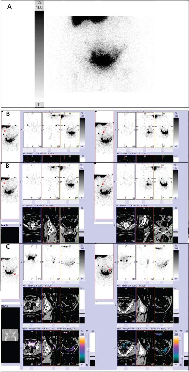
Objective: Assess the role of planar lymphoscintigraphy and fusion imaging of SPECT/CT in sentinel lymph node (SLN) detection in patients with gynecologic tumours.
Material and methods: Planar scintigraphy and hybrid modality SPECT/CT were performed in 64 consecutive women with gynecologic tumours (mean age 53.6 with range 30-77 years): 36 pts with cervical cancer (Group A), 21 pts with endometrial cancer (Group B), 7 pts with vulvar carcinoma (Group C). Planar and SPECT/CT images were interpreted separately by two nuclear medicine physicians. Efficacy of these two techniques to image SLN were compared.
Results: Planar scintigraphy did not image SLN in 7 patients (10.9%), SPECT/CT was negative in 4 patients (6.3%). In 35 (54.7%) patients the number of SLNs captured on SPECT/CT was higher than on planar imaging. Differences in detection of SLN between planar and SPECT/CT imaging in the group of all 64 patients are statistically significant (p<0.05). Three foci of uptake (1.7% from totally visible 177 foci on planar images) in 2 patients interpreted on planar images as hot LNs were found to be false positive non-nodal sites of uptake when further assessed on SPECT/CT. SPECT/CT showed the exact anatomical location of all visualised sentinel nodes.
Conclusion: In some patients with gynecologic cancers SPECT/CT improves detection of sentinel lymph nodes. It can image nodes not visible on planar scintigrams, exclude false positive uptake and exactly localise pelvic and paraaortal SLNs. It improves anatomic localization of SLNs.
Conflict of interest: None declared.
Keywords: SPECT; Sentinel lymph node biopsy; X-Ray computed; gamma camera Imaging; gynecologic neoplasms; scintigraphy; tomography.
Figures
Similar articles
-
Sentinel lymph nodes and planar scintigraphy and SPECT/CT in various types of tumours. Estimation of some factors influencing detection success.Nucl Med Rev Cent East Eur. 2013;16(1):17-25. doi: 10.5603/NMR.2013.0004. Nucl Med Rev Cent East Eur. 2013. PMID: 23677759
-
Localisation of sentinel lymph nodes in patients with melanomas by planar lymphoscintigraphic and hybrid SPECT/CT imaging.Nucl Med Rev Cent East Eur. 2012 Aug 27;15(2):101-7. Nucl Med Rev Cent East Eur. 2012. PMID: 22936502
-
Evaluation of the diagnostic value of preoperative sentinel lymph node (SLN) imaging in penile carcinoma patients without palpable inguinal lymph nodes via single photon emission computed tomography/computed tomography (SPECT/CT) as compared to planar scintigraphy.Urol Oncol. 2018 Mar;36(3):92.e17-92.e24. doi: 10.1016/j.urolonc.2017.11.012. Epub 2017 Dec 14. Urol Oncol. 2018. PMID: 29249274
-
Single-photon emission computed tomography/computed tomographyfor sentinel node mapping in breast cancer.Semin Nucl Med. 2007 Jan;37(1):29-33. doi: 10.1053/j.semnuclmed.2006.08.001. Semin Nucl Med. 2007. PMID: 17161037 Review.
-
Contribution of SPECT/CT imaging to radioguided sentinel lymph node biopsy in breast cancer, melanoma, and other solid cancers: from "open and see" to "see and open".Q J Nucl Med Mol Imaging. 2014 Jun;58(2):127-39. Q J Nucl Med Mol Imaging. 2014. PMID: 24835289 Review.
Cited by
-
The use of SPECT/CT for anatomical mapping of lymphatic drainage in vulvar cancer: possible implications for the extent of inguinal lymph node dissection.Eur J Nucl Med Mol Imaging. 2015 Dec;42(13):2064-71. doi: 10.1007/s00259-015-3127-1. Epub 2015 Jul 30. Eur J Nucl Med Mol Imaging. 2015. PMID: 26219869
-
Lymphoscintigraphy and sentinel lymph node biopsy in vulvar carcinoma: update from a European expert panel.Eur J Nucl Med Mol Imaging. 2020 May;47(5):1261-1274. doi: 10.1007/s00259-019-04650-8. Epub 2020 Jan 2. Eur J Nucl Med Mol Imaging. 2020. PMID: 31897584 Review.
-
Sentinel Lymph Node Mapping in Endometrial Cancer: A Comprehensive Review.Front Oncol. 2021 Jun 29;11:701758. doi: 10.3389/fonc.2021.701758. eCollection 2021. Front Oncol. 2021. PMID: 34268126 Free PMC article. Review.
-
The role of technetium-99m isotope in sentinel lymph node identification in gynecological cancers.Rep Pract Oncol Radiother. 2025 Jun 7;30(2):257-268. doi: 10.5603/rpor.105251. eCollection 2025. Rep Pract Oncol Radiother. 2025. PMID: 40635985 Free PMC article. Review.
-
Sentinel lymph node evaluation in women with cervical cancer.J Minim Invasive Gynecol. 2014 Jul-Aug;21(4):540-5. doi: 10.1016/j.jmig.2013.12.095. Epub 2014 Jan 7. J Minim Invasive Gynecol. 2014. PMID: 24407177 Free PMC article. Review.
References
-
- Burke TW, Levenback C, Tornos C, Morris M, Wharton JT, Gershenson DM. Intraabdominal lymphatic mapping to direct elective pelvic and paraaortic lymphadenectomy in women with high-risk endometrial cancer. Results of a pilot study. Gynecol Oncol. 1996;62:169–173. - PubMed
-
- Nieweg OE, Jansen L, Valdes Olmos RA, Rutgers EJ, Peterse JL, Hoefnagel KA. Kroon BB. Lymphatic mapping and sentinel lymph node biopsy in breast cancer. Eur J Nucl Med. 1999;26:S11–16. - PubMed
-
- Kraft O, Safarcik K, Stepien A. Sentinel Lymph Node Detection and Biopsy in Breast Cancer and Malignant Melanoma. World J Nucl Med. 2004;3:26–32.
-
- Tanaka Y, Sawada S, Murata T. Relationship between lymph node metastases and prognosis in patients irradiated post-operatively for carcinoma of the uterine cervix. Acta Radiol Oncol. 1984;23:455–459. - PubMed
-
- Delgado G, Bundy B, Zaino R, Sevin BU, Creasman WT, Major F. Prospective surgical-pathological study of disease free interval in patients with stage IB squamous cell carcinoma of the cervix: a Gynecologic Oncology Group study. Gynecol Oncol. 1990;38:352–357. - PubMed
LinkOut - more resources
Full Text Sources
Research Materials