. 2013 Apr;140(8):1730-9.
doi: 10.1242/dev.092882. Epub 2013 Mar 13.

Divergent roles for Wnt/β-catenin signaling in epithelial maintenance and breakdown during semicircular canal formation

Affiliations

Divergent roles for Wnt/β-catenin signaling in epithelial maintenance and breakdown during semicircular canal formation

Staci Rakowiecki et al. Development. 2013 Apr.

Abstract

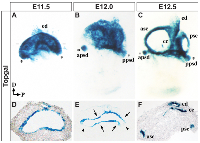
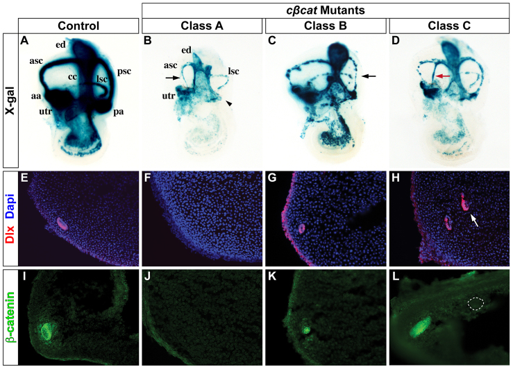
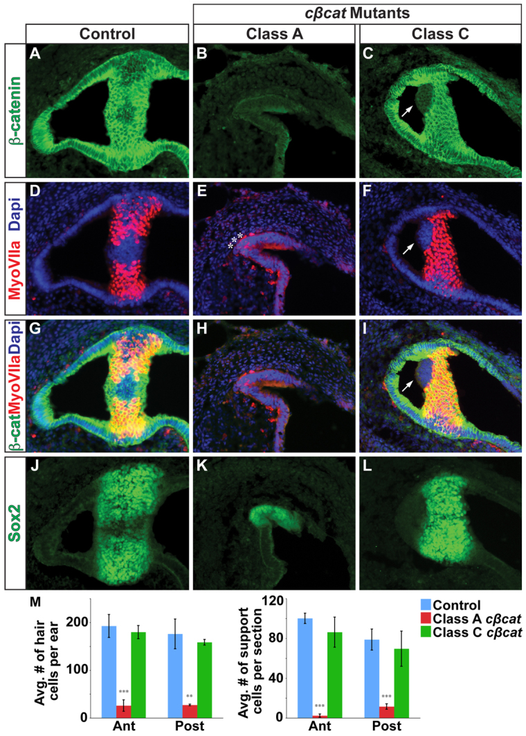
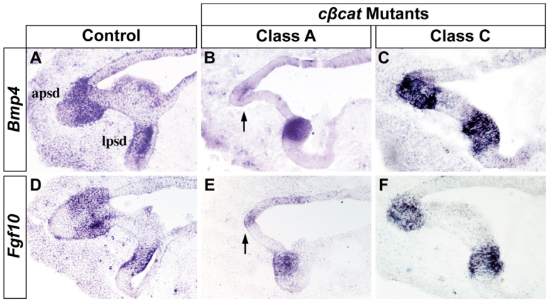
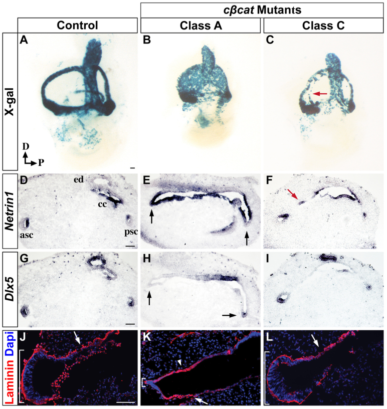
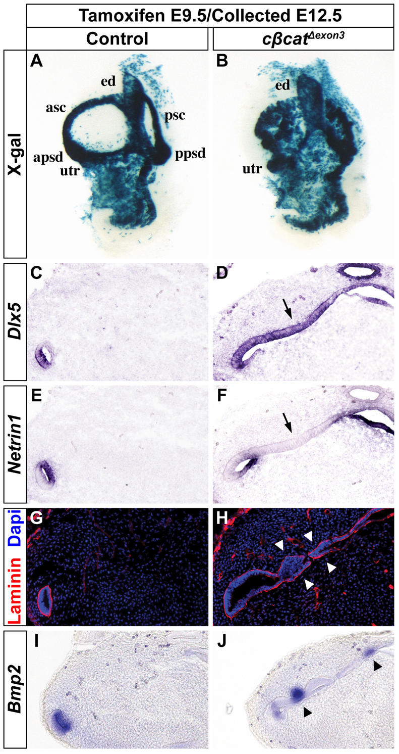
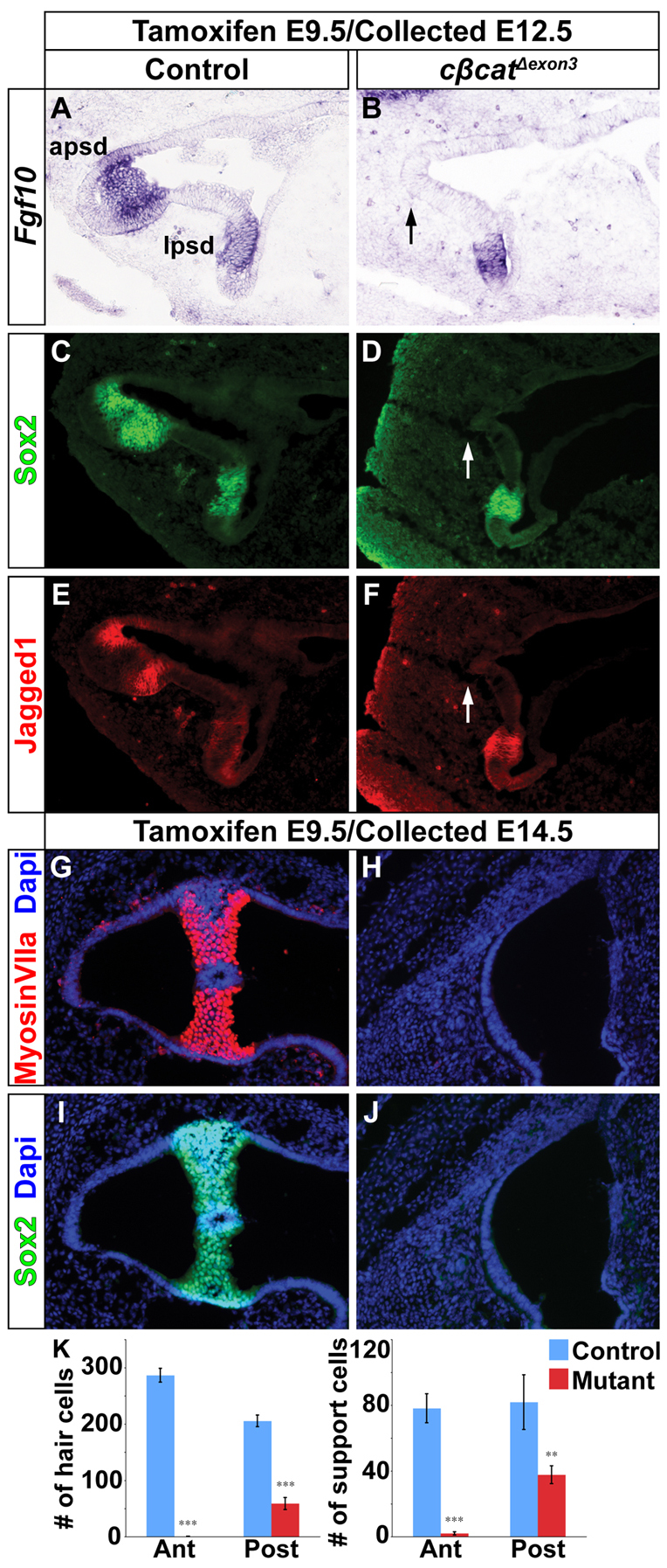
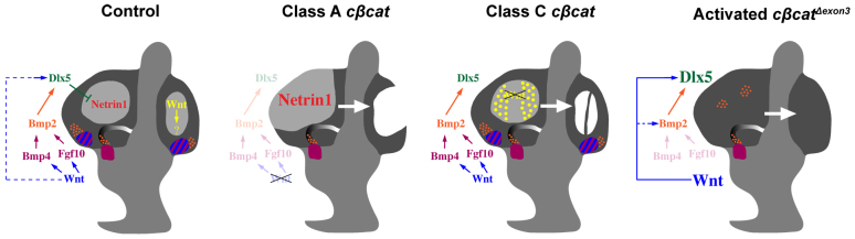
The morphogenetic program that shapes the three semicircular canals (SSCs) must be executed with extreme precision to satisfy their complex vestibular function. The SSCs emerge from epithelial outgrowths of the dorsal otocyst, the central regions of which fuse and resorb to leave three fluid-filled canals. The Wnt/β-catenin signaling pathway is active at multiple stages of otic development, including during vestibular morphogenesis. How Wnt/β-catenin functionally integrates with other signaling pathways to sculpt the SSCs and their sensory patches is unknown. We used a genetic strategy to spatiotemporally modulate canonical Wnt signaling activity during SSC development in mice. Our findings demonstrate that Wnt/β-catenin signaling functions in a multifaceted manner during SSC formation. In the early phase, Wnt/β-catenin signaling is required to preserve the epithelial integrity of the vertical canal pouch perimeter (presumptive anterior and posterior SSCs) by establishing a sensory-dependent signaling relay that maintains expression of Dlx5 and opposes expression of the fusion plate marker netrin 1. Without this Wnt signaling activity the sensory to non-sensory signaling cascade fails to be activated, resulting in loss of vestibular hair and support cells and the anterior and posterior SSCs. In the later phase, Wnt/β-catenin signaling becomes restricted to the fusion plate where it facilitates the timely resorption of this tissue. Mosaic recombination of β-catenin in small clusters of canal pouch cells prevents their resorption, causing instead the formation of ectopic SSCs. Together, these disparate functions of the Wnt/β-catenin pathway in epithelial maintenance and resorption help regulate the size, shape and number of SSCs.


Figures

Fig. 1.
Fig. 1.
Dynamic Wnt/β-catenin signaling during vestibular development. Whole-mount (A-C) and transverse sections (D-F) of mouse inner ears stained for Topgal activity at the specified stages. The dashed line in A indicates the plane of section in D-F. (A,D) Wnt responsiveness is evident throughout the canal pouch at E11.5. (B,E) By E12.0, Topgal is excluded from the outer canal rims (arrowheads in E) and becomes restricted to the prospective fusion plate (arrows in E). (C,F) After the completion of resorption at E12.5, Topgal can be detected in the inner wall of the anterior and posterior SSCs, the common crus and the endolymphatic duct. Topgal expression is evident in anterior and posterior prosensory domains at all three stages (A-C, asterisks). apsd, anterior prosensory domain; asc, anterior semicircular canal; cc, common crus; D, dorsal; ed, endolymphatic duct; P, posterior; ppsd, posterior prosensory domain; psc, posterior semicircular canal.
Fig. 2.
Fig. 2.
Vestibular defects in the absence of Wnt/β-catenin signaling. (A-D) Whole-mount views of control and cβcat mouse embryos at E14.5 (tamoxifen administered at E10.5) showing representative phenotypic classes. Note the absence of the posterior ampulla (arrowhead) and corresponding SSC in Class A mutants (B), as well as the severely truncated anterior SSC (arrow). The black and red arrows in C and D highlight the small and ectopic forming canals typically observed in Class B and Class C mutants, respectively. (E-L) Transverse sections through the vestibulum of control and cβcat mutants immunostained for Dlx (E-H) or β-catenin (I-L) expression. The cβcat mutants show a consistent loss, reduction or gain (arrow in H) in Dlx staining according to their classification. Alterations in β-catenin protein levels correlate with the severity of the phenotypic class (I-L). The formation of an ectopic canal (dotted circle in L) is due to mosaic recombination of Ctnnb1 within the fusion plate. aa, anterior ampulla; asc, anterior semicircular canal; cc, common crus; ed, endolymphatic duct; lsc, lateral semicircular canal; pa, posterior ampulla; psc, posterior semicircular canal; utr, utricle.
Fig. 3.
Fig. 3.
Vestibular hair and support cells are dependent on canonical Wnt signaling. (A-C) Immunostaining for β-catenin on sections through the crista ampullaris (anterior or posterior) in control (A) and cβcat mutant (B,C) mice at E14.5 (tamoxifen administered at E10.5). β-catenin expression is greatly reduced in Class A mutants and only occasionally affected in Class C mutants, as compared with controls. (D-L) The numbers of myosin VIIa+ hair cells and Sox2+ support cells (J-L) are greatly reduced in Class A mutants but only occasionally affected in Class C mutants. Asterisks (E) mark examples of the few myosin VIIa+ cells detected in Class A mutants. Arrows (C,F,I) highlight the cell-autonomous loss of myosin VIIa staining in β-catenin-deficient cells in Class C mutants. (M) Quantification of hair and support cells in control (blue), Class A (red) and Class C (green) mutants. Error bars indicate s.e.m. ***P<0.01, **P<0.05 (unpaired Student’s t-test).
Fig. 4.
Fig. 4.
The prosensory signals Bmp4 and Fgf10 are dependent on Wnt/β-catenin signaling. Transverse sections through the prosensory regions of control and cβcat mutant mice at E12.5 (tamoxifen administered at E10.5) stained for (A-C) Bmp4 and (D-F) Fgf10 by RNA in situ hybridization. The expression of Bmp4 and Fgf10 is lost in the anterior (shown) and posterior prosensory domains of Class A, but not Class C, mutants (arrows in B,E). The lateral prosensory domain is unaffected in all mutants. apsd, anterior prosensory domain; lpsd, lateral prosensory domain.
Fig. 5.
Fig. 5.
Preservation of canal rim epithelium through Wnt-dependent regulation of Dlx5 and restriction of Ntn1. (A-C) Whole-mount X-gal staining of control and cβcat mutant mouse inner ears at E12.5 (tamoxifen administered at E10.5) demonstrates varying degrees of perduring canal pouch epithelium. The arrow (C) indicates perduring canal pouch epithelium. (D-I) Adjacent transverse sections analyzed for Ntn1 (D-F) and Dlx5 (G-I) mRNA expression. The inactivation of Wnt/β-catenin signaling in Class A mutants results in the downregulation of Dlx5 in the canal rim (arrows in H) and subsequent expansion of Ntn1 into this domain (arrows in E). The arrow in F indicates a confined region of epithelial maintenance. (J-L) Laminin immunostaining is continuous around the canal rim of control embryos (bracket in J) and punctate at the position of the fusion plate (arrow in J) indicative of basement membrane breakdown. In Class A mutants, the epithelial integrity around the canal rim is lost, as evidenced by the reduction in laminin (small bracket in K), whereas the fusion plate shows areas of continuous laminin staining, consistent with the delayed resorption of this tissue (arrowhead in K). Arrows (J-L) indicate regions of basement membrane breakdown. Scale bar: 100 μm.
Fig. 6.
Fig. 6.
Forced activation of Wnt/β-catenin signaling expands the Dlx5-expressing canal rim identity at the expense of Ntn1-expressing fusion plate. (A,B) Whole-mount X-gal staining of control and cβcatΔexon3 mouse inner ears at E12.5 (tamoxifen administered at E9.5). (C-J) Transverse sections analyzed for Dlx5 (C,D), Ntn1 (E,F), laminin (G,H) and Bmp2 (I,J) expression. The inner ears of cβcatΔexon3 mutants show an expansion in Dlx5 staining throughout the canal pouch (arrow in D) at the expense of Ntn1 (arrow in F) resulting in an epithelialization of the fusion plate, as indicated by the maintained laminin expression (arrowheads in H). Arrowheads in J indicate ectopic Bmp2 expression.
Fig. 7.
Fig. 7.
Ectopic canal rim epithelium in cβcatΔexon3 mouse mutants occurs in the absence of prosensory development. (A-F) The expression of Fgf10, Sox2 and Jag1 was downregulated in the anterior (arrows in B,D,F) and posterior prosensory domains of cβcatΔexon3 mutants at E12.5 (tamoxifen administered at E9.5). The lateral prosensory domain was unaffected. (G-J) Hair (myosin VIIa) and support (Sox2) cell markers were reduced in cβcatΔexon3 mutants compared with controls at E14.5. (K) Quantification of hair and support cells in control and cβcatΔexon3 embryos. Error bars indicate s.e.m. ***P<0.01, **P<0.05 (Student’s t-test).
Fig. 8.
Fig. 8.
The dual roles of Wnt/β-catenin signaling in SSC formation in the mouse inner ear. Initially, canonical Wnt signaling (blue) regulates the epithelial identity of the vertical canal pouch rim (prospective anterior and posterior SSCs) through the activation of Dlx5 expression (dashed blue line) and restriction of Ntn1 to the fusion plate (see Control). As vestibular development progresses, the Dlx5+ canal rim loses its responsiveness to Wnt/β-catenin and instead becomes dependent on a sensory to non-sensory signaling relay that involves Bmp4 and Fgf10 from the prosensory domain (blue and purple hatched region) and Bmp2 from the canal genesis zone (orange), all of which is initiated by Wnt/β-catenin. The inactivation of Ctnnb1 throughout the canal pouch epithelium leads to a downregulation in the sensory-dependent and sensory-independent regulation of Dlx5, an expansion in the fusion plate marker Ntn1, and consequent loss of anterior and posterior SSCs and corresponding cristae (Class A cβcat). The mosaic inactivation of Ctnnb1 in small clusters of canal pouch cells reveals a second, independent role of Wnt/β-catenin signaling (yellow) in mediating the timely resorption of the fusion plate (Class C cβcat). Perturbation of this later function of Wnt/β-catenin signaling results in the ectopic formation of canal-like structures, which are supported by prosensory signals. The forced activation of a stabilized form of β-catenin leads to the expansion of Dlx5+, and some Bmp2+, canal rim progenitors at the expense of Ntn1+ fusion plate and prosensory signals (activated cβcatΔexon3).

References

    1. Abraira V. E., Del Rio T., Tucker A. F., Slonimsky J., Keirnes H. L., Goodrich L. V. (2008). Cross-repressive interactions between Lrig3 and netrin 1 shape the architecture of the inner ear. Development 135, 4091–4099 - PMC - PubMed
    1. Bok J., Chang W., Wu D. K. (2007). Patterning and morphogenesis of the vertebrate inner ear. Int. J. Dev. Biol. 51, 521–533 - PubMed
    1. Brault V., Moore R., Kutsch S., Ishibashi M., Rowitch D. H., McMahon A. P., Sommer L., Boussadia O., Kemler R. (2001). Inactivation of the beta-catenin gene by Wnt1-Cre-mediated deletion results in dramatic brain malformation and failure of craniofacial development. Development 128, 1253–1264 - PubMed
    1. Chang W., Brigande J. V., Fekete D. M., Wu D. K. (2004). The development of semicircular canals in the inner ear: role of FGFs in sensory cristae. Development 131, 4201–4211 - PubMed
    1. Chang W., Lin Z., Kulessa H., Hebert J., Hogan B. L., Wu D. K. (2008). Bmp4 is essential for the formation of the vestibular apparatus that detects angular head movements. PLoS Genet. 4, e1000050 - PMC - PubMed

Publication types

MeSH terms