. 2013 May;87(10):5678-96.
doi: 10.1128/JVI.00840-12. Epub 2013 Mar 13.

Systematic analysis of enhancer and critical cis-acting RNA elements in the protein-encoding region of the hepatitis C virus genome

Affiliations

Systematic analysis of enhancer and critical cis-acting RNA elements in the protein-encoding region of the hepatitis C virus genome

Derrick Chu et al. J Virol. 2013 May.

Abstract

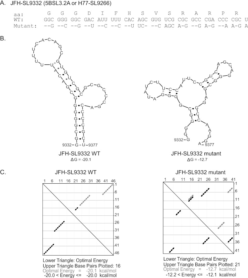
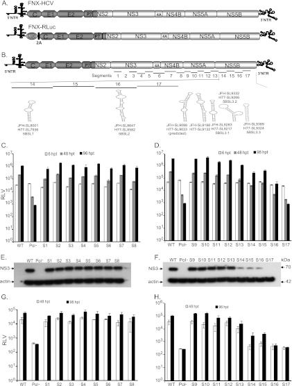
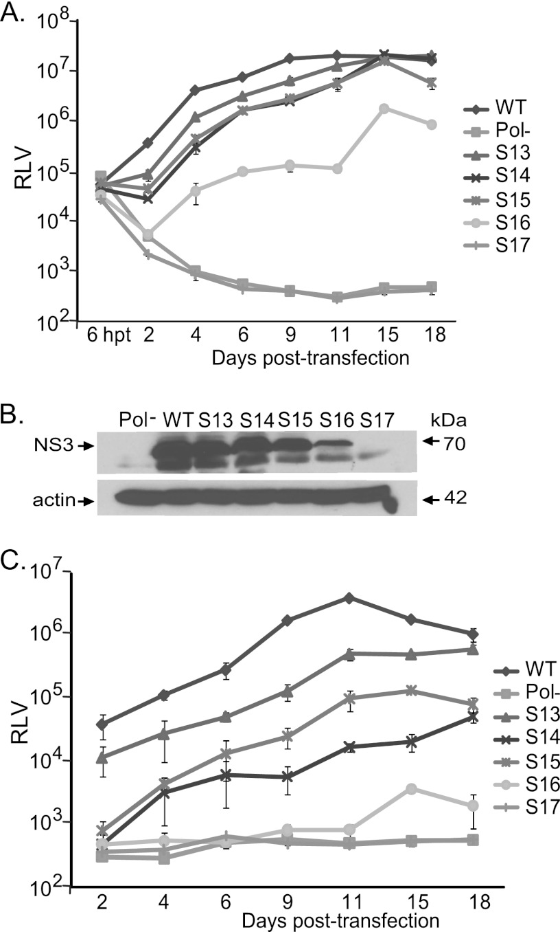
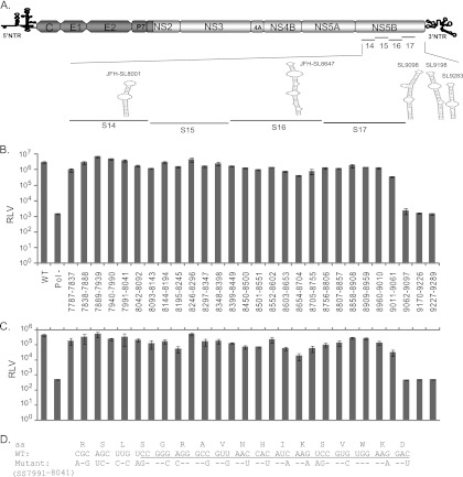
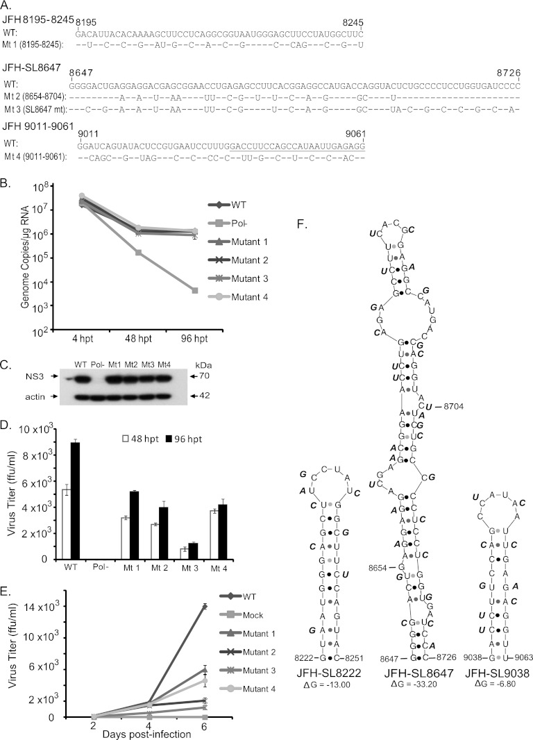
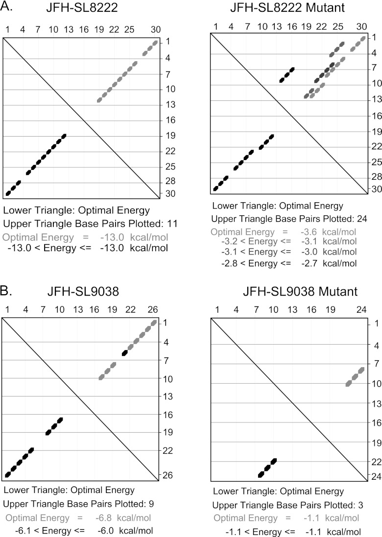
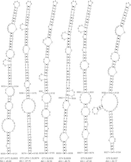
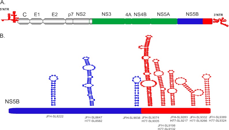
Hepatitis C virus (HCV) causes chronic hepatitis, cirrhosis, and liver cancer. cis-acting RNA elements of the HCV genome are critical for translation initiation and replication of the viral genome. We hypothesized that the coding regions of nonstructural proteins harbor enhancer and essential cis-acting replication elements (CRE). In order to experimentally identify new cis RNA elements, we utilized an unbiased approach to introduce synonymous substitutions. The HCV genome coding for nonstructural proteins (nucleotide positions 3872 to 9097) was divided into 17 contiguous segments. The wobble nucleotide positions of each codon were replaced, resulting in 33% to 41% nucleotide changes. The HCV genome containing one of each of 17 mutant segments (S1 to S17) was tested for genome replication and infectivity. We observed that silent mutations in segment 13 (S13) (nucleotides [nt] 7457 to 7786), S14 (nt 7787 to 8113), S15 (nt 8114 to 8440), S16 (nt 8441 to 8767), and S17 (nt 8768 to 9097) resulted in impaired genome replication, suggesting CRE structures are enriched in the NS5B region. Subsequent high-resolution mutational analysis of NS5B (nt 7787 to 9289) using approximately 51-nucleotide contiguous subsegment mutant viruses having synonymous mutations revealed that subsegments SS8195-8245, SS8654-8704, and SS9011-9061 were required for efficient viral growth, suggesting that these regions act as enhancer elements. Covariant nucleotide substitution analysis of a stem-loop, JFH-SL9098, revealed the formation of an extended stem structure, which we designated JFH-SL9074. We have identified new enhancer RNA elements and an extended stem-loop in the NS5B coding region. Genetic modification of enhancer RNA elements can be utilized for designing attenuated HCV vaccine candidates.


Figures

Fig 1
Fig 1
Computationally modeling the effects of synonymous mutations in a well-characterized CRE, 5BSL3.2a (H77-SL9266 or JFH-SL9332). (A) JFH-1 SL9332 wild-type (WT) and mutant sequences are shown. aa, amino acids. (B) Predicted stem-loop structures of wild-type and mutated sequences using RNA mfold analysis. Note the altered Watson-Crick base pair interactions in the mutant structure and lowered folding energy, resulting in a destabilized RNA structure. The folding Gibbs free energy (ΔG, in kcal/mol) for the predicted stem-loops is shown under each structure. (C) Energy dot plots for optimal and suboptimal folding of stem-loop wild-type and mutant structures. The upper triangle displays possible base pair combinations at various energy levels. The nucleotide positions of each base are displayed on the top axis and right axis of the upper triangle. The paired bases are shown as diamond dots in the plot. For example, wild-type base 46 (top axis) pairs with base 1 (right axis). The lower triangle shows the paired bases with optimal folding energy at 37°C to form a stem-loop structure.
Fig 2
Fig 2
RNA cis element analysis of the HCV nonstructural-protein-coding region. (A) Schematic diagram of hepatitis C viruses used in this study. FNX-HCV is a synthetic version of a genotype 2a J6CF and JFH-1 chimera (the J6CF strain region is shown in dark gray, including the 5′ NTR, and the JFH-1 strain region is shown in light gray). A monocistronic Renilla luciferase (luc) reporter virus (FNX-Rluc) based on the J6/JFH chimeric virus is shown. The luciferase gene is fused in frame with the core gene through foot and mouth disease virus 2A sequence. (B) Schematic representation of the HCV genome showing the locations of 17 mutated segments in the nonstructural-protein-coding region. The stem-loop structures described in the NS5B region corresponding to the genome positions of JFH-1 and H77 are noted. (C and D) Analysis of viral genome replication of mutant reporter viruses. Huh-7.5.1 cells were electroporated with 10 μg of in vitro-transcribed genomic RNA of wild-type FNX-Rluc reporter virus and individual S1 to S17 mutant reporter viruses. A control reporter virus with nonfunctional polymerase activity (Pol) is included. The transfected cells were lysed at 6, 48, and 96 h p.t. using Promega Renilla luciferase assay lysis buffer, and the levels of Renilla luciferase were quantified. The experiment was performed in triplicate. The mean Renilla luciferase values (RLV) with standard deviations are shown in log10 scale as a bar graph. The replication-deficient mutant S17 showed luciferase activity similar to that of the Pol control virus. At 96 h p.t., mutant viruses S14, S15, S16, and S17 had significant reductions in genome replication compared to that of wild-type virus (P value < 0.0001 by unpaired t test). (E and F) Western blot of the expression of HCV protein. The 70-kDa NS3 antigen was detected from the protein lysates obtained at 96 h posttransfection by primary mouse monoclonal antibody and secondary goat-anti mouse IgG conjugated with HRP. As a loading control, β-actin was included. (G and H) Infectivities of mutant viruses. Naïve Huh-7.5.1 cells were infected with the cell-free supernatants harvested at 48 and 96 h posttransfection. Renilla luciferase activities were measured from the lysates harvested at 48 h postinfection. The mean RLV with standard deviations are depicted in the bar graph. Mutant reporter viruses S13, S14, S15, S16, and S17 exhibited significant reductions in infectivity at 48 h p.t. (P value ≤ 0.005) and 96 h p.t. (P value < 0.005) compared to that of wild-type virus. The experiment was repeated three times, and data from a representative experiment are shown.
Fig 3
Fig 3
Genome replication and infectious virus production kinetics during long-term culturing of CRE mutant reporter viruses. (A) Genome replication kinetics of wild-type reporter virus, as well as mutant reporter viruses S13, S14, S15, S16, and S17, at the indicated time points posttransfection. Cell lysates were harvested at various time points to measure Renilla luciferase activity. The mean and standard deviation calculated from triplicate RLV for each mutant are shown in the graph. (B) Western blots showing the expression of NS3 antigen at 9 days p.t. and control β-actin. The Pol and S17 mutants lack NS3 expression. (C) Analysis of virus production kinetics. Huh-7.5.1 cells were inoculated with the supernatant harvested from each virus-infected culture at the indicated time points. The Renilla luciferase activities of infected cells were measured. Mean values with standard deviations are depicted in the graph.
Fig 4
Fig 4
Confirming the growth phenotypes of CRE mutant viruses in the NS5B region. (A) Immunofluorescence assay to detect viral antigen. The in vitro-transcribed RNA genomes of FNX wild-type virus and individual mutant viruses S13, S14, S15, S16, and S17 were electroporated into Huh-7.5.1 cells. At 48 and 96 h posttransfection, the cells were fixed and immunostained for HCV NS5A antigen (red). Hoechst staining was used to visualize cell nuclei (blue). (B) Genome replication kinetics of mutant viruses. RNA was harvested from transfected cells at 4, 48, and 96 h posttransfection and subjected to RT-quantitative PCR. The genome copy numbers per microgram of RNA were quantified and are depicted in the graph. The FNX-HCV mutant lacking polymerase activity was included as a control for the genome replication-deficient phenotype. (C) Analysis of HCV antigen expression. Western blotting was performed to detect NS3 antigen and β-actin. (D) Infectivities of various mutant viruses. The virus titer (focus-forming units/ml) was measured by infecting naïve Huh-7.5.1 cells with the cell-free supernatants collected at 48 and 96 h posttransfection from each mutant-virus-transfected cell culture. The mean values and standard deviations of the titers are shown in the graph. All the mutants had significantly reduced infectivity at 96 h posttransfection compared to that of the wild type (P value < 0.05 by unpaired t test). To assess infectivity, we included an FNX mutant virus lacking envelope genes (Env-) as an additional control. The experiment was done three times, and data from a representative experiment are shown.
Fig 5
Fig 5
High-resolution mutational analysis for identification of additional cis-acting replication elements in the NS5B protein-encoding region. (A) Schematic diagram of HCV genome depicting the mutated subsegments in NS5B. JFH-1 CRE stem-loops present in and adjacent to the mutated subsegments are presented. SL9332 and SL9389 are not shown. (B) Genome replication of mutant reporter viruses. A total of 28 contiguous mutant reporter viruses were constructed in NS5B (covering nucleotide positions 7787 to 9289), and a full-length RNA genome was generated for each mutant reporter virus by in vitro transcription. Huh-7.5.1 cells were electroporated with individual mutant RNA genomes, and 72 h posttransfection, cell lysates were harvested for Renilla luciferase assay. Means of triplicate values were calculated and are presented in the bar graph with standard deviations. The nucleotide positions of each mutant are given below the bar graph. FNX-Rluc wild-type and polymerase activity-null mutant reporter viruses were included as controls. (C) The corresponding infectivities of mutant reporter viruses presented in panel B are shown in the bar graph as mean values with standard deviations. The Renilla luciferase activities were measured from the Huh-7.5.1 cell lysates harvested 48 h postinfection. Mutants SS8195-8245, SS8654-8704, SS9011-9061, SS9062-9097, and SS9170-9226, exhibiting over 5-fold reductions (P value < 0.005) in infectivity compared to that of the wild-type virus, were selected for detailed study. (D) Nucleotide sequence of subsegment SS7991-8041. The nucleotide substitutions engineered in the mutant virus are depicted. The sequence forming the predicted stem-loop JFH-SL8001 is underlined. The screening experiment was done twice, and data from a representative experiment are shown.
Fig 6
Fig 6
Detailed analysis of JFH-SL8647 (H77-SL8582) and RNA elements in nucleotide positions 8195 to 8245 and 9011 to 9061. (A) Nucleotide sequence alignments of JFH-SL8647, various subsegments, and mutants. The nucleotide substitutions engineered in the individual FNX-HCV mutant viruses are depicted. The underlined nucleotide sequence in JFH 9011-9061 forms part of JFH-SL9038. Mt, mutant. (B) Replication kinetics of mutant viral genomes. The in vitro-transcribed RNA genomes were electroporated into Huh-7.5.1 cells. Total RNAs harvested at 4, 48, and 96 h posttransfection from cells were subjected to RT-qPCR. The genome copy numbers are depicted in the graph. Mutant 3 (the SL8647 mutant) had significant reduction in genome replication at 96 h p.t. (P value < 0.05 by unpaired t test) compared to that of the wild-type virus. (C) Western blotting of HCV antigen expression. Protein lysates obtained at 96 h posttransfection were used. HCV NS3 antigen and loading control β-actin are shown. (D) Titers of various mutant viruses at 48 and 96 h posttransfection. The cell-free supernatants collected at the indicated time points were inoculated onto naïve Huh-7.5.1 cells, and viral titers (focus-forming units/ml) were quantified as described in Materials and Methods. Mean values and standard deviations of the titers are shown in the graph. All the mutants had significantly reduced infectivities at 48 and 96 h posttransfection compared to that of the wild type (P value < 0.005 by unpaired t test). (E) Growth kinetics of mutant viruses. For multistep growth curve data, the Huh-7.5.1 cells were infected with the indicated viruses at an MOI of 0.1. Cell-free supernatants were harvested at 2, 4, and 6 days postinfection, and the virus titers were measured. Mean values and standard deviations of the titers are shown in the line graph. Compared to wild-type virus growth, all the mutants showed significantly attenuated growth at 4 days postinfection (P value < 0.05 by unpaired t test) and 6 days postinfection (P value < 0.0001 by unpaired t test). (F) Predicted stem-loop structures of JFH-SL8222, JFH-SL8647, and JFH-SL9038. Each nucleotide substitution engineered in the mutant viruses is shown beside the wild-type nucleotide sequence in the stem-loop. The folding Gibbs free energy (ΔG, in kcal/mol) for the predicted stem-loops is depicted under each structure. The experiment was repeated three times, and data from a representative experiment are presented.
Fig 7
Fig 7
Energy dot plot analysis of predicted stem-loops JFH-SL8222 and JFH-SL9038. mfold analysis was performed to generate energy dot plots for SL8222 and SL9038 using the respective wild-type and mutant nucleotide sequences shown in Fig. 6F. (A) Energy dot blots for SL8222 wild-type and mutant sequences. The wild-type SL8222 has an optimal energy of −13.0 kcal/mol, and the mutant SL8222 has a significantly lower free energy of −3.6 kcal/mol. Also note that mutant SL8222 could form various base-pairing combinations, as shown in the upper triangle. (B) Energy dot plots for wild-type and mutant SL9038 sequences. Mutations destabilize the stem-loop by altering base pairing, resulting in a lower optimal free-energy level.
Fig 8
Fig 8
Genetic characterization of extended stem structure formed by JFH-SL9098 (H77-SL9033). (A) JFH-SL9098 stem-loop flanking nucleotide sequences of WT and mutant subsegments (9062 to 9097 and 9170 to 9226). The underlined nucleotide sequence forms part of JFH-SL9198. Sequence forming the long-range interaction with JFH-SL9332 is in boldface and underlined. Asterisks indicate conserved nucleotides. (B) Predicted extended stem structure formed by JFH-SL9098. The previously reported stem-loop structure is formed by nucleotide positions 9098 to 9170. The silent complementing nucleotide substitutions engineered on the left and right sides of the extended stem are shown. Sequences predicted to form long-range interactions between JFH-SL9074 and JFH-SL9332 are shown in the stem-loop. (C) Analysis of genome replication of mutant reporter viruses at the indicated times posttransfection. Mutants 5′ UCC and 3′ GAA exhibited significant reductions in genome replication at 48 h p.t. (P value = 0.0093 by unpaired t test) and 96 h p.t. (P value < 0.05) compared to that of wild-type virus. (D) Infectivity of each virus. The infectivities of mutants 5′ UCC and 3′ GAA were significantly reduced at 48 h p.t. (P value = 0.0018) and 96 h p.t. (P value = 0.0021) compared to that of wild-type virus. (E) Western blotting of HCV antigen expression at 96 h p.t. Western blotting was performed to detect NS3 antigen and β-actin. The detailed methodology for panels C, D, and E is given in the legend to Fig. 1. (F) Predicted extended stem structures formed by mutant sequences. Due to space constraints, only the lower halves of the stems are shown. Note that the 5′ UCC and 3′ GAA mutant sequences have altered base pairing in the nucleotide-substituted stem region. In the UUC/GAA combination mutant, the stem structure is restored. The folding Gibbs free energy (ΔG, in kcal/mol) for the stem-loops is given under each structure. The data presented are from one of three independent experiments.
Fig 9
Fig 9
Predicted stem-loop structures homologous to extended JFH-SL9074. The extended stem-loop structures formed by all six genotypes are depicted. The genome positions of the respective genotypes are displayed for each stem-loop. Previously predicted genotype 1 (GT1) stem-loop H77 SL9033 nucleotide positions, 9033 to 9104, are indicated. The folding Gibbs free energy (ΔG, in kcal/mol) for each stem-loop structure is shown.
Fig 10
Fig 10
Schematic diagram of enhancer and critical RNA cis elements in the HCV genome. (A) HCV genome regions are color coded based on the nature of the CREs. Both 5′ and 3′ NTRs and the 3′ end of NS5B (the red region) harbor essential CREs. The 5′ end of NS5B (blue) contains cis elements with enhancer functions. NS3, NS4A, NS4B, and NS5A coding regions (green) may not harbor RNA structures involved in viral genome replication. The C, E1, E2, p7, and NS2 coding regions (gray) are dispensable for genome replication. (B) The NS5B coding region harbors both enhancer (blue) and critical (red) RNA cis elements. The enhancer elements are clustered in the 5′ two-thirds of the NS5B coding region and may be involved in stabilizing RNA-RNA and RNA-protein interactions, as well as efficient packaging of the viral genome. Predicted stem-loop structures in the NS5B enhancer subsegments SS8195-8245 (JFH-SL8222) and SS9011-9061 (JFH-SL9038) are depicted. We observed that stem-loop JFH-SL8647 is required for efficient genome replication. The location of the extended stem-loop JFH-SL9074 (homologous to H77-SL9005) is shown in the context of upstream enhancer structures and the downstream critical CREs. Not drawn to scale.

References

    1. Armstrong GL, Wasley A, Simard EP, McQuillan GM, Kuhnert WL, Alter MJ. 2006. The prevalence of hepatitis C virus infection in the United States, 1999 through 2002. Ann. Intern. Med. 144:705–714 - PubMed
    1. Alter MJ. 1997. Epidemiology of hepatitis C. Hepatology 26:62S–65S - PubMed
    1. Wasley A, Grytdal S, Gallagher K. 2008. Surveillance for acute viral hepatitis—United States, 2006. MMWR Surveill Summ. 57:1–24 - PubMed
    1. Perz JF, Alter MJ. 2006. The coming wave of HCV-related liver disease: dilemmas and challenges. J. Hepatol. 44:441–443 - PubMed
    1. El-Serag HB. 2002. Hepatocellular carcinoma: an epidemiologic view. J. Clin. Gastroenterol. 35:S72–S78 - PubMed

Publication types

Substances

LinkOut - more resources