. 2013 May;87(10):5895-903.
doi: 10.1128/JVI.03469-12. Epub 2013 Mar 13.

Dissociation between transmissible spongiform encephalopathy (TSE) infectivity and proteinase K-resistant PrP(Sc) levels in peripheral tissue from a murine transgenic model of TSE disease

Affiliations

Dissociation between transmissible spongiform encephalopathy (TSE) infectivity and proteinase K-resistant PrP(Sc) levels in peripheral tissue from a murine transgenic model of TSE disease

Karen Dobie et al. J Virol. 2013 May.

Abstract

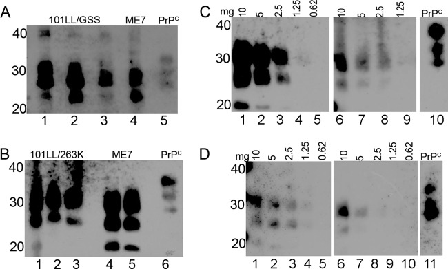
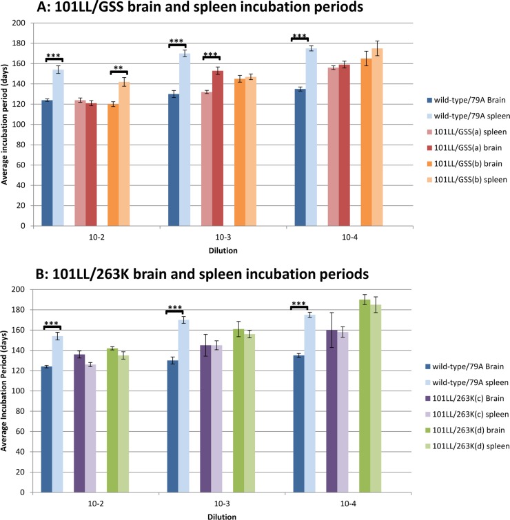
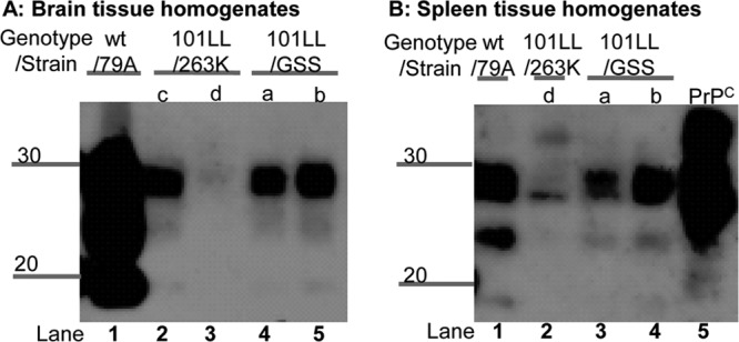
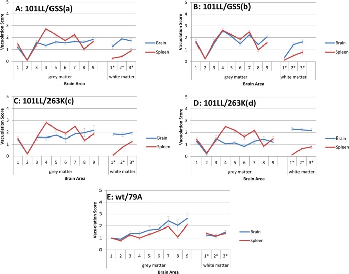
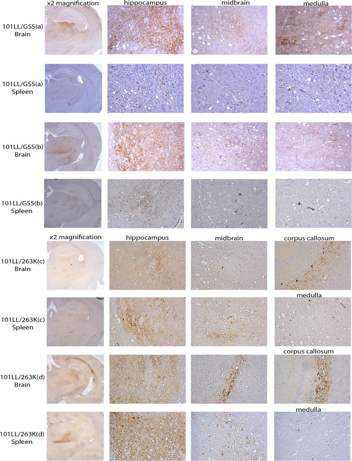
Most current diagnostic tests for transmissible spongiform encephalopathies (TSE) rely on the presence of proteinase K (PK)-resistant PrP(Sc) (PrP-res) in postmortem tissues as an indication of TSE disease. However, a number of studies have highlighted a discrepancy between TSE infectivity and PrP-res levels in both natural and experimental cases of TSE disease. Previously, we have shown high TSE infectivity levels in the brain tissue of mice that have a clinical TSE disease with associated vacuolar pathology but little or no detectable PrP-res. Here, the levels of TSE infectivity and PrP-res within a peripheral tissue of this mouse model were investigated. Biochemical analysis showed that low levels of PrP-res were present in the spleen tissue in comparison to the levels observed in the spleen of mice infected with ME7 or 79A. However, upon subpassage of brain and spleen tissue from clinically ill mice with little or no PrP-res detectable, similar short incubation periods to disease were observed, indicating that infectivity levels were similarly high in both tissues. Thus, the discrepancy between PrP-res and TSE infectivity was also present in the peripheral tissues of this disease model. This result indicates that peripheral tissues can contain higher levels of infectivity given the correct combination of host species, PrP genotype, and TSE agent. Therefore, the assumption that the levels of peripheral infectivity are lower than those in the central nervous system is not always correct, and this could have implications for current food safety regulations.


Figures

Fig 1
Fig 1
PrP-res levels in 101LL/GSS and 101LL/263K spleens compared to PrP-res levels in wild-type/ME7 spleens. Spleen homogenates were PK digested and NaPTA precipitated, and the samples loaded at a tissue equivalent of 15 mg (A and B) or at different tissue weights (mg) as indicated (C and D). PrP-res was present in 101LL/GSS (A) and 101LL/263K (B) spleens at levels that varied between three individual spleens (lanes 1 to 3). ME7 spleen samples were loaded as a control (A, lane 4; B, lanes 4 and 5). The dilution series (C and D) allowed estimation of the difference in PrP-res levels between 101LL/GSS (C, lanes 6 to 9) or 101LL/263K (D, lanes 6 to 10) spleens and wild-type/ME7 spleens (C and D, lanes 1 to 5). Normal brain homogenate was loaded on each blot to provide a PrPC band as an internal control. Blots were probed with MAb 8H4 and exposed for 5 min. Numbers to the left of the panels indicate molecular weight markers (in kilodaltons).
Fig 2
Fig 2
Similar average incubation periods to disease from brain and spleen transmissions from 101LL/GSS and 101LL/263K. In comparison, the average incubation periods for transmissions of brain and spleen tissue from wild-type mice were statistically different, with longer incubation periods produced from inoculation with spleen tissue (blue bars). Statistical calculations with Student's two-tailed t test determined P values of 2.88 × 10⁻⁵, 3.66 × 10⁻⁶, and 1.08 × 10⁻⁸ for the 10−2, 10−3, and 10−4 dilution group, respectively, for wild-type/79A transmission. One dilution of 101LL/GSS(a), 10−3, produced a significant difference (P value, 8.3 × 10⁻⁵), and one dilution of 101LL/GSS(b), 10−2, produced a significant difference (P value, 5.6 × 10⁻⁴).
Fig 3
Fig 3
Low PrP-res levels were present in brain (A) and spleen (B) residual inocula as identified by NaPTA precipitation. In contrast, PrP-res was present in wild-type/79A brain and spleen residual homogenates. Residual homogenates were PK digested and NaPTA precipitated. Samples were loaded at a wet weight tissue equivalent of 10 mg, and proteins separated by SDS-PAGE. Immunoblots were probed with MAb 8H4 and exposed for 10 min. Numbers to the left of the panels indicate molecular weight markers (in kilodaltons).
Fig 4
Fig 4
Similar lesion profiles, with slight variations in vacuolation levels depending on the tissue type inoculated. Lesion profiles were similar for both brain and spleen tissue inocula in 101LL/GSS(a) (A), 101LL/GSS (b) (B), 101LL/263K(c) (C), and 101LL/263K(d) (D), but a higher vacuolation level was observed after inoculation of the spleen tissue than after inoculation of the brain tissue. In comparison, the lesion profiles from inoculation of brain and spleen tissue from wild-type/79A mice (E) followed the same pattern, with inoculation of the brain tissue producing a slightly higher vacuolation level than the spleen tissue. Numbers 1 to 9 represent nine areas of grey matter scored and 1* to 3* represent three areas of white matter scored (see Materials and Methods).
Fig 5
Fig 5
Lighter PrPSc deposition was observed from inoculation of spleen homogenates than from inoculation of brain homogenates from 101LL/GSS. Similar levels of PrPSc deposition from inoculation of 101LL/263K homogenates were observed irrespective of the tissue type. Tissues were taken from mice inoculated with the 10−2 dilution of inocula. Immunohistochemistry performed with MAb 6H4. Hippocampus regions are shown at ×2 magnification in the left-hand column. Brain areas containing PrPSc deposition are shown at ×20 magnification.
Fig 6
Fig 6
PrP-res levels in recipient mouse brains varied depending upon the tissue type inoculated from 101LL/GSS mice. This phenomenon was observed from both 101LL/GSS(a) and 101LL/GSS(b) mice (A). In contrast, 101LL/263K(c) and 101LL/263K(d) had varvarious levels of PrP-res independent of the tissue type inoculated (B), while wild-type/79A recipient brains had the same level of PrP-res independent of the tissue type inoculated (A and B). Tissue homogenates were PK digested and NaPTA precipitated. Lanes 1, 2, 5, 6, 9, and 10, brain tissue; lanes 3, 4, 7, 8, 11, and 12, spleen tissue. Samples were loaded at the equivalent of a wet tissue weight of 33 mg. An uninfected brain homogenate was loaded at 10 mg/ml (wt/vol) wet weight tissue in lane 13 as an internal control. Blots were probed with MAb 8H4. Numbers to the left of the panels indicate molecular weight markers (in kilodaltons).

References

    1. Prusiner SB. 1982. Novel proteinaceous infectious particles cause scrapie. Science 216:136–144 - PubMed
    1. Lasmezas CI, Deslys JP, Robain O, Jaegly A, Beringue V, Peyrin JM, Fournier JG, Hauw JJ, Rossier J, Dormont D. 1997. Transmission of the BSE agent to mice in the absence of detectable abnormal prion protein. Science 275:402–405 - PubMed
    1. Race R, Meade-White K, Raines A, Raymond GJ, Caughey B, Chesebro B. 2002. Subclinical scrapie infection in a resistant species: persistence, replication, and adaptation of infectivity during four passages. J. Infect. Dis. 186(Suppl 2):S166–S170 - PubMed
    1. Andréoletti O, Berthon P, Marc D, Sarradin P, Grosclaude J, van Keulen L, Schelcher F, Elsen JM, Lantier F. 2000. Early accumulation of PrP(Sc) in gut-associated lymphoid and nervous tissues of susceptible sheep from a Romanov flock with natural scrapie. J. Gen. Virol. 81:3115–3126 - PubMed
    1. Mohri S, Farquhar CF, Somerville RA, Jeffrey M, Foster J, Hope J. 1992. Immunodetection of a disease specific PrP fraction in scrapie-affected sheep and BSE-affected cattle. Vet. Rec. 131:537–539 - PubMed

Publication types

LinkOut - more resources