. 2012 Jun 1;5(2):143-154.
doi: 10.1007/s12195-012-0225-z.

Coordinated Mechanosensitivity of Membrane Rafts and Focal Adhesions

Affiliations

Coordinated Mechanosensitivity of Membrane Rafts and Focal Adhesions

Daniela E Fuentes et al. Cell Mol Bioeng. .

Abstract

Endothelial cells sense mechanical forces of blood flow through mechanisms that involve focal adhesions (FAs). The mechanosensitive pathways that originate from FA-associated integrin activation may involve membrane rafts, small cholesterol- and sphigolipid-rich domains that are either immobilized, by virtue of their attachment to the cytoskeleton, or highly mobile in the plane of the plasma membrane. In this study, we fluorescently labeled non-mobile and mobile populations of GM1, a ganglioside associated with lipid rafts, and transfected cells with the red fluorescent protein-(RFP-) talin, an indicator of integrin activation at FAs, in order to determine the kinetics and sequential order of raft and talin mechanosensitivity. Cells were imaged under confocal microscopy during mechanical manipulation of a FA induced by a fibronectin (FN)-functionalized nanoelectrode with feedback control of position. First, FA deformation led to long range deformation of immobile rafts followed by active recoil of a subpopulation of displaced rafts. Second, initial adhesion between the FN-probe and the cell induced rapid accumulation of GM1 at the probe site with a time constant of 1.7 s. Talin accumulated approximately 20 s later with a time constant of 0.6 s. Third, a 1 μm deformation of the FA lead to immediate (0.3 s) increase in GM1 fluorescence and a later (6 s) increase in talin. Fourth, long term deformation of FAs led to continual GM1 accumulation at the probe site that was reversed upon removal of the deformation. These results demonstrate that rafts are directly mechanosensitive and that raft mobility may enable the earliest events related to FA mechanosensing and reinforcement upon force application.

Keywords: Endothelial cells; Focal adhesion; Mechanotransduction; Membrane rafts; Scanning ion conductance microscopy; Talin.


Figures

FIGURE 1
FIGURE 1
The membrane raft and FA connection: membrane rafts, rich in sphingolipids and cholesterol are identified by ganglioside marker GM1. Transmembrane proteins in some membrane rafts render a raft anchored via linkage to the cystoskeleton. In contrast, mobile rafts have no direct or indirect connection to the cytoskeleton. Focal adhesion sites, areas of clustered activated integrins linked to the cysto-skeleton via talin, are known mechanotransducers. The connection between membrane rafts and FAs may involve both the cytoskeleton and the membrane.
FIGURE 2
FIGURE 2
Experimental setup and procedure. (a) Experimental instrumentation enables precise timing of probe-cell contact via a nanoelectrode probe positioning system consisting of a nanoelectrode, piezoelectric stage, and a confocal microscope. (b) Probe-cell contact timing is obtained by noting the current drop of the nanoelectrode as it approaches the cell. (c) Experimental procedure: left: functionalized probe-cell contact and binding; right: subsequent manipulation of the FA by the probes on the apical surface of the cell.
FIGURE 3
FIGURE 3
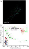
Passive membrane raft response. (a) Rafts from multiple cells were tracked and overlaid to create a composite image of raft trajectories (n = 4 cells), in response to a 1 μm displacement of a FA. The probe (circle) was displaced in the positive x direction which corresponds to a FA translocation away from the nucleus approximately perpendicular to the main axis of the cell. The probe was initially located approximately halfway between the nucleus and the outer edge of the cell. Individual trajectories initiated at the point furthest from the probe location (circle) and terminate closer to the probe. (b) Distance to probe vs. displacement magnitude of individual rafts. Green group (hexagons) corresponds to rafts that are located on the same side as the probe (relative to the nucleus); the red group (pentagons) corresponds to rafts located behind the nucleus (relative to the probe); control rafts are in blue (diamonds).
FIGURE 4
FIGURE 4
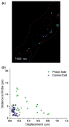
Active response: Recoil. (a) After deformation the probe was stationary and membrane rafts moved in the direction opposite to deformation, indicative of active recoil. Individual trajectories initiated closer to the probe location and terminate with subsequent continued remodeling as indicated by the overlapped tracked displacements resulting in a clustered trajectory endpoint. (b) Distance to probe vs. displacement of individual active rafts is plotted with the green group (pentagons) corresponding to rafts that are located on the same side as the probe (relative to the nucleus), and control rafts in blue (diamonds).
FIGURE 5
FIGURE 5
Directional dependence of passive and active membrane raft response. (a) Passive response aligned with the direction of FA displacement (aligned with 0°). (b) Active response was in the opposite direction of displacement ranging from angles between 90° and 270°, and covering a range of displacements up to 0.8 μm. (c) Control rafts do not exhibit a particular directionality as their displacements cover the full 360° range, and their magnitudes of displacement are less than 0.2 μm.
FIGURE 6
FIGURE 6
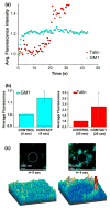
Kinetic response of membrane rafts and talin upon contact. (a) GM1 fluorescence accumulated with a time constant of 1.68 s, reached a plateau at 10 s. This increase was followed by accumulation of talin at 20 s that reached a plateau at 30 s. (b) On average, GM1 accumulation increased 14 ± 4.6% whereas talin increased by 2.26 ± 1.14% (n = 3). (c) (Top) GM1 accumulation around the probe (circle) is represented by 3-D intensity maps (Bottom).
FIGURE 7
FIGURE 7
Kinetic response of membrane rafts and talin upon deformation. (a) Upon FA-deformation, GM1 accumulated with a characteristic time constant of 0.3 s and reached a plateau at 3 s followed by accumulation of talin at 7 s reaching a plateau at 10 s. (b) GM1 accumulation increased 15 ± 3.8% whereas talin increased by 11 ± 3.1% (n = 3).
FIGURE 8
FIGURE 8
Deformation induces long term accumulation of membrane rafts. (a) Selected frames of time-lapsed images of GM1 display six time points starting at t = 0 with initial and final locations of the probe (circles). GM1 accumulation continued to increase first around the probe site (t = 163, 357, and 1419 s) and continued to grow radially outward (away from the probe) and inward (inside the probe). (b) Long term kinetic response of GM1 accumulation exhibited a continuous increase; insets represent 3-D displaying of intensity and radial geometry of accumulation. (c) An average increase of 7.9 ± 1.6% (n = 3) in intensity from the initial value was observed at time points ranging from 10 to 15 min after deformation.
FIGURE 9
FIGURE 9
GM1 fluorescence decreases upon reversal of deformation. (a) Reversal of GM1 accumulation was continuous and leveled off after 10 min. The radial decrease is depicted by the insets showing 3-D representation of GM1 fluorescence intensity around the probe site. (b) Average decrease in accumulation upon reversal was 41.4 ± 7.4% (n = 3).

References

    1. Anthis NJ, Wegener KL, Ye F, Kim C, Goult BT, Lowe ED, Vakonakis I, Bate N, Critchley DR, Ginsberg MH, Campbell ID. The structure of an integrin/ talin complex reveals the basis of inside-out signal trans-duction. EMBO J. 2009;28:3623–3632. - PMC - PubMed
    1. Askari JA, Buckley PA, Mould AP, Humphries MJ. Linking integrin conformation to function. J Cell Sci. 2009;122:165–170. - PMC - PubMed
    1. Bini L, Pacini S, Liberatori S, Valensin S, Pellegrini M, Raggiaschi R, Pallini V, Baldari CT. Extensive temporally regulated reorganization of the lipid raft proteome following T-cell antigen receptor triggering. Biochem J. 2003;369:301–309. - PMC - PubMed
    1. Bouaouina M, Lad Y, Calderwood DA. The N-terminal domains of talin cooperate with the phosphotyrosine binding-like domain to activate beta1 and beta3 integrins. J Biol Chem. 2008;283:6118–6125. - PubMed
    1. Brown DA, London E. Structure and function of sphingolipid- and cholesterol-rich membrane rafts. J Biol Chem. 2000;275:17221–17224. - PubMed