. 2013 Mar 19;110(12):4610-5.
doi: 10.1073/pnas.1214887110. Epub 2013 Mar 4.

Crucial role of the hydrophobic pocket region of Munc18 protein in mast cell degranulation

Affiliations

Crucial role of the hydrophobic pocket region of Munc18 protein in mast cell degranulation

Na-Ryum Bin et al. Proc Natl Acad Sci U S A. .

Abstract

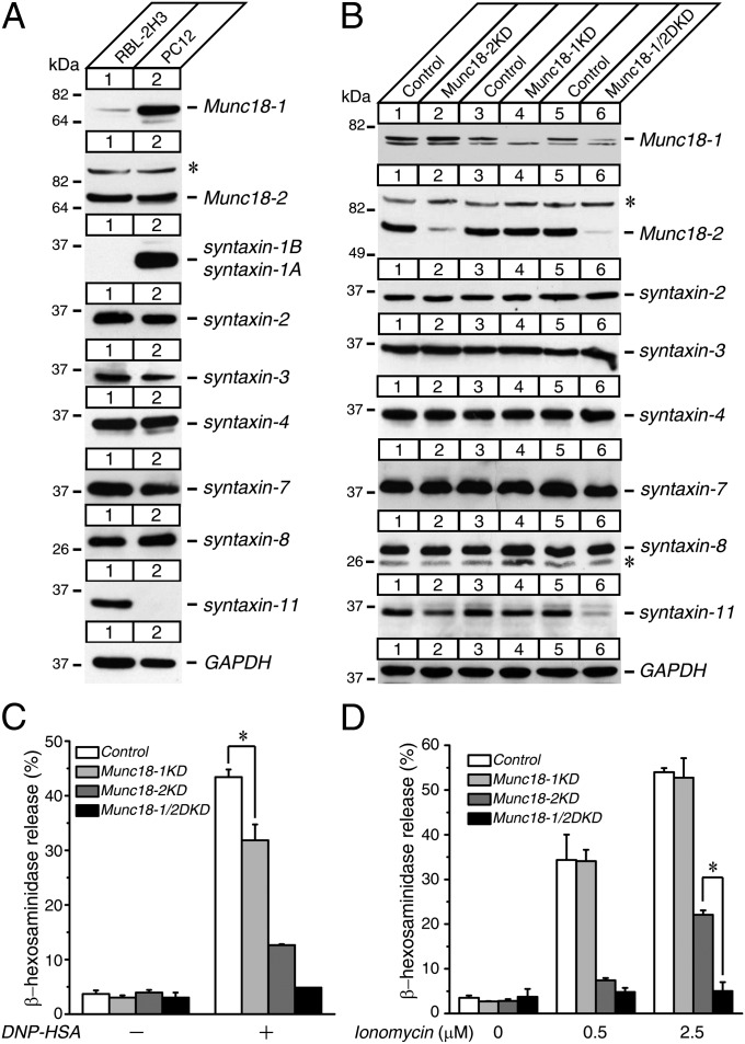
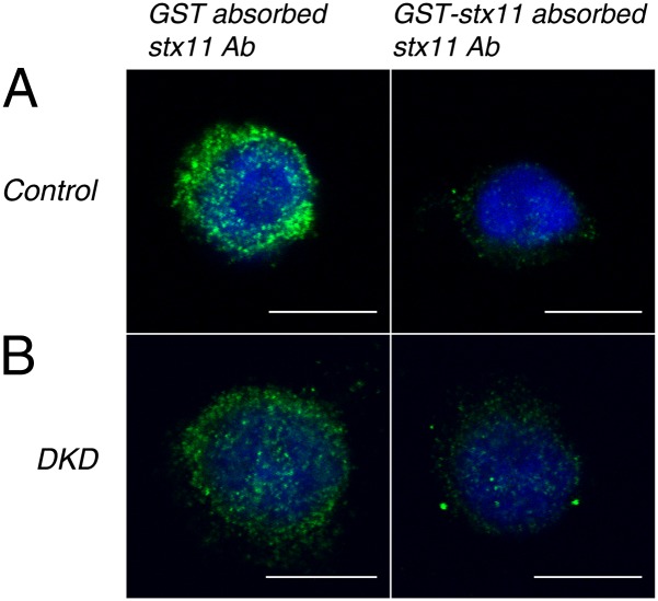
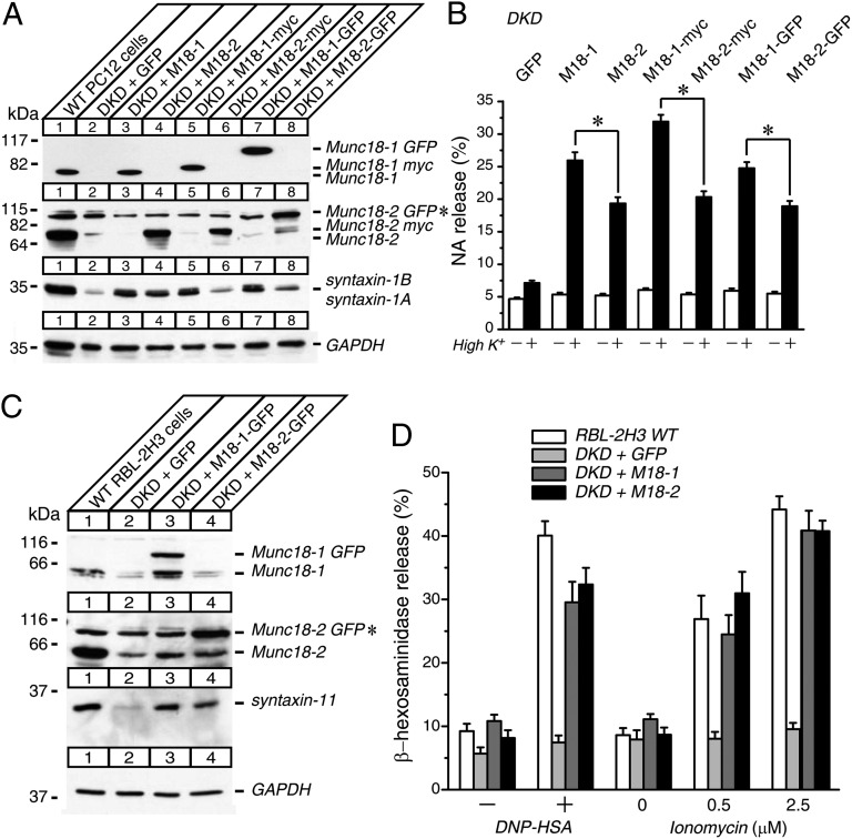
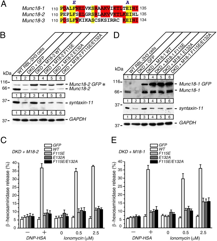
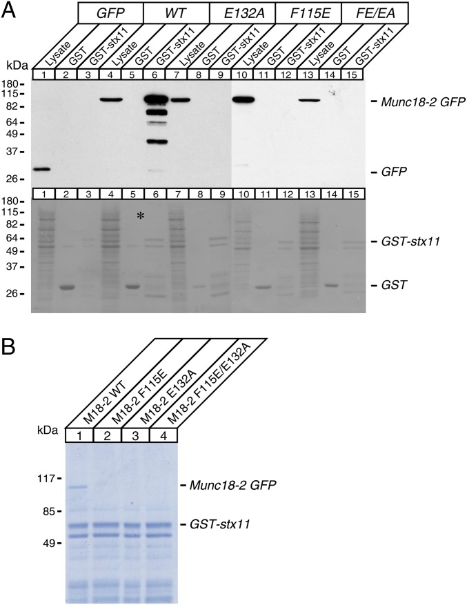
The function of the Munc18-1 protein hydrophobic pocket, which interacts with the syntaxin-1 N-terminal peptide, has been highly controversial in neurosecretion. Recent analysis of patients with familial hemophagocytic lymphohistiocytosis type 5 has identified the E132A mutation in the hydrophobic pocket of Munc18-2, prompting us to examine the role of this region in the context of immune cell secretion. Double knockdown of Munc18-1 and Munc18-2 in RBL-2H3 mast cells eliminates both IgE-dependent and ionomycin-induced degranulation and causes a significant reduction in syntaxin-11 without altering expressions of the other syntaxin isoforms examined. These phenotypes were effectively rescued on reexpression of wild-type Munc18-1 or Munc18-2 but not the mutants (F115E, E132A, and F115E/E132A) in the hydrophobic pocket of Munc18. In addition, these mutants show that they are unable to directly interact with syntaxin-11, as tested through protein interaction experiments. Our results demonstrate the crucial roles of the hydrophobic pocket of Munc18 in mast cell degranulation, which include the regulation of syntaxin-11. We also suggest that the functional importance of this region is significantly different between neuronal and immune cell exocytosis.


Conflict of interest statement

The authors declare no conflict of interest.

Figures

Fig. 1.
Fig. 1.
KD of both Munc18-1 and Munc18-2 results in dramatic reductions of syntaxin-11 expressions and striking degranulation defects in RBL-2H3 cells. (A) Expression profiles of Munc18-1 and Munc18-2, as well as various syntaxin proteins present in wild-type RBL-2H3 and PC12 cells. Twenty micrograms of homogenates from wild-type RBL-2H3 and PC12 cells were analyzed by SDS/PAGE and immunoblotting, using the antibodies indicated. (B) Stable Munc18-1 KD, Munc18-2 KD, and Munc18-1/2 DKD RBL-2H3 cells were generated by lentivirus-mediated shRNA. Twenty micrograms of cell homogenates were analyzed by SDS/PAGE and immunoblotting, using the antibodies indicated. (C and D) β-hexosaminidase release was stimulated by applying 0.01 μg/mL DNP-IgE and 50 ng/mL dinitrophenyl-human serum albumin (DNP-HSA) (C) and 0.5 μM and 2.5 μM ionomycin (D) for 1 h from control, Munc18-1 KD, Munc18-2 KD, and Munc18-1/2 DKD cells. Error bars, SEM (n = 5). The statistical significance of the differences in β-hexosaminidase release between control and Munc18-1 KD (C), Munc18-2 KD, and Munc18-1/2 DKD (D) is indicated. *P < 0.05 (Student t test). *Nonspecific band observed with rabbit polyclonal anti-Munc18-2 and anti-syntaxin-8 antibodies.
Fig. 2.
Fig. 2.
Confocal immunofluorescence microscopy revealing that the subcellular localization of syntaxin-11 is unaltered on Munc18-1/2 DKD. Control and stable Munc18-1/2 DKD RBL-2H3 cells were permeabilized and stained with either GST or GST-syntaxin-11–absorbed rabbit polyclonal anti-syntaxin-11 antibody followed by Alexa488-conjugated goat anti-rabbit antibody (Invitrogen) and DAPI (see SI Materials and Methods for more detail). Green indicates syntaxin-11 and blue indicates DAPI. (A) Control and (B) Munc18-1/2 DKD cells. Note that there is substantially diminished green intensity (Right) where GST-syntaxin-11 absorbed the anti-syntaxin-11 antibody was used to stain. (Scale bar, 10 μm.)
Fig. 3.
Fig. 3.
Stable reexpression of wild-type Munc18-1 or Munc18-2 restores the syntaxin-11 expressions and the degranulation defects of Munc18-1/2 DKD RBL-2H3 cells while functional specificities exist in PC12 cells. (A) Twenty micrograms of homogenates of Munc18-1/2 DKD PC12 cells rescued with EmGFP, wild-type Munc18-1, and Munc18-2 (without tag, with myc, and with EmGFP) were analyzed by SDS/PAGE and immunoblotting, using the antibodies indicated. (B) NA release was stimulated by 70 mM KCl for 15 min in the rescued cells. Error bars, SEM (n = 9). The statistical significance of the differences in NA release between wild-type Munc18-1 and Munc18-2 rescued PC12 cells in each tag configuration are indicated. *P < 0.05 (paired t test). (C) Munc18-1/2 DKD RBL-2H3 cells were infected with lentiviruses that express EmGFP, wild-type Munc18-1-EmGFP, and wild-type Munc18-2-EmGFP. Twenty micrograms of cell homogenates were analyzed by SDS/PAGE and immunoblotting, using the antibodies indicated. (D) β-Hexosaminidase release was stimulated by applying 0.01 μg/mL DNP-IgE and 50 ng/mL DNP-HSA, as well as 0.5 μM and 2.5 μM ionomycin for 1 h from wild-type RBL-2H3 cells and rescued cells. Error bars, SEM (n = 9). *Nonspecific band observed with rabbit polyclonal anti-Munc18-2 antibody that comigrated with Munc18-2-EmGFP.
Fig. 4.
Fig. 4.
Hydrophobic pocket mutants of either Munc18-1 or Munc18-2 do not restore the syntaxin-11 expressions or the degranulation defects of Munc18-1/2 DKD RBL-2H3 cells. (A) Sequence alignment among Munc18-1, Munc18-2, and Munc18-3, indicating that F115 and E132 are conserved residues in Munc18 isoforms. (B and D) Munc18-1/2 DKD RBL-2H3 cells were infected with lentiviruses that express EmGFP, wild-type Munc18-EmGFP, and hydrophobic pocket mutant Munc18-EmGFP (F115E, E132A, and F115E/E132A) (B) Munc18-2 and (D) Munc18-1. Infected cells were selected with blasticidin (20 μg/mL). Twenty micrograms of homogenates of surviving cells were analyzed by SDS/PAGE and immunoblotting, using the antibodies indicated. *Nonspecific band observed with rabbit polyclonal anti-Munc18-2 antibody that comigrated with Munc18-2-EmGFP. (C and E) β-hexosaminidase release was stimulated by applying 0.01 μg/mL DNP-IgE and 50 ng/mL DNP-HSA, as well as 0.5 μM and 2.5 μM ionomycin for 1 h from rescued cells (C) Munc18-2 and (E) Munc18-1. Error bars, SEM (n = 5 for C, n = 6 for E).
Fig. 5.
Fig. 5.
Hydrophobic pocket mutants of Munc18-2 abolish the direct interaction with syntaxin-11. (A, Upper) Solubilized lysates of HEK-293FT cells that were transfected with EmGFP, wild-type Munc18-2-EmGFP, F115E, E132A, and F115E/E132A of Munc18-2-EmGFP were incubated with 10 μg GST or GST-syntaxin-11 attached to glutathione agarose beads. The mixtures were washed extensively, and the proteins were analyzed on SDS/PAGE and immunoblotting, using mouse monoclonal anti-EmGFP antibody. Solubilized lysates of HEK-293FT cells were loaded in lysate lanes. (A, Lower) Ponceau S staining of the above membrane showing similar loadings between lanes. *Wild-type Munc18-2-EmGFP band that was pulled down by GST-syntaxin-11. (B) The binding mixture between solubilized lysates of HEK-293FT cells that were transfected with wild-type Munc18-2-EmGFP, F115E, E132A, F115E/E132A, and 10 μg of GST-syntaxin-11 was analyzed on SDS/PAGE, followed by Coomassie Blue staining. Note that only wild-type Munc18-2-EmGFP was pulled down by GST-syntaxin-11, not any of the mutants.

References

    1. Söllner T, et al. SNAP receptors implicated in vesicle targeting and fusion. Nature. 1993;362(6418):318–324. - PubMed
    1. Hata Y, Slaughter CA, Südhof TC. Synaptic vesicle fusion complex contains unc-18 homologue bound to syntaxin. Nature. 1993;366(6453):347–351. - PubMed
    1. Verhage M, et al. Synaptic assembly of the brain in the absence of neurotransmitter secretion. Science. 2000;287(5454):864–869. - PubMed
    1. Augustin I, Rosenmund C, Südhof TC, Brose N. Munc13-1 is essential for fusion competence of glutamatergic synaptic vesicles. Nature. 1999;400(6743):457–461. - PubMed
    1. Jahn R, Fasshauer D. Molecular machines governing exocytosis of synaptic vesicles. Nature. 2012;490(7419):201–207. - PMC - PubMed

Publication types

MeSH terms

Supplementary concepts

LinkOut - more resources