. 2013 Mar 19;110(12):E1152-61.
doi: 10.1073/pnas.1218991110. Epub 2013 Mar 4.

Retrograde monosynaptic tracing reveals the temporal evolution of inputs onto new neurons in the adult dentate gyrus and olfactory bulb

Affiliations

Retrograde monosynaptic tracing reveals the temporal evolution of inputs onto new neurons in the adult dentate gyrus and olfactory bulb

Aditi Deshpande et al. Proc Natl Acad Sci U S A. .

Abstract

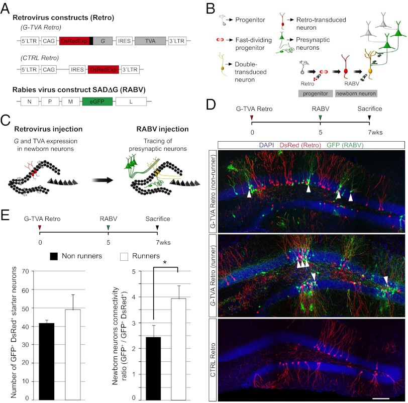
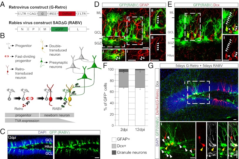
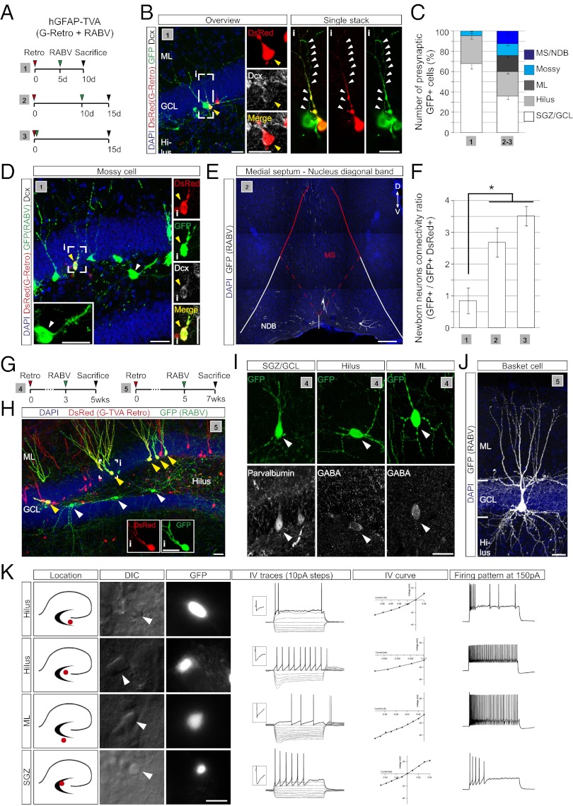
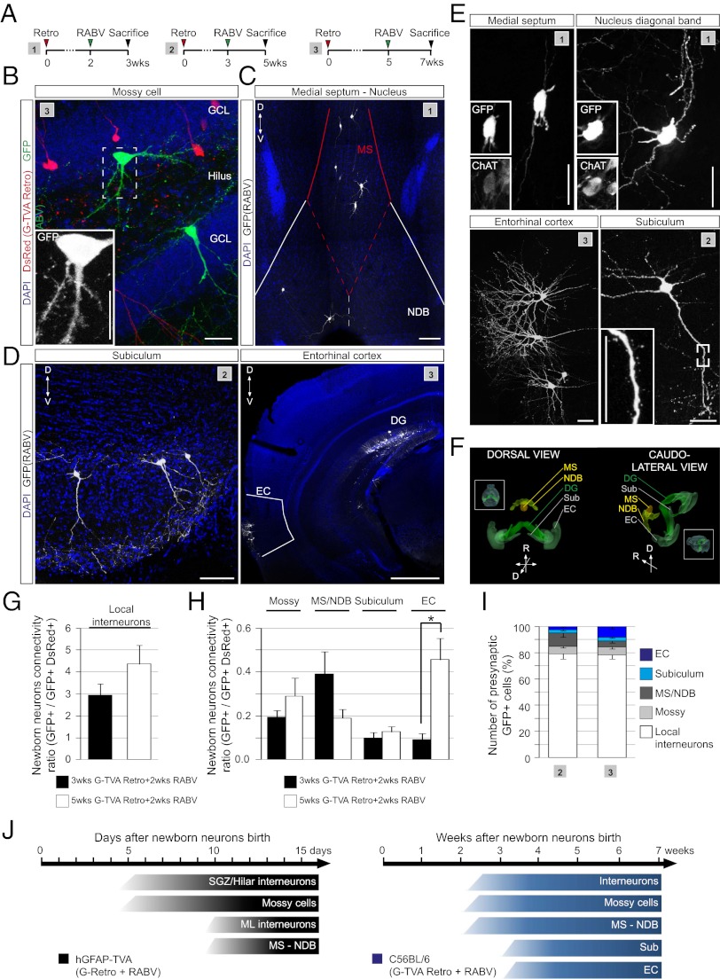
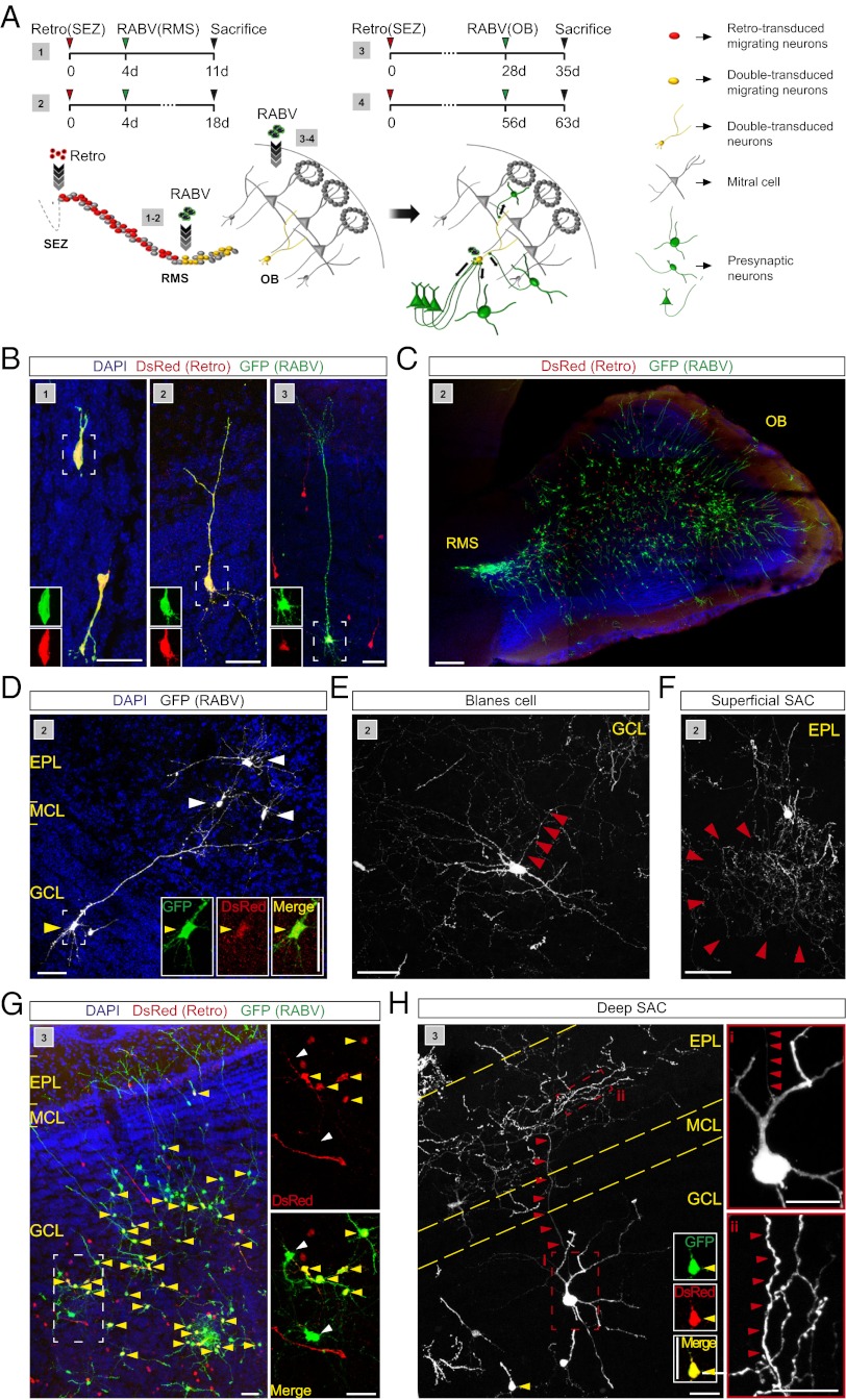
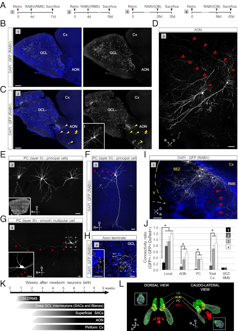
Identifying the connectome of adult-generated neurons is essential for understanding how the preexisting circuitry is refined by neurogenesis. Changes in the pattern of connectivity are likely to control the differentiation process of newly generated neurons and exert an important influence on their unique capacity to contribute to information processing. Using a monosynaptic rabies virus-based tracing technique, we studied the evolving presynaptic connectivity of adult-generated neurons in the dentate gyrus (DG) of the hippocampus and olfactory bulb (OB) during the first weeks of their life. In both neurogenic zones, adult-generated neurons first receive local connections from multiple types of GABAergic interneurons before long-range projections become established, such as those originating from cortical areas. Interestingly, despite fundamental similarities in the overall pattern of evolution of presynaptic connectivity, there were notable differences with regard to the development of cortical projections: although DG granule neuron input originating from the entorhinal cortex could be traced starting only from 3 to 5 wk on, newly generated neurons in the OB received input from the anterior olfactory nucleus and piriform cortex already by the second week. This early glutamatergic input onto newly generated interneurons in the OB was matched in time by the equally early innervations of DG granule neurons by glutamatergic mossy cells. The development of connectivity revealed by our study may suggest common principles for incorporating newly generated neurons into a preexisting circuit.


Conflict of interest statement

The authors declare no conflict of interest.

Figures

Fig. 1.
Fig. 1.
RABV-mediated tracing of synapses onto adult-generated neurons in the DG. (A) Retroviral and RABV constructs. (B) Scheme of sequential virus delivery. (C) Implementation of the method in the DG of the hippocampus. Proliferating neural progenitors in the adult SGZ are transduced with the retrovirus, thus rendering them infectable by the RABV and their neuronal progeny capable of transsynaptic RABV transfer, followed by a second injection of RABV. Once synaptogenesis has taken place, RABV spreads from the primary infected neurons to their presynaptic partners. (D) Injection scheme used in the DG. Representative examples demonstrating the specificity of the system. Reconstructed confocal images from multiple fields depict cells labeled after injecting G and TVA encoding retrovirus in runner and nonrunner mice or control DsRed-only retrovirus, followed by RABV injection. White arrowheads: double transduced newborn neurons. (Scale bar, 50 μm.) (E) Absolute number of double-transduced starter neurons per mouse and ratio of RABV-traced local presynaptic neurons versus double-transduced neurons in running mice compared with nonrunners (n = at least three mice per condition; *P < 0.05).
Fig. 2.
Fig. 2.
Implementation of the RABV-mediated synaptic tracing in hGFAP-TVA mice. (A) Retroviral and RABV constructs. (B) Adaptation of the method to the hGFAP-TVA mouse line. Temporal profile of TVA expression is indicated by the lower bar. (C) Distribution of the cells targeted by EnvA-pseudotyped RABV in the DG of hGFAP-TVA mice 12 d postinjection (dpi). (Scale bar, 50 μm.) (D) Representative example of RABV-targeted cells in hGFAP-TVA mice comprising of GFAP+ horizontal glia (i) and radial glia (ii). Enlargements show single and merged channels of the boxed areas, with arrowheads pointing to the colocalizing immunoreactive signal. (Scale bars, 20 μm.) (E) Example depicting Dcx+ newborn neurons targeted by RABV. Enlargement of the boxed area (i) shows the colocalization between GFP and Dcx (arrowheads). (Scale bars, 20 μm.) (F) Quantification of the identity of RABV-targeted cells in hGFAP-TVA mice at 2 and 12 dpi (n = 3 mice). (G) Example of RABV-traced presynaptic neurons at 10 d following injection of G-encoding retrovirus. Enlargements of the boxed area (i) show presynaptic neurons (white arrowheads) surrounding a double-transduced newborn neuron (yellow arrowhead). (Scale bars, 20 μm.)
Fig. 3.
Fig. 3.
Temporal development of the presynaptic connectome of newborn DG granule neurons. (A) Injection schemes (1–3) used in hGFAP-TVA mice. (B) Example of a presynaptic GFP-only positive neuron contacting a double-transduced newborn neuron (yellow arrowhead, enlarged in Insets). An individual stack of the boxed area (i) showing single and merged channels is depicted on the right. Arrowheads point to the axon of the presynaptic neuron. (Scale bars, 20 μm.) (C) Quantification of RABV-traced cells obtained following injection schemes (1–3) based on their morphology and location (n = 3–5). (D) Example of a RABV-traced mossy cell (white arrowhead, enlarged in the inset) located in the hilus of hGFAP-TVA mice following injection scheme 1. Enlargements of single and merged channels of a nearby double-transduced newborn neuron (yellow arrowhead, i) are shown on the right. (Scale bars, 30 μm.) (E) Example of RABV-traced neurons in the MS and NDB. (Scale bar, 70 μm.) (F) Ratio of RABV-traced neurons versus double-transduced newborn neurons following injection paradigms 1–3. (n = 3–5 mice per experimental condition; *P < 0.05). (G) Injection schemes (4 and 5) used in C57BL/6 mice. (H) Example of presynaptic tracing, 7 wk after retrovirus injection; double-transduced adult-generated granule neurons (yellow arrowheads) and putative presynaptic neurons (white arrowheads) are indicated. (Insets) Single channel images of the boxed cell. (Scale bars, 30 µM.) (I) Phenotypic characterization of RABV-traced local interneurons in the DG. (J) Example of a reconstructed RABV-traced basket cell profusely innervating the GCL. (Scale bars, 20 μm) (K) Visual and electrophysiological identification of presynaptic neurons in the DG and subiculum. GFP-only positive neurons (RABV-traced) were classified based on the location of their cell body (see micrographs) and the voltage responses following current injections. Several examples of presynaptic neurons differing in their IV traces and firing pattern are shown. (Scale bar, 20 μm.)
Fig. 4.
Fig. 4.
RABV-mediated tracing of long-range connectivity. (A) Injection schemes (1–3) used in the adult hippocampus of C56BL6 mice. (B) Overview depicting the anatomical location of RABV-traced mossy cell (Inset shows enlarged image). (Scale bar, 20 μm.) (C) RABV-traced neurons in the MS and NDB. (Scale bar, 50 μm.) (D) Examples of RABV-traced neurons in the subiculum and the EC. [Scale bars, 50 μm (Subiculum) and 1 mm (EC).] (E) High magnification view of RABV-traced neurons in MS/NDB, EC, and subiculum. (Insets) Colocalization of choline acetyltransferase (ChAT) with GFP in MS/NDB and the presence of spines on neurons in the subiculum. (Scale bars, 20 μm.) (F) Three-dimensional reconstruction of the anatomical locations of RABV-traced long-distance projection neurons; (Insets) An entire brain view of the reconstructed anatomical regions. (G) Ratio of RABV-traced local interneurons versus double-transduced neurons following injection paradigms 2–3. (n = 4–6 mice per experimental condition). (H) Ratio of different types of presynaptic neurons versus double-transduced neurons (n = 4–6 mice per experimental condition; *P < 0.05). (I) Quantification of the identity of RABV-traced neurons obtained following injection paradigms 2–3 (n = 4–6 mice). (J) Summary of the identity and location of RABV-labeled presynaptic neurons appearing during the course of maturation of adult-born DG neurons in hGFAP-TVA and C57BL/6 mice.
Fig. 5.
Fig. 5.
RABV-mediated tracing of local presynaptic partners of adult-generated neurons in the OB. (A) Scheme of sequential virus delivery in two different injection paradigms (1 and 2). Injection of retrovirus into the SEZ, followed by RABV infection of migrating neuroblasts in the RMS (3 and 4). Injection of retrovirus into the SEZ, followed by RABV infection in the OB. (B) Double-transduced newborn granule cells at different stages of maturation obtained following injection schemes 1, 2, and 3. (Scale bar, 30 μm.) (C) Overview of RABV-labeled neurons in the OB following injection scheme 2. (Scale bar, 200 μm.) (D) Example of a double-transduced granule cell and RABV-traced superficial short axon cells (SACs) following RABV injection in the RMS. (Insets) Enlarged images of single and merged channels. (Scale bars, 50 μm.) (E) Example of a RABV-traced Blanes cell in the GCL. Arrowheads point to the emerging axon. (Scale bar, 50 μm.) (F) Example of a RABV-traced superficial SAC in the EPL. (Scale bar, 50 μm.) (G) Overview of double-transduced (yellow arrowheads) and RABV-traced local neurons following RABV injection into the OB. A 3× digital zoom of the boxed area shows double-transduced and RABV-only transduced neurons (white arrowheads). (Scale bars, 50 μm.) (H) RABV-traced deep SAC following RABV injection in the OB. Note the double-transduced cell (yellow arrowhead; Insets show single and merged channels); red arrowheads point to the ascending axon of the SAC. Boxed area (i) shows the cell body, with the emerging axon indicated by the arrowheads; area (ii) shows part of the axonal arborisation in the EPL. (Scale bars, 20 μm.)
Fig. 6.
Fig. 6.
RABV-mediated tracing of long distance projections of adult-generated neurons in the OB. (A) Injection schemes (1–4) used in adult C57BL/6 mice. (B and C) Overview of RABV-labeled neurons in the OB following injection scheme 1 (B) and 2 (C). Inset in C shows an enlargement of the boxed area (i) in the AON. (Scale bar, 200 μm.) (D) Examples of RABV-traced neurons in the AON. Red arrowheads point to the emerging axons. (Scale bar, 30 μm.) (E) RABV-traced long-distance projecting neurons located in layer II of the piriform cortex (PC) revealed following injection scheme 2; Inset shows the anatomical position of the neurons. (Scale bar, 20 μm.) (F) RABV-traced deep pyramidal neurons located in layer III of the PC. (Scale bar, 20 μm.) (G) RABV-traced smooth multipolar cell located in layer III of the PC. (Inset) Enlarged image of the boxed area. Red arrowheads point to the axon directed toward the OB. (Scale bars, 20 μm.) (H) Axonal terminals of long-range projection neurons. Inset shows the numerous axonal boutons in the GCL. (Scale bars, 50 μm.) (I) RABV-traced neurons in the vicinity of the SEZ obtained following injection paradigms 1 and 2. Red arrowheads point to the axons of GFP-labeled neurons directed toward the RMS. (Scale bar, 50 μm.) (J) Ratio of presynaptic neurons in different locations versus double-transduced neurons in the OB. (n = 3–4 mice per experimental condition; *P < 0.05). (K) Summary of the identity and location of RABV-labeled presynaptic neurons appearing during the course of maturation of adult-born granule cells in the OB. (L) Three-dimensional reconstruction of the anatomical locations of RABV-traced neurons.

References

    1. Nakashiba T, et al. Young dentate granule cells mediate pattern separation, whereas old granule cells facilitate pattern completion. Cell. 2012;149(1):188–201. - PMC - PubMed
    1. Aimone JB, Deng W, Gage FH. Resolving new memories: A critical look at the dentate gyrus, adult neurogenesis, and pattern separation. Neuron. 2011;70(4):589–596. - PMC - PubMed
    1. Alonso M, et al. Activation of adult-born neurons facilitates learning and memory. Nat Neurosci. 2012;15(6):897–904. - PubMed
    1. Marín-Burgin A, Mongiat LA, Pardi MB, Schinder AF. Unique processing during a period of high excitation/inhibition balance in adult-born neurons. Science. 2012;335(6073):1238–1242. - PMC - PubMed
    1. Bardy C, Alonso M, Bouthour W, Lledo PM. How, when, and where new inhibitory neurons release neurotransmitters in the adult olfactory bulb. J Neurosci. 2010;30(50):17023–17034. - PMC - PubMed

Publication types

LinkOut - more resources