. 2013 Mar 19;110(12):4685-90.
doi: 10.1073/pnas.1219260110. Epub 2013 Mar 4.

Dimerization of complement factor H-related proteins modulates complement activation in vivo

Affiliations

Dimerization of complement factor H-related proteins modulates complement activation in vivo

Elena Goicoechea de Jorge et al. Proc Natl Acad Sci U S A. .

Abstract

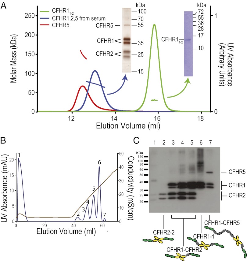
The complement system is a key component regulation influences susceptibility to age-related macular degeneration, meningitis, and kidney disease. Variation includes genomic rearrangements within the complement factor H-related (CFHR) locus. Elucidating the mechanism underlying these associations has been hindered by the lack of understanding of the biological role of CFHR proteins. Here we present unique structural data demonstrating that three of the CFHR proteins contain a shared dimerization motif and that this hitherto unrecognized structural property enables formation of both homodimers and heterodimers. Dimerization confers avidity for tissue-bound complement fragments and enables these proteins to efficiently compete with the physiological complement inhibitor, complement factor H (CFH), for ligand binding. Our data demonstrate that these CFHR proteins function as competitive antagonists of CFH to modulate complement activation in vivo and explain why variation in the CFHRs predisposes to disease.


Conflict of interest statement

The authors declare no conflict of interest.

Figures

Fig. 1.
Fig. 1.
CFHR1, CFHR2, and CFHR5 contain an identical unique dimerization motif. (A) Alignment of the SCR domains of CFHR1, CFHR2, and CFHR5 with CFH. These proteins are comprised of subunits termed SCR domains and domains have been aligned according to the CFH domain with which they share the highest amino acid similarity, percentage identity indicated. Red boxing denotes domains for which unique X-ray structures are presented in this article. The complement regulatory domains of CFH reside within the first four amino-terminal domains (cyan). None of the CFHR proteins contain domains similar to these. CFH surface-recognition domains, which contain C3b/C3d and glycosaminoglycan binding sites reside within the carboxyl-terminal two domains (CFH19–20) and all three CFHR proteins contain highly similar domains. Mapping of the conserved residues onto the existing structure of CFH19–20 suggests that glycosaminoglycan but not C3b/C3d binding is altered or lost within CFHR23–4 (Figs. S1 and S2). The first two amino-terminal domains of CFHR1, CFHR2, and CFHR5 are highly conserved and have previously been described as CFH67-like, although the level of identity is less than 40%. (B) X-ray crystal structure of CFHR11–2. The two copies of CFHR112 that form the head-to-tail dimer are shown as gray cartoons with a semitransparent surface. Residues Tyr34, Ser36 and Tyr39 that are critical in stabilizing the dimer are shown in a ball-and-stick representation (Figure drawn using program PyMol, www.pymol.org). (C) Sequence alignment of CFHR11–2, CFHR21–2, and CFHR51–2 with CFH6–7. The dimerization interface is conserved between these CFHR proteins but not in CFH [interface residues determined using PISA; residues Tyr34, Ser36, and Tyr39 indicated by an asterisk (*); other interface residues by a dot (•)]. Red boxed residues, nonconservative; green boxed residues, conservative variation; and yellow boxed residues, residues unique to CFH67. (D) Mapping sequence variation onto the molecular surface of one copy of CFHR112. This analysis confirmed that the dimerization interface is conserved among CFHR112, CFHR212, and CFHR512 but not in CFH67 (positions of Tyr34, Ser36, and Tyr-39 indicated with an asterisk).
Fig. 2.
Fig. 2.
CFHR1, CFHR2, and CFHR5 are dimeric in serum. (A) MALS of a serum fraction containing CFHR1, CFHR2, and CFHR5, recombinant CFHR5 and recombinant CFHR11–2. MALS analysis of the serum fraction (green trace and mass profile) demonstrates that this mixture contains a mass range between 65 and 80 kDa. MALS using recombinant CFHR5 and CFHR11–2 (red and blue traces and mass profiles, respectively) demonstrates that both form dimers in solution. (B) Purification of homo- and heterodimers formed between CFHR1, CFHR2, and CFHR5. CFHR1, CFHR2, and CFHR5 were copurified from serum and the different species separated according to their affinity for heparin, eluting with a salt gradient. (C) Western blot analysis of each peak from the heparin elution in B using anti-CFHR1/2/5. Because our analyses above demonstrate that CFHR1/2/5 circulate in dimeric forms, we interpret peaks containing more than one CFHR coeluting as containing heterodimeric species.
Fig. 3.
Fig. 3.
Dimerization enhances the interaction of CFHR5 with complement C3 in vivo. (A) Generation of a CFHR5 protein lacking critical amino acids within the dimerization motif. Monomeric CFHR5 (CFHR5dimer mutant) was generated by mutating the three stabilizing amino acids (Tyr34Ser, Ser36Tyr, Tyr39Glu) within the dimerization motif to the corresponding amino acids within CFH. (B) Analysis of recombinant CFHR5 and CFHR5dimer mutant using SDS/PAGE. Both the wild-type and dimer mutants were purified to single homogenous species as visualized by denaturing electrophoresis. (C) Analysis of recombinant CFHR5 and CFHR5dimer mutant using size-exclusion chromatography. Size-exclusion chromatography was performed on a Superdex200 10/30 column (GE Healthcare) equilibrated in 50 mM Tris•HCl, pH 7.5, 150 mM NaCl at 0.4 mL/min. The column was followed in-line by an Optilab-Rex refractive index monitor (Wyatt Technologies). The CFHR5 dimer elutes early from the column (blue trace) but the monomeric CFHR5dimer mutant protein elutes at a larger column volume (red trace). (D) Interaction of CFHR5 and CFHR5dimer mutant with renal-bound mouse C3 in vivo. When recombinant CFHR5dimer mutant was injected at identical concentration to that of CFHR5, CFHR5dimer mutant binding to glomerular C3 was significantly reduced compared with that of wild-type CFHR5. Original magnification, ×40.
Fig. 4.
Fig. 4.
CFHR1 and CHFR5 deregulate complement activation by competitively inhibiting of the interaxtion of CFH with C3b. (A) CFH binding to C3b is inhibited by either recombinant CFHR5 or serum-derived CFHR1. ELISA wells were coated with C3b and 0.07 μM CFH was incubated with increasing amounts of either CFHR1 (0.014–1.8 μM) or CFHR5 (0.005–0.6 μM). Both proteins reduced the CFH–C3b interaction in a dose-dependent manner. Similar results were obtained when recombinant CFHR1345 (0.14–18 μM) and CFHR234 (0.13–16 μM) were used. (B) CFH-dependent alternative pathway hemolytic assay. Using a CFH dose that reduced lysis of guinea pig erythrocytes to 50%, the addition of increasing concentrations of CFHR135, CFHR234, serum-derived CFHR1, and recombinant CFHR5 resulted in a dose-dependent increase in lysis. Full-length, dimeric, CFHR1, and CFHR5 were orders-of-magnitude more potent with respect to the monomeric CFHR1 and CFH2 fragments lacking the dimerization motif. (C) Enhanced de-regulation by plasma-derived preparations containing CFHR1, CFHR2, and CFHR5 from individuals with familial C3 glomerulopathy. Using the hemolytic assay described in B, serum-derived preparations from patients with either a CFHR5 mutation (CFHR51212–9) or a CFHR3-1 hybrid protein associated with C3 glomerulopathy showed significantly greater hemolysis than controls.
Fig. 5.
Fig. 5.
Modulation of complement in vivo by CFHR1, CFHR2, and CFHR5. These proteins compete with CFH for interaction with C3b (–20). Unlike CFH, these proteins are devoid of intrinsic complement regulatory activity under physiological conditions. However, their interaction with C3b prevents the binding of C3b to CFH and thereby prevents inactivation of C3b by CFH. This process we term deregulation. Whether or not C3b interacts with CFH or components of the CFHR family will be influenced by factors such as C3b density, surface polyanions, and the local concentrations of CFH and CFHR proteins (see text). In this way, CFHR proteins provide a sophisticated means through which complement activation can be modulated in vivo. (Inset) A general schematic for the functionally important portions of CFHR1, CFHR2, and CFHR5.

References

    1. Pickering MC, Cook HT. Translational mini-review series on complement factor H: renal diseases associated with complement factor H: Novel insights from humans and animals. Clin Exp Immunol. 2008;151(2):210–230. - PMC - PubMed
    1. de Cordoba SR, Tortajada A, Harris CL, Morgan BP. Complement dysregulation and disease: From genes and proteins to diagnostics and drugs. Immunobiology. 2012;217(11):1034–1046. - PubMed
    1. Hageman GS, et al. AMD Clinical Study Group Extended haplotypes in the complement factor H (CFH) and CFH-related (CFHR) family of genes protect against age-related macular degeneration: characterization, ethnic distribution and evolutionary implications. Ann Med. 2006;38(8):592–604. - PMC - PubMed
    1. Abarrategui-Garrido C, Martínez-Barricarte R, López-Trascasa M, de Córdoba SR, Sánchez-Corral P. Characterization of complement factor H-related (CFHR) proteins in plasma reveals novel genetic variations of CFHR1 associated with atypical hemolytic uremic syndrome. Blood. 2009;114(19):4261–4271. - PubMed
    1. Gharavi AG, et al. Genome-wide association study identifies susceptibility loci for IgA nephropathy. Nat Genet. 2011;43(4):321–327. - PMC - PubMed

Publication types

Substances