. 2013 Mar 19;110(12):E1162-8.
doi: 10.1073/pnas.1219777110. Epub 2013 Mar 4.

Xerocytosis is caused by mutations that alter the kinetics of the mechanosensitive channel PIEZO1

Affiliations

Xerocytosis is caused by mutations that alter the kinetics of the mechanosensitive channel PIEZO1

Chilman Bae et al. Proc Natl Acad Sci U S A. .

Abstract

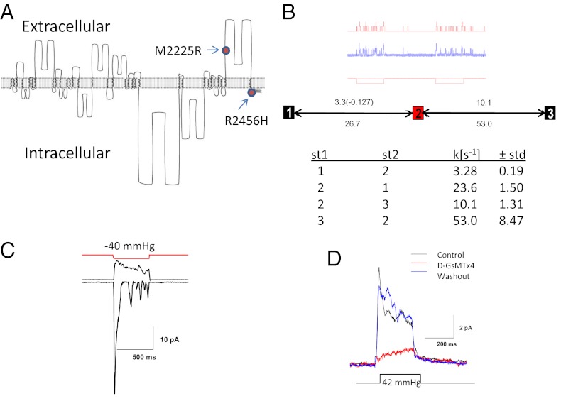
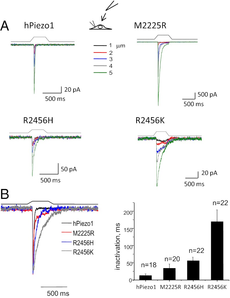
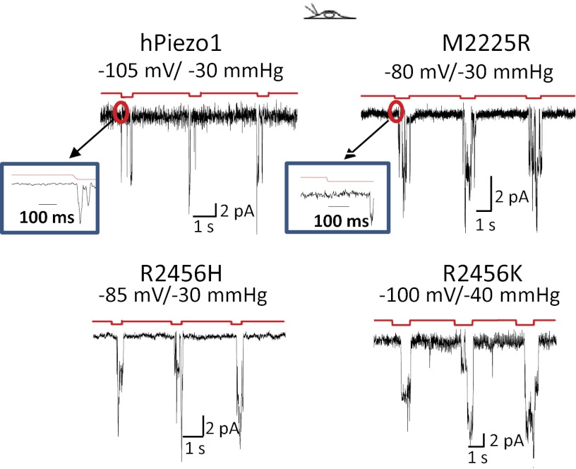
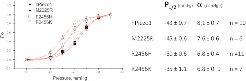
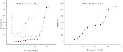
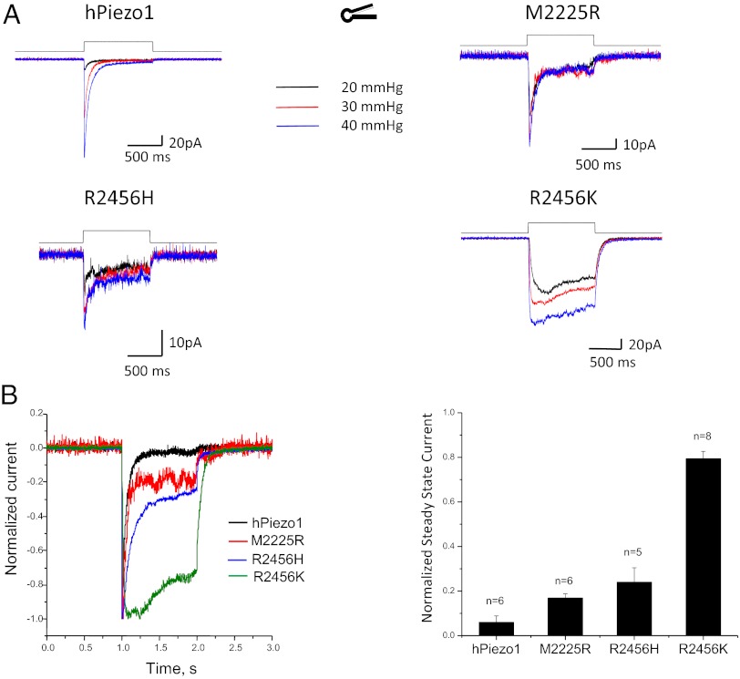
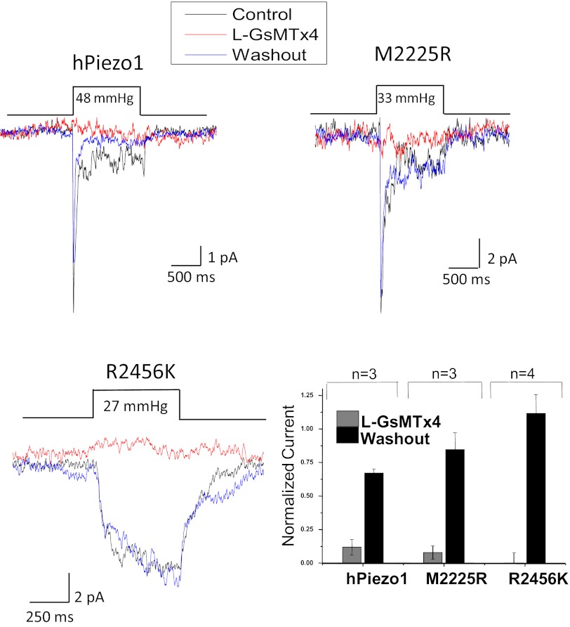
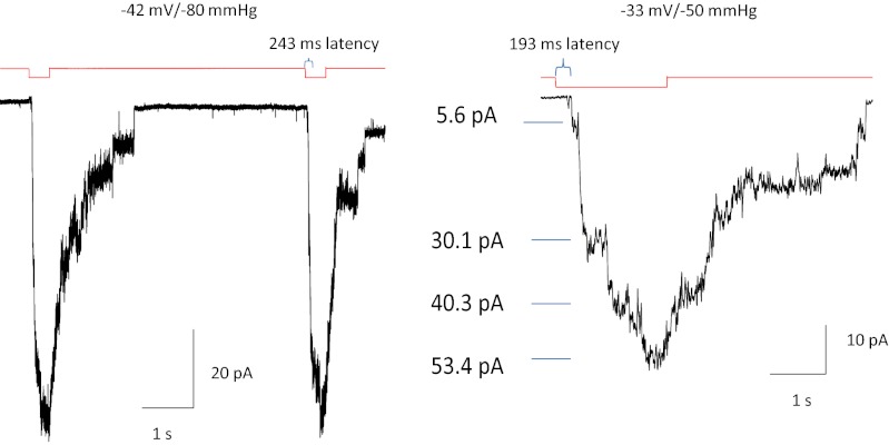
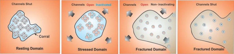
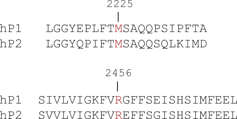
Familial xerocytosis (HX) in humans is an autosomal disease that causes dehydration of red blood cells resulting in hemolytic anemia which has been traced to two individual mutations in the mechanosensitive ion channel, PIEZO1. Each mutation alters channel kinetics in ways that can explain the clinical presentation. Both mutations slowed inactivation and introduced a pronounced latency for activation. A conservative substitution of lysine for arginine (R2456K) eliminated inactivation and also slowed deactivation, indicating that this mutant's loss of charge is not responsible for HX. Fitting the current vs. pressure data to Boltzmann distributions showed that the half-activation pressure, P1/2, for M2225R was similar to that of WT, whereas mutations at position 2456 were left shifted. The absolute stress sensitivity was calibrated by cotransfection and comparison with MscL, a well-characterized mechanosensitive channel from bacteria that is driven by bilayer tension. The slope sensitivity of WT and mutant human PIEZO1 (hPIEZO1) was similar to that of MscL implying that the in-plane area increased markedly, by ∼6-20 nm(2) during opening. In addition to the behavior of individual channels, groups of hPIEZO1 channels could undergo simultaneous changes in kinetics including a loss of inactivation and a long (∼200 ms), silent latency for activation. These observations suggest that hPIEZO1 exists in spatial domains whose global properties can modify channel gating. The mutations that create HX affect cation fluxes in two ways: slow inactivation increases the cation flux, and the latency decreases it. These data provide a direct link between pathology and mechanosensitive channel dysfunction in nonsensory cells.


Conflict of interest statement

The authors declare no conflict of interest.

Figures

Fig. 1.
Fig. 1.
Characterization of cloned hPIEZO1. (A) A diagram showing the putative transmembrane domains of hPiezo1 and intracellular and extracellular regions. The HX mutation sites are shown where M2455R is thought to be extracellular and H2456 is intracellular. (B) Kinetic data and a fitted model for hPIEZO1 with three states (closed-open-inactivated). The theoretical fit to the model shown in red provided the kinetic rate constants shown in the table. The pressure dependence is shown in parenthesis. (C) Voltage-dependent inactivation. At depolarized voltages (+40 mV) there is slow inactivation, and at hyperpolarized voltages (−110 mV) there is rapid inactivation. (D) hPIEZO1 is inhibited by the D enantiomer of GsMTx4 (2 μM, +50 mV holding potential).
Fig. 2.
Fig. 2.
The effect of HX mutations on whole-cell currents. The mutations were introduced at positions 2225 or 2456 (see sequence in Fig. S1). HEK293 cells were transfected with ∼1 μg of DNA and measured 24–48 h later. (A) Whole-cell currents as a function of depth of the indenting probe. The stimulus waveform is shown above the current trace. (B) (Left) Average of repeated current traces showing the slowing of inactivation for both M2225R and R2456H. These traces have been normalized for kinetic comparison. The conservative mutation that replaced arginine with lysine at position 2456 (R2456K) was intended to measure the effect of residue charge. Despite maintaining charge, the mutation completely removed inactivation, suggesting that this site may be part of a hinge domain. (Right) The bar graph shows the mean response ± SD.
Fig. 3.
Fig. 3.
hPIEZO1 mutations slow inactivation in cell-attached patches at single-channel resolution. M2225H and R2456H slow inactivation, and R24565 removes inactivation. (Insets) All three mutants introduced a profound latency for activation not seen in the WT.
Fig. 4.
Fig. 4.
Comparison of mechanical sensitivities via peak current vs. suction and fit to a Boltzmann distribution. The mean values ± SD for P1/2 and α are shown in the table. The α is approximately the same for the four channels, indicating they have a similar dimensional change between closed and open orientations. Mutations at position 2456 shifted P1/2 but the mutation at 2225 did not.
Fig. 5.
Fig. 5.
Comparison of the bacterial channel MscL with Piezo1 or TREK using cotransfection. (Left) Current vs. suction for hPIEZO1 and MscL in the same patch. (Inset) An expanded view of the hPIEZO1 response curve. (Right) The same measurement but using TREK-1. In contrast to hPIEZO1, TREK-1 had a lower ratio of α channel/MscL indicating either that TREK-1 has smaller dimensional changes or that the local tension around TREK-1 is smaller, as might occur if TREK-1 were in a different domain from PIEZO1.
Fig. 6.
Fig. 6.
Response of O/O patches. (A) The difference between WT and mutants. M2225R and R2456H have partially restored inactivation; R2456H remains slow; R2456K does not inactivate at all. (B) The traces normalized for kinetic comparison. The ratio of peak to steady-state current (bar graph with SD) illustrates that, even with the partial restoration of inactivation, M2225R and R2456H transfer more permeant charge than WT during an opening because of the longer open time. Deactivation (closure upon release of stress) was fast in patches from WT but was slow in mutants.
Fig. 7.
Fig. 7.
The peptide L-GsMTx4 (10 μM) reversibly inhibits mutant hPIEZO1 channels in O/O patches from cells transfected with the indicated channels. The bar graph shows the average ± SEM from the indicated number of patches. Note that R2456K did not inactivate but was inhibited, implying that the peptide does not act upon the inactivation domain.
Fig. 8.
Fig. 8.
Removal of the C terminus with a stop codon at position 2218 of hPIEZO1 leads to a non-inactivating form of the channel with pronounced latency, loss of inactivation, and extremely slow deactivation. The slow deactivation might represent the rate for mutant monomers to reassociate. A and B show the currents from many channels in cell-attached patches, and B shows the loss of inactivation in an expanded view. The unitary conductance at −33mV was 73 pS (0.5 mM Mg2+ in pipette).
Fig. 9.
Fig. 9.
Cartoon model of how domains might affect channel behavior. We arbitrarily modeled the domain as a cytoskeletal corral. The panel at the far left shows closed WT channels (black) in a domain with a flexible boundary that can transmit external stress without delay. In this domain the channels exist as tetramers. The second panel from the left shows the expansion of the flexible boundary with external tension causing channels to open (red) and to inactivate (blue). The panel third from the left shows what happens when high forces rupture the domain boundary. The channels diffuse outward into the bulk bilayer where the tension is sufficient to activate them, but the tetramers can dissociate. We postulate that inactivation requires channel–channel interactions so the diffusing channels do not inactivate quickly. The right panel shows deactivation in a ruptured domain after tension is removed. In this model, HX mutations would decrease the interchannel-binding energy, allowing easier dissociation. Although the cartoon shows the domain as a corral, the domain may be a lipid phase, caveolae, cytoskeletal structures, or even channel aggregates.
Fig. 10.
Fig. 10.
Sequence comparison between human Piezo1 and Piezo2. The amino acids in Piezo1 involved in xerocytosis also are found in Piezo2. This homology suggests that mutations at these sites in Piezo2 lead to a loss or slowing of inactivation and potential increase in nociception.

Similar articles

Cited by

References

    1. Zarychanski Y, et al. (2012) Mutations in the mechanotransduction protein PIEZO1 are associated with hereditary xerocytosis. Blood 120(9):1908–1915. - PMC - PubMed
    1. Coste B, et al. Piezo1 and Piezo2 are essential components of distinct mechanically activated cation channels. Science. 2010;330(6000):55–60. - PMC - PubMed
    1. Coste B, et al. Piezo proteins are pore-forming subunits of mechanically activated channels. Nature. 2012;483(7388):176–181. - PMC - PubMed
    1. Gottlieb PA, Bae C, Sachs F. Gating the mechanical channel Piezo1: A comparison between whole-cell and patch recording. Channels (Austin) 2012;6(4):282–289. - PMC - PubMed
    1. Bae C, Sachs F, Gottlieb PA. The mechanosensitive ion channel Piezo1 is inhibited by the peptide GsMTx4. Biochemistry. 2011;50(29):6295–6300. - PMC - PubMed

Publication types

MeSH terms

Supplementary concepts

Associated data